Sequences Analysis of a Gene Encoding Extracellular Xylanase
in
Streptomyces costaricanus
45I-3
Sipriyadi
1, Aris Tri Wahyudi
2, Maggy Thenawidjaja Suhartono
3, Anja Meryandini
2*1 Microbiology Study Program, Graduate School of Bogor Agricultural University, Bogor, West Java, Indonesia, 16680
2 Biology Department, Faculty of Mathematics and Natural Sciences, Bogor Agricultural University, Bogor, West Java, Indonesia, 16680
3 Food Science and Technology Department, Bogor Agricultural University, Bogor, West Java, Indonesia, 16680
Abstract
Streptomyces costaricanus 45I-3 is a bacterial strain belongs to actinomycetes group isolated from peat soil. The bacterium is known to produce extracellular xylanase. The aims of this study were to analyze DNA sequence and sub-clone gene involved in the synthesis of extracellular xylanase. Complete DNA sequence predicted to encode xylanase genes was isolated from bacterial genome using Inverse Polymerase Chain Reaction (I-PCR). Total DNA sequence of 1664 bp in size obtained from I-PCR consisted of two open reading frames (ORF) in opposite direction. ORF1 was 1029 bp and ORF2 (partial sequence) was 309 bp. Analysis sequence using BlastX indicated that ORF1 was homologous with xylanase bacterium enrichment culture clone Xyl8B8 (GenBank accession No. AFH35005.1), i.e. 95% in identity and 99% in similarity. In addition, ORF2 was homologous with glyoxalase bacterium enrichment culture clone Xyl8B8 (GenBank accession No. AFH35007.1), i.e. 95% in identity and 98% in similarity. Analysis of amino acid sequence revealed that ORF1 consisted of 2 domains, i.e. glyco-hydrolase 11 (GH11) and Carbohydrate Binding Type 2(CBM2). Active site was found at 130th amino acid on GH11 domain. Visualization of 3-dimension structure showed that 1029 bp fragment is of 19 areas.
Keywords: Streptomyces costaricanus 45I-3, xylanase extracellular, Inverse-PCR
*Corresponding author :
Anja Meryandini.
Biology Department, Faculty of Mathematics and Natural Sciences, Bogor Agricultural University, Bogor, West Java, 16680 Indonesia. Email: [email protected]. Tel: 0251-8622833) Introduction
Endo- -1,4-xylanase is one of the important enzymes that hydrolyze xylan into xylose perfectly (Esteves et al., 2004). This enzyme can be produced by several organisms such as bacteria, algae, fungi, actinomycetes (Beg et al., 2001; Li et al., 2000), yeast, protozoa, gastropods, and arthropods (Collins et al., 2005). Endoxylanase has wide applications in industry. Xylanase is used to improve lignin extraction and to release chromophore in early stages of
pulp bleaching. The use of xylanase in pulp and paper industry has increased due to the discovery of microbes that producing xylanase and their applications in the industry (Beg et al., 2001; Kulkarni et al., 1999). Other applications are including xylan conversion on agriculture and food industry and raw material production for fuels and chemicals (Sunna and Antranikian 1997). In addition, this enzyme is used as bio-bleaching material in paper industry, purifi cation and enhance juice and grape aroma. This enzyme is also used to improve bread quality and animal feeds quality.
endoxylanase activity was dominant at optimum temperature of 50ºC and pH 5 (Meryandini, 2007), making this isolate potential to be applied in industry. However, the production of endoxylanase from S. costaricanus 45I-3 was less efficient and not cost-wise commercially because the growth of which was slower than other bacteria in general (Paul and Chlark, 1996). DNA recombinant technique through gene cloning could be one of the alternatives to maximize the potential of xylanase from S. costaricanus 45I-3 in the industry. Initial steps critical prior to gene cloning were isolation and characterization of genes involved in extracellular xylanase production.
Steps to isolate and characterize genes encoding endoxylanase from S. costaricanus 45I-3 through genomic library approach had been reported by Satria (2008). Recombinant colonies with xylanase genes could be detected with the presence of clear zone on xylan media. However, further verifi cation showed that the DNA inserts failed to be maintained for a long term and might even be lost. Other approach also had been reported by Aziz (2010) through Polymerase Chain Reaction (PCR). The gene fragments of family 11 endoxylanase were successfully amplifi ed, but they only covered 40% of the intact genes. Because of such problems, it is necessary to carry out further study using primer whose extension direction is outward, known as inverse-PCR (i-PCR). Using i-PCR method, gene fragments reported by Aziz (2010) served as template for primer extension to both gene’s upstream and downstream parts simultaneously, making endoxylanase genes from S. costaricanus 45I-3 were acquired entirely. The objective of this study was to analyze DNA sequences that play a role in the synthesis of xylanase enzyme from Streptomyces costaricanus 45I-3.
Materials and Methods
Media and culture condition
Streptomyces costaricanus 45I-3 used in this study was re-cultured in xylan agar
media (0.5% yeast extract; 10.3% sucrose; 0.5% NaCl; 0.5% xylan Birchwood; 2% agar) and Yeast Malt Agar (YMA) media. The cultured was incubated at room temperature for 4 days.
Endoxylanase gene amplification using inverse-PCR
The genomic DNA was cut using restriction enzyme that does not cut target fragment namely HinfI to amplify DNA flanking both upstream and downstream fragments. A total of 10 μl genomic DNA was added to 2 μl 10x RE buffer, 1 μl restriction enzyme and 6 μl sterile distilled water prior to re-suspension and incubation at 37°C overnight. And then the DNA was extracted using phenol-chloroform, precipitated using 100% ethanol and washed using 70% ethanol. Pellets acquired were dried at room temperature and suspended in 5 μl sterile ddH2O. Target fragments were then ligated using ligase enzyme (T4 DNA ligase, ligation kit ver.2 solution I). Ligation process was carried out overnight at 15ºC. Ligated DNA samples were precipitated using 100% ethanol and DNA pellets were dried and re-suspended into 5 μl sterile ddH2O.
Amplification of complete open reading frame (ORF1) encoding endoxylanase
Primers for xylanase gene isolation were designed and synthesized based on alignment between xyn11 with enrichment culture clone Xyl8B8 bacterium from Genbank. Xylanase gene was amplifi ed using forward primer pET-15b (f) NdeI-gggggaCATATGAAGATCCG
CAGCCGAAGA (underlined is NdeI
restriction site) and reverse primer
pET-15b (r) BamHI-gggGGATCCTCAGTTGG
CCGTGCAGCTGAACGT (underlined is BamHI restriction site). DNA was amplifi ed using Gene Amp System 2400, thermocycler (Perkin Elmer). PCR reactions comprised 25 μl 2x Buffer with GC (Takara, Japan), 1 μl of each primer (10 μM), 1 μl LATaq polymerase, 3 μl DNA sample and sterile distilled water up to 50 μl. PCR amplifi cation was performed in 30 cycles and each comprised pre-denaturation (94 °C, 5 minutes), denaturation (94 °C, 2 minutes), annealing (59 °C, 1 minute), elongation (72 °C, 1 minute) and final elongation (72 °C, 10 minutes). Other alternative primers used in this study referred to study by Cristel et al. (2012).
Sub-cloning of endoxylanase gene from S. costaricanus 45I-3
Complete ORF amplified using PCR machine was purifi ed using gel extraction kit (Genaid) following procedures recommended by the producing company. Purifi ed complete ORF was then sub-cloned into plasmid pGEMT-Easy at MCS site. Ligation reaction consisted of 2.5 μl DNA fragments (15 ng/μl), 1 μl pGEMT-Easy vector (50 ng in concentration), 5 μl ligation buffer, 1 μl T4 DNA ligase and 0.5 μl nuclease free water. Ligation process was performed by incubating the sample at 10 °C for 16 hours. The result of recombinant DNA ligation was transformed into competent cell suspension (Escherichia coli DH5 ). Samples were incubated in ice for 45 minutes and fl icked slowly every 15 minutes. Heat shock on the samples was carried out at 42 °C for 45 seconds prior to incubation in ice for 15 minutes. Samples
were re-suspended in 250 μl SOC media (2% tryptone, 0.5% yeast extract, 10 mM NaCl, 2.5 mM KCl, 10 mM MgCl2, 10 mM MgSO4, and 20 mM glucose), incubated for 1 hour at 37 °C, centrifuged at 3000 rpm for 5 minutes, re-suspended in 100 μl LB media and then grown on LA media. LA media contained of 50 mg/mL ampicillin and 40 μg / mL X-Gal. Cultures were incubated at 37 °C for 24 hours (Sambrook and Russell 2001).
Analysis on complete ORF1 sequence
Escherichia coli DH5 carring recombinant plasmid (PGEMT-Easy + ORF1) was selected through white blue screening and then confi rmed using plasmid isolation and double digest techniques using EcoRI restriction enzyme. The recombinant plasmid was then used for sequensing by using the service of PT Genetika Science Indonesia. Nucleotide sequence acquired was edited using Genetyx Win version 4.0 software and aligned using ClustalX and Mega 5.0 (Tamura et al. 2007). Amino acid was analyzed using BlastX program, Open Reading Frame (ORF) was analyzed using ORF Finder program on the website of National Center for Biotechnology Information (NCBI), promoter and Ribosome Binding Site (RBS) were analyzed using program from softberry (www. Softberry. com), domain and active sites of enzyme were analyzed using the Conserved Domain Database (CDD) available on NCBI website. In addition, three-dimensional structure was visualized using Cn3D program (Claviere and Notredame, 2003).
Results and Discussion
sources, and incapable of using L-arabinose, raffinose, and L-ranossa. The growth of which was inhibited by NaCl of above 5% in concentration, 100 g/ml thallium acetate, streptomycin, and phenol (Esnard et al., 1995).
Analysis of endoxylanase gene fragment using Inverse-PCR
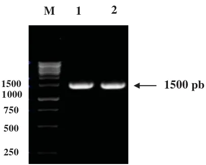
Approach used to isolate endoxylanase gene from S. costaricanus 45I-3 was previously done by amplifi cation using i-PCR is 1500 bp in size (Figure 1).
Figure 1. Amplifi cation using Inverse-PCR on family 11
endoxylanase gene with primer I (F-SC1) and primer II (R-SCI) which extension direction of which is outward. M = marker 1 Kb; 1 and 2 = i-PCR products after being cut by HinfI restriction enzyme.
I-PCR method is principally almost the same with regular PCR method. The fundamental difference between both is outward direction of gene amplifi cation. In this study, i-PCR amplifi cation used three different primer pairs (data not shown), among which only one pair (Sc-Sc-F1 and R1) produced target band of 1500 bp in size. I-PCR product was then sequenced and restriction enzyme’s cutting point was analyzed using alignment (contiq) to determine the length of amplicon generated. Alignment result indicated that HinfI restriction enzyme cut the sequence at nucleotide 694 at the downstream direction of partial gene (Aziz,
2010) and at nucleotide 562 at the upstream direction of partial gene, indicated with overlapping area on the nucleotides. The total amplicon acquired was 1664 bp in size.
Gene structure analysis of inverse PCR product
Analysis result using Basic Local Alignment Tools X (BLASTX) revealed that fragment of 1664 bp in size obtained by incorporating sequence from this study and sequence from Aziz (2010) expressed protein Glyco_hydro and Gloxylase. ORF Finder analysis was performed to study both proteins further. Result of ORF analysis provided information on the absence/ presence of start codon (ATG) and stop codon (TAA/TAG/TGA).
Analysis showed that ORF was found at nucleotide sequence 316 to 1344. The sequence’s start codon (ATG) was at nucleotide 316 and the stop codon (TGA) was at nucleotide 1344, meaning that the ORF was a complete sequence with 342 amino acids. BLASTP result revealed that the ORF was assumed to express Glyco_hydro 11 which belongs to protein family 11glycoside hydrolase (GH11). Other enzyme belonging to the GH11 family was endo-1,4-beta-xylanase (Christel et al., 2012).
In addition, another ORF was also found at nucleotide 1355 to 1663 with stop codon (TAG) at nucleotide 1355 and 102 amino acids but without start codon. BLASTP result showed that this ORF expresses glyoxalase/bleomycin resistance protein (Gly_BRP). Based on ORF Finder analysis, protein Glyco_Hidro 11 and Gly_BRP were not of the same operon, indicated with their stop codons’ opposite position. Because there were two ORFs with different operons, it is necessary to observe the RBS or Shine Delgarno and promoter of each operon (Tang, 1991; Baldini et al. ,1999). Promoter analysis result indicated that the two genes were of opposite transcription direction.
1500 pb
1500
1000
750
500
250
Amplification and sub-cloning of ORF encoding endoxylanase
The result of gene structure analysis on inverse PCR product indicated that there were two open reading frames (ORF), one of which expressed protein Glyco_hydro 11 from family 11 glycoside hydrolase (GH11), known as endoxylanase. The ORF was a complete gene with start codon and stop codon, meaning that this gene could be expressed to produce functional endoxylanase enzymes. Using the fact, further study and analysis were made focus on this ORF. Gene sequences incorporated in the ORF were amplifi ed using forward primer 15b (f) and reverse primer pET-15b (r) with PCR technique and sub-cloned into pGEMT-Easy plasmid. Amplifi cation using a pair of gene mentioned above produced PCR product of around 1000 bp in size. This result was in line with previous ORF analysis stating that based on inverse PCR, ORF sequence expressing GH11 from S. costaricanus 45I-3 is 1029 bp in size.
Results from several studies showed that endoxylanase ORF genes among bacteria are of different size one to another. Endoxylanase gene from bacterium enrichment culture clone Xyl8B8 is of 1025 bp nucleotide in size (Christel et al., 2012), from Streptomyces sp.9 is of 1023 bp (Li et al., 2008), while from Streptomyces sp. SWU10 is of 1011 bp (Deesukon et al. 2011), from Streptomyces lividans is of 1700 bp (Jun et al., 2013) and while from Streptomyces olivaceoviridis A1 is 576 bp (Yaru, 2007).
Amino acid analysis using BlastX
Program Basic Local Alignment Search Tools X (BLASTX) was used to determine the
homology of amino acid sequence from this study with amino acid sequence from NCBI (Table 1).
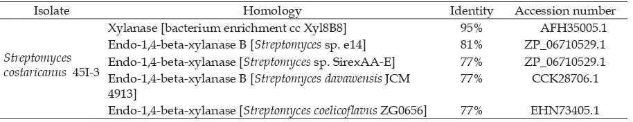
Highest similarity between amino acid sequence for endoxylanase was found between S. costaricanus 45I-3 and that of bacterium enrichment culture clone Xyl8B8 NCBI, i.e. 95% similarity; isolate cc Xyl8B8 is a Gram-positive bacterium isolated from the gut of termites (Reticulitermes santonensis) (Christel et al., 2012). Other homology found was endo-1,4-beta-xylanase B from Streptomyces sp. E14 with 81% similarity and endo-1,4-beta-xylanase from three isolates, namely Streptomyces davawensis JCM 4913, Streptomyces coelicofl avus ZG0656, and Streptomyces sp. SirexAA-E with 77% similarity. Higher similarity indicated the more accurate endoxylanase gene sequences from S. costaricanus 45I-3. Two DNA fragments could be determined homologous where 70% base sequence or 25% amino acid sequence of which was identical (sequence of at least 100 base pairs in size) (Claviere and Notredame, 2003).
Expectation value (E-value) described statistically calculated probability value of sequence similarity between endoxylanase genes from S. costaricanus 45I-3 with its affi liations in the GenBank (www.ncbi.nlm. nih.gov). Table 4 showed that xyalanase gene from bacterium enrichment culture clone Xyl8B8 had the lowest E-value (4e-175), followed with endo-1,4-beta-xylanase B from Streptomyces sp. E14 (2e-145) and endo-1,4-beta-xylanase from Streptomyces davawensis JCM 4913, Streptomyces coelicofl avus ZG0656, and Streptomyces sp. SirexAA-E (2e-134, 4e-136, and 2e-141, respectively). BLASTP
Table 1. Analysis of gene homology of Endoxylanase S. costaricanus 45I-3 using BLASTX
Isolate Homology Identity Accession number
Streptomyces costaricanus 45I-3
Xylanase [bacterium enrichment cc Xyl8B8] 95% AFH35005.1 Endo-1,4-beta-xylanase B [Streptomyces sp. e14] 81% ZP_06710529.1 Endo-1,4-beta-xylanase [Streptomyces sp. SirexAA-E] 77% ZP_06710529.1 Endo-1,4-beta-xylanase B [Streptomyces davawensis JCM
4913]
77% CCK28706.1
analysis for gene encoding amino acid of endoxylanase from S. costaricanus 45I-3 generated signifi cant; E-value from BLAST analysis is considered signifi cant if the value of which is 1x10⁻¹⁰ or lower (Altschul et al., 1990).
Analysis of domain, active site, and 3-dimensional structure
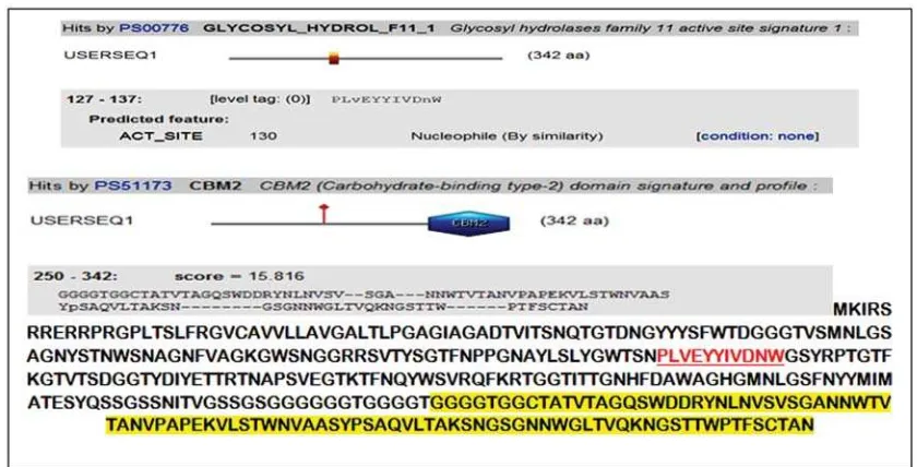
Result of analysis on endoxylanase gene domain from S. costaricanus 45I-3 using Conserved Domains Database (CDD) program available at NCBI website revealed that the gene fragments were consisted of two domains, i.e. family 11 glycosyl hydrolase (GH11) and, at the end of the fragment, carbohydrate binding domain type 2 (CBM2) which is xylan substrate binding site. Analysis using Scan Prosite revealed that the active site of the enzyme is at 130th amino acid (Figure 3).
Successfully amplified glycosyl hydrolase gene fragments are consisted of two domains, i.e. family 11 glycosyl
hydrolase (GH11) and, at the end of the fragment, carbohydrate binding domain type 2 (CBM2) as xylan and cellulose binding site, depending on the type of CBM2. CBM2 itself is consisted of CBM2a and CBM2b. In this study, the one found was CBM2a that specifi cally binds cellulose substrate. Like any other CBM domain, CBM2 domain contains -sheet structure that interacts with ligand through aromatic residue hydrophobic strip. Hydrophobic surface of CBM2a family is consisted of three tryptophan residues which are needed to bind cellulose, while CBM2b has only 2 tryptophan residues with different orientation one another CBM2a forms a wide -sheet structure with -barrel fold. Titration using cellohexaose [beta-D-glucopyranosyl-(1.4)] 5-D-glucose suggested that Trp 54 and 72 play a role in binding cellulose (Xu et al. 1995). CBM increases enzyme concentration on substrate surface (Shoseyov et al. 2006). CBM location in conserved domains can be found at the N-terminal or C-terminal linked to catalytic domain (CD) with various
S treptom y c es c os taric anus 45I-3
x y lanas e bac terium enric hm ent c ulture c lone Xy l8B 8
endo-14-beta-x y lanas e B S treptom y c es s p. e14
endo-14-beta-x y lanas e S treptom y c es s p. S irex A A -E
endo-14-beta-x y lanas e B S treptom y c es gris eoaurantiac us M 045
x y lanas e II S treptom y c es therm oviolac eus
x y lanas e B prec urs or S treptom y c es therm oc y aneoviolac eus
endo-14-beta-D-x y lanas e prec urs or S treptom y c es s p. THW 31
4-x y lanas e S treptom y c es c oelic oflavus ZG 0656
endo-beta-14-x y lanas e S treptom y c es s p. S W U10
E ndo-14-beta-x y lanas e
endo-14-beta-x y lanas e B S treptom y c es c oelic olor A 3(2)
endo-14-beta-x y lanas e B S treptom y c es lividans TK 24 100
100
80
49 33
100
65
85
59 100
0.02
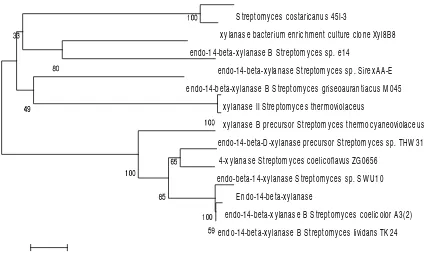
Figure 2. Phylogenetic tree based on comparison of amino acid sequences from several strains of Streptomyces
size linker sequences. Generally it is rich in serine or threonine amino acids (Gilbert and Hazlewood, 1993). Family 11 endoxylanase belongs to glycoside hydrolase family with glutamate (Glu) as the nucleophile/base or proton donor. The topology of this enzyme is overlapping -folds (Christel et al., 2012).
Based on an analysis using Scan Prosite, the active site of this enzyme was at amino acid 130 (Figure 3). In enzyme structure, one or more substrate binding sites (active site)
can be found. Xylan molecule (substrate) will attach onto active site to form enzyme-substrate complex and produce bond formation or destruction by releasing one or several products. The reaction process will be faster to inhibit lower enzyme activation by decreasing activation energy. Without enzyme activity, cell reaction will be too slow to sustain call’s life. According to the hypothesis of ‘lock and key’ shape, an active site only fits to one substrate shape. This
Figure 3. Domain analysis using Scan Prosite program. Red letters indicate the position of the active site domain of family 11 Glyco_Hydrolase (GH11). Yellow letters indicate amino acids that make up CBDII domain. The active site is at amino acid 130.
(a) (b)
prevents random reactions as well as gives control on non-specifi c substrates.
T h r e e - d i m e n s i o n a l s t r u c t u r e visualization of endoxylanase 11 fragment using Cn3D showed that this fragment shape was like a closed right hand, which is a common characteristic of family 11endoxylanase. Törrönen and Rouvinen, (1997) stated that to be an active enzyme, endoxylanase family 11 should have 19 areas consisted of N, B1, B2, A2, A3, B3, A5, B5, B6, Cord, B9, B8, Thumb, B7, A6, Helix, B4, A4 and C terminal. Based on the results of Cn3D visualization in Figure 4 above, all the 19 areas were successfully amplifi ed so that this enzyme is assumed can be expressed.
Three-dimensional structure of family 11 endoxylanase has been determined in several enzymes from bacteria and fungi. The fold of catalytic domain is divided into two sheets (A and B) that are antiparallel strands with one short alpha helix that resembles closed right hand. Loop between strand B7 and B8 forms a ‘thumb’ while loop connecting strand B6 and B9 forms ‘cord’. Two glutamic acid residues that were found respectively in strand B4 and B6 are important catalysts in the activation of extracellular xylanase enzyme (Törrönen and Rouvinen, 1997).
Analysis of molecular weight and isoelectric point
Molecular weight (MW) is relative molecular mass of xylanase GH11, while isoelectric point (pI) is the degree of acidity or pH when a macromolecule is zero in charge due to the increasing proton or loss charge because of acid-base reaction. Molecular weight and isoelectric point were calculated by inputting the data of amino acids that make up GH11 domain to website http:// web.expasy.org/compute_pi/.
Based on the analysis on molecular weight and isoelectric point, was known that family 11 endoxylanase enzyme (GH11) from S. costaricanus 45I-3 had molecular weight of 35.67 kDa with isolectric point of 9.26 A. These results were in line with that of Wang et al.
(1997) who stated that endoxylanase enzyme belongs to glycoside hydrolase family 10 and 11 (also called family F and G) based on its pshyco-chemical components including molecular weight and isoelectric point. Family 10 was an endoxylanase group with relatively high molecular mass (>40 kDa) and low pH (acidic); while endoxylanase family 11 is of relatively low molecular mass (<40 kDa) and high pH (Collins et al., 2005).
Molecular weight and pH of family 11 xylanase enzyme from S. costaricanus 45I-3 are almost identical to that of from bacterium enrichment culture clone Xyl8B8 reported by Cristel et al. (2012) with molecular weight of 35.4 kDa and pI of 9.2 A. Result of molecular weight prediction using website http://web.expasy.org/compute_pi/ showed that the MW of the enzyme was not that different with molecular weight of isolate Streptomyces sp. 45I-3 reported by Meryandini et al. (2007) who stated that the purification of the xylanase enzyme from Streptomyces sp. 45I-3 using anionic Eudragit S100 polymer resulted in 2 bands of protein enzyme that are close one another with molecular weight of 39.2 kDa and 43.2 kDa, respectively.
Conclusion
References
Altschul, S.F. Gish, W., Myers, E.W., Lipman, D.J. 1990. Basic local alignment search tool. J. Mol. Biol., 215, 403-410.
Aziz S. 2010. Isolasi fragmen gen penyandi endoxylanase Streptomyces costaricanus 45I-3 melalui pendekatan PCR [tesis]. Bogor: Program Pascasarjana Institut Pertanian Bogor.
Baldini, R. L., Tahara, S. T., Rosato, Y. B. 1999. A rolling circle miniplasmid on Xhanthomonas campestris pv campestris. the nucleotida sequence and its use as a cloning vector. Plasmid., 42, 126-133. Beg, Q. K., Kapoor, M., Mahajan, L., Hoondal,
G.S. 2001. Microbial xylanase and their industrial application. Appl. Microbiol. Biotechnol., 56, 326-338.
Christel M, Julien B, Catherine B, Cédric T, Philippe T, Jacqueline D. 2012. Identifi cation and characterization of a new xylanase from Gram-positive bacteria isolated from termite gut (Reticulitermes santonensis). Prot. Expres. Purif., 83, 117–127.
Collins, T., Gardy, C., Feller, G. 2005. Xylanase, xylanase families and extremophilic xylanase. FEMS. Microbiol. Rev., 29, 3-23.
Claviere J. M. and Notredame J. 2003. Bioinformatics for Dummies. Indianapolis: Willey Publishing Inc.
Deesukon, W., Yuichi N., Naoki H., Tatsuji, S., and Wasana, S. 2011. Purification, charac-terization and gene cloning of two forms of a thermostable endoxylanase from Streptomyces sp. SWU10. Proc. Biochem.,46, 2255–2262.
Esnard, J., Potter, T. L., Zuckerman, B.M. 1995. Streptomyces costaricanus sp. nov isolated from nematode-suppressive soil. Int. J. Sys. Bacteriol., 45, 775-779.
Esteves, F. D., Reulle, V., Lamotte-Braseur, J. 2004. Acidophilic adaption of family 11 endo-β-1,4-xilanase: Modeling and mutational analysis. Prot. Sci., 13, 1209-1218.
Gilbert, H. J., and Hazlewood, G.P. 1993. Bacterial cellulase and xylanase. J. Gen.. Microbiol., 139, 187-194.
Jun, X. L., Long, M. Z., Ru, J. W., Zhao, J. Z and Ri, J. Z. 2013. High-level overproduction of thermobifi da enzyme in Streptomyces lividans using a novel expression vector. Int. J. Mol. Sci., 14, 18629-18639.
Kulkarni, N., Shendye, A,, Rao, M. 1999. Molecular and biotechnological aspects of xilanases. FEMS. Microbiol. Rev., 23, 411-418.
Li, N., Kun. M., Yaru, W., Pengjun, S., Huiying, L., Yingguo, B. 2008. Cloning, expression, and characterization of a new xylanase with broad temperature adaptability from Streptomyces sp. S9. Appl. Microbiol. Biotechnol., 80, 231–240.
Li, Y. K., You, H. J., Pan, H. 2000. Mechanistic study of -xylosidase from Trichoderma koningii G-39. J. Biochem., 127, 315-320. Meryandini, A. 2007. Characterization of
Streptomyces sp. 45I-3 Xylanase. Biotropia., 14, 32-42.
Paul, E. A, Chlark, F.E. 1996. Soil Microbiology and Biochemestry. 2nd ed. California: Academic Press.
Sambrook, J., Russell, D. W. 2001. Molecular Cloning a Laboratory Manual. 3rd ed. New York. Cold Spring Harbor Laboratory Press.
Satria, H. 2008. Kloning gen xilanase termozim dari Streptomyces costaricanus 45I-3 [tesis]. Bogor: Program Pascasarjana Institut Pertanian Bogor.
Shoseyov, O. Ziv, Z., Ilan L. 2006. Carbohydrate binding modules: biochemical properties and novel applications. Microbiol. And Mol. Biol. Reviews., 112, 283–295.
Sunna, A., and Antranikian, G. 1997. Characterization of the xilanase from the new isolated thermophilic xylan-degrading Bacillus thermoleovarans galur K-3d and Bacillus fl avothermus galur LB3A. FEMS. Microbial. Lett., 148, 209-216. Tamura, K, Dudley, J., Nei, M., Kumar,
version 5.0. Mol Biol and Evolution., 153, 311-320.
Tang, J. 1991. Genetic and analysis of a cluster of rpf genes involved in positive regulation of synthesis of extracellular enzymes and polysaccharides in Xanthomonas campestris pv campestris. Mol. Gen. Geneat., 199, 338-343.
Törrönen, R., Rouvinen, T. 1997. Structural and functional properties of low molecular weight endo-1,4-xylanases. J. Biotechnol., 57, 137–149.
Wahyudi, A. T., Takeyama, H., Matsunaga, T. 2001. Isolasi of Magnetospirillum magneticum AMB-1 mutans detective in bacterial magnetic particle synthesis by transoson mutagenesis. Appl. Biochem. Biotechnol., 91, 147-154.
Wang, D. I. C., Cooney, C. L., Denail, A. L., Dunnail, P., Lilly, M. D. 1997. Fermentation and Enzyme Technology. New York: John Wiley and Sons.
Xu, G.Y., Ong, E., Gilkes, N. R., Kilburn, D. G., Muhandiram, D. R., Harris-Brandts, M., Carver, J. P, Kay, L. E., Harvey, T.S.,1995. Solution structure of a cellulose-binding domain from Cellulomonas fi mi by nuclear magnetic resonance spectroscopy. Biochemistry., 21, 6993-7009.