Basic immunology
The immune system is the body’s natural defence in combating organisms.
Immunology has developed rapidly over the past decade owing to the refinements made in the molecular tests employed in this area of research. Therefore, the keen reader is encouraged to peruse the ophthalmic and immunological literature in order to keep abreast of the latest developments in this field.
The College of Optometrists has awarded this article 2 CET credits. There are 12 MCQs with a pass
mark of 60%.
Owing to the complex nature of this subject, it is far beyond the scope of this article to cover all aspects of immunology. Rather, the aims of the article are twofold: first to acquaint the busy practitioner with the basic concepts of the immune system; and second, to introduce the reader to the more specific topic of ocular immunology –the study of the ocular immune
system.
Finally, since it is envisaged that optometrists will one day prescribe therapeutic agents, the discussion is limited to the anterior segment and anterior uvea.
Innate & adaptive immune systems The immune system can be thought of as having two “lines of defence”: the first, representing a non-specific (no memory) response to aannttiiggeenn (substance to which the body regards as foreign or potentially harmful) known as the iinnnnaattee iimmmmuunnee ssyysstteemm; and the second, tthhee aaddaappttiivvee iimmmmuunnee ssyysstteemm, which displays a high degree of memory and specificity. The innate system represents the first line of defence to an intruding pathogen. The response evolved is therefore rapid, and is unable to “memorise” the same said pathogen should the body be exposed to it in the future. Although the cells and molecules of the adaptive system possess slower temporal dynamics, they possess a high degree of specificity and evoke a more potent response on secondary exposure to the pathogen.
The adaptive immune system frequently incorporates cells and molecules of the innate system in its fight against harmful pathogens. For example, ccoommpplleemmeenntt (molecules of the innate system - see later) may be activated by aannttiibbooddiieess (molecules of the adaptive system) thus providing a useful addition to the adaptive system’s armamentaria.
A comparison of the two systems can be seen in TTaabbllee 11..
o
t
www.optometry.co.uk
Gregory Heath BSc (Hons), MCOptom, Dip. Clin. Optom
ABDO has awarded this article 2 CET credits (GD).
February 8, 2002 OT
26
Sponsored by
a
Cells of the innate
immune system
Phagocytes
Although sub-divided into two main types, namely neutrophils and macrophages, they both share the same function - to engulf microbes (phago - I eat, Latin).
Neutrophils
Microscopically, these cells possess a characteristic, salient feature - a multilobular nucleus ((FFiigguurree 22)). As such, these cells have been referred to as polymorphonuclear leukocytes (PMNs) and have a pivotal role to play in the development of acute inflammation. In addition to being phagocytic, neutrophils contain granules and can also be classed as one of the granulocytes. The granules contain acidic and alkaline phosphatases, defensins and peroxidase - all of which
represent the requisite molecules required for successful elimination of the unwanted microbe(s).
Macrophages
Macrophages (termed monocytes when in the blood stream) have a horseshoe-shaped nucleus and are large cells. Properties of macrophages include phagocytosis and antigen presentation to T cells (see later). Unlike neutrophils (which are short-lived cells), they are seen in chronic inflammation as they are long-lived cells.
Mononuclear phagocytic system The cells comprising the monocyte phagocytic system are tissue bound and, as a result, are further sub-divided depending on their location. A list of the cells together with their
corresponding location can be found in TTaabbllee 22. Table 1: Cells and molecules of the innate and adaptive immune systems
Immunity Cells Molecules
Innate Natural killer (NK) cells Cytokines
Mast cells Complement
Dendritic cells Acute phase proteins Phagocytes
Adaptive T and B cells Cytokines
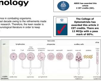
Antibodies Components of the immune system can be seen in Figure 1.
Figure 1 The principle components of the immune system are listed, indicating which cells produce which soluble mediators. Complement is made primarily by the liver, with some synthesised by mononuclear phagocytes. Note that each cell only produces a particular set of cytokines, mediators etc
Table 2:Examples of cells of the mononuclear phagocytic system and their respective locations
Cells Location
Monocytes Blood stream
Alveolar macrophages Lungs Sinus macrophages Lymph nodes
and spleen
Kupffer cells Liver
Dendritic cells
Dendritic cells consist of Langerhans’ and interdigitating cells and form an important bridge between innate and adaptive immunity, as the cells present the antigenic peptide to the T helper cell (adaptive immunity). Such cells are therefore known as professional antigen presenting cells ((AAPPCCss)). TTaabbllee 33 illustrates the various types of dendritic cells together with an example of their location.
Eosinophils
Eosinophils (so called because their granules stain with eosin – FFiigguurree 44) are granulocytes that possess phagocytic properties. Despite the fact that they represent only 2-5 % of the total leukocyte population, they are instrumental in the fight against parasites that are too big to be phagocytosed.
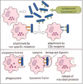
Phagocytosis - the process
Phagocytosis is the process by which cells engulf microorganisms and particles ((FFiigguurree 33)). Firstly, the phagocyte must move towards the microbe under the influence of chemotactic signals, e.g. complement (see later). For the process to continue, the phagocyte must attach to the microbe either by recognition of the microbial sugar residues (e.g. mannose) on its surface or complement/antibody, which is bound to the pathogen. Following attachment, the phagocyte’s cell surface invaginates and the microbe becomes internalised into a
phagosome. The resultant pphhaaggoossoommee fuses with multiple vesicles containing O2 free radicals and other toxic proteins known as lysosomes to form a phagolysosome. The microbe is subsequently destroyed.
Opsonisation (“to make tasty” - Greek)
O
Oppssoonniinnss are molecules, which enhance the efficiency of the phagocytic process by coating the microbe and effectively marking them for their destruction. Important opsonins are the complement component C3b and antibodies.
Natural killer (NK) cells N
NKK cells are also known as “large granular lymphocytes” (LGLs) and are mainly found in the circulation. They comprise between 5-11% of the total lymphocyte fraction. In addition to possessing receptors for immunoglobulin type G (IgG), they contain two unique cell surface receptors known as killer activation receptor and killer inhibition receptor. Activation of the former initiates cytokine (“communication”) molecules from the cell whilst activation of the latter inhibits the aforesaid action.
NK cells serve an important role in attacking virally-infected cells in addition to certain tumour cells. Destruction of infected cells is achieved through the release of perforins and granyzymes from its granules, which induce apoptosis (programmed cell death). NK cells are also able to secrete interferon-γ(IFN-γ). This interferon serves two purposes: first, to prevent healthy host cells from becoming infected by a virus; and second, to augment the T cell response to other virally infected cells (see later).
Mast cells and basophils
Morphologically, mast cells and basophils are very similar in that both contain electron dense granules in the cytoplasm. Basophils are so-called owing to the fact that their granules
stain with a basic dye. Unlike mast cells, which are present in close proximity to blood vessels in connective tissue, basophils reside in the circulation.
Both cell types are instrumental in initiating the acute inflammatory response. Degranulation is achieved either by binding to components of the complement system or by cross-linking of the IgE antibody which results in the release of pro-inflammatory mediators including histamine and various cytokines. The former induces vasodilation and augments vascular permeability whilst the latter are important in attracting both neutrophils and eosinophils.
Table 3:Dendric cells and location
Cells Location
Langerhans cell Limbus, skin Interdigitating cell T cell areas in
lymph nodes
Figure 4 Morphology of the eosinophil. The multilobed nucleus is stained blue and the cytoplasmic granules are stained red. Leishman stain, x 1800
Figure 3
o
t
February 8, 2002 OT www.optometry.co.uk
for the successful eradication of an invading virus by the innate immune system.
Type II IFN, IFN-γ, is produced by T Helper cells and NK cells and is able to augment both the antigen presenting properties together with the phagocytic properties of the APCs (e.g. macrophages and dentritic cells).
Adaptive immunity
As mentioned previously, there is a great deal of synergy between the adaptive immune system and its innate counterpart. The adaptive immune system comprises two main types of leukocyte known as B and T lymphocytes. Before describing these important cell types, it is necessary to acquaint the reader with both the primary and secondary lymphoid organs and tissues in the body. These are summarised in TTaabbllee 44. The bone marrow represents the dominant site for haemopoiesis (production of blood cells and platelets). Although most of the haemopoietic cells maturate in this region, T lymphocytes do so in the thymus. In the thymus, premature T cells undergo a process of positive and negative selection whereby the former are allowed to progress to maturity whilst the latter are marked for termination via apoptosis (see central tolerance).
Lymphocytes
Morphologically, there are three types of lymphocytes: T, B and NK cells. However, only T and B lymphocytes exhibit memory and specificity and, as such, are responsible for the unique quality of the adaptive immune system. Resting B lymphocytes are able to react with free antigen directly when it binds to their cell surface immunoglobins which act as receptors. T lymphocytes do not react with free antigen and instead make use of APCs to phagocytose the antigen and then to express its component
28
Molecules of the innate
immune system
There are many molecules, which work in concert with the cells of the innate immune system and which also foster close functional links with their adaptive counterpart. The three major molecules are:
• Complement
• Acute phase proteins (APP) • Interferons (IFNs)
Complement
The complement system represents a large group of independent proteins (denoted by the letter C and followed by a number), secreted by both hepatocytes (liver cells) and monocytes. Although these proteins maybe activated by both the adaptive immune system (ccllaassssiiccaall ppaatthhwwaayy) or innate immune system (aalltteerrnnaattiivvee ppaatthhwwaayy), the nomenclature is derived from the fact that the proteins help (“complement”) the antibody response.
Activation of complement via the microbe itself is known as the alternative pathway. The classical pathway requires the interaction of antibody with specific antigen. The C3 component is the pivotal serum protein of the complement system. Binding of the antigen to C3 results in two possible sequelae. In either case, C3 component becomes enzymatically converted to C3b. The bacterial cell wall can either remain bound to C3b and become opsonised (since phagocytes have receptors for C3b) or act as a focus for other complement proteins (namely C5, 6, 7, 8 and 9). The latter form the membrane attack complex (MAC), which induces cellular lysis.
The functions of the complement system may be summarised as follows:
• Opsonisation
• Lysis (destruction of cells through damage/ rupture of plasma membrane)
• Chemotaxis (directed migration of immune cells)
• Initiation of active inflammation via direct activation of mast cells
It is important that complement is regulated to protect host cells from damage and/or their total destruction. This is achieved by a series of regulatory proteins, which are expressed on the host cells themselves.
Acute phase proteins
These serum proteins are synthesised by hepatocytes and are produced in high numbers in response to cytokines released from macrophages.
Interferons (IFNs)
IFNs are a group of molecules, which limit the spread of viral infections ((FFiigguurree 55)). There are two categories of IFNs, namely type I and type II. Type I IFNs maybe sub-divided further into IFN-α and β. IFN-γis the sole type II interferon. Type I IFNs are induced by viruses, pro-inflammatory cytokines and endotoxins from gram negative bacterial cell walls. Their presence remains vital
proteins on the cell surface adjacent to special host proteins called major histocompatibility complex (MHC) class II molecules. As discussed, antigen presenting cells which express MHC class II molecules include dendritic cells and macrophages. This “afferent” phase must occur in order for the T cell to recognise the antigen. The “efferent” phase occurs when activated lymphocytes enter the tissue and meet antigen again. This results in multiplication and secretion of cytokines or immunoglobins in order to destroy the antigen.
T cells
T cells can be broadly divided into both T helper (TH) and cytotoxic T cells (Tc). Furthermore, TH cells may be sub-divided into TH1and TH2. The former are pro-inflammatory T cells and stimulate macrophages whilst the latter orchestrate B cell differentiation and maturation and hence are involved in the production of humoral immunity (antibody mediated). T cells express cell surface proteins, described by cluster determination (CD) numbers. THcells express CD4 molecules on their cell surface, which enable the lymphocyte to bind to a MHC class II molecule. The T cell receptor is unique in that it is only able to identify antigen when it is associated with a MHC molecule on the surface of the cell.
Cytotoxic T cells are primarily involved in the destruction of infected cells, notably viruses. Unlike THcells, cytotoxic cells possess CD8 cell surface markers, which bind to antigenic peptides expressed on MHC class I molecules.
B cells and antibodies (immunoglobulins - Ig)
B cells are lymphocytes that produce antibodies (immunoglobulins) and can recognise free antigen directly. They are produced in the bone marrow and migrate to secondary lymphoid organs. B cells are responsible for the
Figure 5
When host cells become infected by virus, they may produce interferon. Different cell types produce interferon-α (IFN-α ) or interferon-β (IFN-β); interferon-γ (IFN-γ) is produced by some types of lymphocyte (TH) after activation by antigen. Interferons act on other host cells to induce a state of resistance to viral infection. IFN-γ has many other effects as well
Table 4:
Primary and secondary lymphoid organs
Primary lymphoid organs Secondary lymphoid organs
Bone marrow Lymph nodes
Module 4 Part 2
Sponsored by
a
development of antibody mediated immunity known as hhuummoorraall mmeeddiiaatteedd iimmmmuunniittyy.
When activated by foreign antigen, B cells undergo proliferation and mature into antibody secreting ppllaassmmaa cceellllss. The latter are rich in organelles such as rough endoplasmic reticulum and mitochondria, which confer their ability to secrete soluble proteins (antibodies). Not all proliferating B cells develop into plasma cells. Indeed, a significant proportion remain as memory B cells through a process known as clonal selection. This process is vital in eliminating the antigen should the body become re-exposed to it in the future. T cells are also clonally selected and this confers to the production of T memory cells.
Although T and B cells behave differently, both are able to recirculate around the body migrating from blood to tissue and vice versa. The ability to recirculate obviously increases the efficiency with which cells of the immune system can home onto the invading antigen.
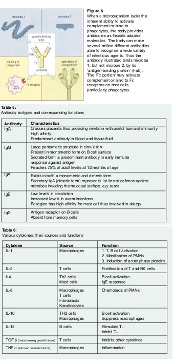
Antibodies
Antibodies have two roles to play - the first is to bind antigen and the second is to interact with host tissues and effector systems in order to ensure removal of the antigen
((FFiigguurree 66)).
There are five different types (known as iissoottyyppeess) of antibody in the human immune system - namely IgM, IgG, IgA, IgE and IgD. In addition, there are four sub classes of IgG (IgG1-4). The basic antibody unit consists of a glycosylated protein consisting of two heavy and two light, polypeptide chains. The region which binds to the antigen is known as the FFaabb region, while the constant region, FFcc, not only determines the isotype but is the region responsible for evoking effector systems, e.g. mast cell activation. The term immune complex refers to the combination of antigen and antibody and will be discussed later in the article (see type III hypersensitivity).
The antibody isotypes together with their corresponding function are illustrated in TTaabbllee 55..
MHC
Major histocompatability complex (MHC) are cell surface proteins classified as class I (also termed human leucocytic antigen [HLA] A, B and C), found on all nucleated cells and class II (termed HLA, DP, DQ and DR), found on all antigen presenting cells (APCs). MHC molecules are the
sine qua nonof T cell induced immunity. Clinically, there is a strong association between HLA and certain systemic and ocular diseases (see later).
Cytokines
Cytokines (also termed interleukins [IL] meaning “between white blood cells”) are small molecules that act as a signal between cells and have a variety of roles including chemotaxis, cellular growth and cytotoxicity. Owing to their ability to control immune activity, they have been described as the “hormones” of the immune system.
Characteristics
Crosses placenta thus providing newborn with useful humoral immunity High affinity
Predominant antibody in blood and tissue fluid Large pentameric structure in circulation Present in monometric form on B cell surface
Secreted form is predominant antibody in early immune response against antigen
Reaches 75% of adult levels at 12 months of age Exists in both a monometric and dimeric form
Secretory IgA (dimeric form) represents 1st line of defence against microbes invading the mucosal surface, e.g. tears
Low levels in circulation
Increased levels in worm infections
Fc region has high affinity for mast cell thus involved in allergy Antigen receptor on B cells
Absent from memory cells
Table 5:
Antibody isotypes and corresponding functions
Antibody
IgG
IgM
IgA
IgE
IgD
Table 6:
Various cytokines, their sources and functions
Cytokine Source Function
IL-1 Macrophages 1. T, B cell activation
2. Mobilisation of PMNs
3. Induction of acute phase proteins
IL-2 T cells Proliferation of T and NK cells
Il-4 Th2 cells B cell activation
Mast cells IgE response
IL-8 Macrophages Chemotaxis of PMNs
T cells Fibroblasts Keratinocytes
IL-10 TH2 cells B cell activation
Macrophages Suppress macrophages
IL-12 B cells Stimulate TH1
Inhibit TH2
TGF β(transforming growth factor) T cells Inhibits other cytokines TNF α(tumour necrosis factor) Macrophages Inflammation
Figure 6
TTaabbllee 66 summarises some of the cytokines pertinent to ocular immunology, their functions and their progenitors. Since interferons have been discussed earlier in the article, they have been omitted from the table.
Central and peripheral
tolerance: nature’s way of
containing the immune
response
Since various cells of the immune system are capable of reacting with self-antigens, it is therefore essential that the human body has mechanisms to suppress/eliminate autoreactive cells. Failure to do so, can, in some cases, lead to the development of autoimmune diseases (see later).
Central tolerance
Central tolerance refers to the process whereby both immature B and T cell lymphocytes, which react against normal, healthy cells
(self-antigens), are eliminated via apoptosis.
Peripheral tolerance
This involves the removal of mature lymphocytes, which are not tolerant to healthy cells.
Ocular immune privilege
There are numerous sites in the body whereby tissue may be grafted with minimal risk of rejection. Such regions include, inter alia, the testis, thyroid lens, anterior chamber, cornea, iris and ciliary body1,2.It is important that immune privilege is not simple interpreted as the host’s inability to initiate an immune response to a transplanted tissue. Rather, it is an area of the body in which there exists a paucity of various elements of the human immune system in response to an antigen.
Factors
The factors purported by investigators that contribute to the phenomenon of ocular immune privilege include:
• Isolation from a vascular supply • Isolation from a lymphatic supply • Presence of a vascular barrier
• Ability to suppress the immune response • Anterior chamber associated immune
deviation (ACAID)
Vascular supply
The healthy cornea is a good example of an ocular site devoid of a vascular network. The evidence to support the role a vascular network has to play in the mechanism of graft rejection is unequivocal since the risk of failure correlates positively with the degree of host
vascularisation3 .
Vascular barrier
There is a plethora of evidence in the
ophthalmic literature to support the existence of a blood-ocular barrier. Furthermore, the same said barrier encompasses different elements including tight junctions between retinal
o
t
February 8, 2002 OT
30
endothelial cells and the presence of junctional complexes linking retinal pigment epithelial cells4
.
Lymphatic role
The fact that skin allografts were not rejected following lymph node removal5
led investigators to hypothesise that immune privilege was solely due to the absence of the same said system at a particular anatomical site. However, although certain immune privileged sites do indeed lack lymphatic drainage, others such as the testes6 and eye7
do possess such a system. It appears that a proportion of the aqueous humour drains via the uveoscleral pathway into the lymphatic vessels in the head and neck.
The eye, APCs & MHCs
As mentioned previously, APCs, through their ability to express MHC class II molecules, are potent progenitors of the immune response. Moreover, such cells are capable of activating T cells within the tissue itself. It is therefore not unreasonable to assume that a paucity of APCs may play an important role in immune privilege. In addition, failure to express MHC class I molecule would make a tissue immune against the lytic action of the cytotoxic T cells.
Although the aforementioned mechanisms are theoretically plausible, cells expressing both MHC class I and II molecules have been detected in the eye. TTaabbllee 77 illustrates the relationship between histocompatability class and ocular cell type.
It is noteworthy that the epithelial cells of the crystalline lens are devoid of class I expression14
and that the Langerhans’ cells (class II expression) are absent from the central cornea15
.
It is interesting that not all cells, which express MHC class II act as professional APCs in the eye. Indeed, it has been shown that such cells reside in the iris and ciliary body and not only fail to present alloantigens to T cells, but have the ability to suppress mixed lymphocyte reactions16
.
The failure to incite the inflammatory
response has attracted a great deal of interest amongst ophthalmologists and immunologists alike. It appears such suppression is achieved by various factors present in the aqueous humour (e.g. transforming growth factor - β).
Anterior chamber associated immune deviation (ACAID)
As a result of experiments with rats,
investigators discovered that antigens placed in the anterior chamber resulted in systemic inhibition of delayed type hypersensitivity (DTH or type IV hypersensitivity) reactions to the same said antigens17. This phenomenon has been coined anterior chamber associated immune deviation (ACAID). The anterior chamber is thus able to suppress delayed type hypersensitivity reactions and inhibit the production of complement fixing antibodies18,19. However, it has no inhibitory effect on cytotoxic T cell activity and has a minimal influence on the production of non-complement fixing antibodies.
With respect to the endothelium, two adaptations prevent it from immunological injury: first, avoidance of cytotoxic T lymphocyte (CTL)-mediated lysis; and second, inhibition of DTH responses in the anterior chamber22. It achieves the former through the cells inability to express MHC class I molecules21. The corollary of this, however, is that virally infected cells may persist in this region. The râison d’etre of ACAID is to protect the eye from the DTH response to pernicious antigens. As a result of its location in relation to the anterior chamber, the corneal endothelium seems well placed to reap the benefits of ACAID.
ACAID is beneficial in reducing the incidence of stromal keratitis in herpes simplex virus infection. It therefore seems reasonable to assume that such an unwanted corneal side-effect occurs as a result of a DTH response rather than the toxic effect of the virus per se22.
Corneal graft rejection may be due, in part, to failure to invoke ACAID. Streilein at al23not only discovered that the immunosuppressive effects of the cornea were abolished in corneas
www.optometry.co.uk
Table 7:
MHC and ocular cell type8-13
Ocular cell type MHC class I MHC class II
Corneal epithelium •
Corneal stroma •
Corneal endothelium •
Trabecular meshwork •
Pigmented and non pigmented •
cells of ciliary body
Anterior iris •
RPE cells •
Corneal limbus •
Iris and ciliary body •
Uveoscleral pathway •
Module 4 Part 2
Sponsored by
a
that had ACAID removed via cauterisation or keratoplasty but also abolished in corneas that had been denervated.
Ocular immunology
Anterior segment immunology may be sub-divided into the following aspects: • Tear filmThis layer is produced both by the conjunctival goblet and epithelial cells. The glycocalyx synthesised by the corneal epithelial cells serves to attach the mucous layer and in doing so binds to immunoglobulin in the aqueous24
. It has been suggested that the latter immunological sign may have antiviral effects. The evidence to support this is that certain intestinal mucosa, which have similar binding properties to those seen in the eye, are able to exert an inhibitory action against viral replication25
.
b) Aqueous layer
The aqueous layer contains the following antimicrobial factors:
Lactoferrin is produced via the acinar cells of the lacrimal gland. Its main function is to bind iron, which is required for bacterial growth. It therefore possesses both bacteriostatic and bacteriocidal properties. Furthermore, it is able to enhance the effects of certain
immunoglobulins.
2. Lyszome
Produced by type A cells in the lacrimal gland, lysozome constitutes up to 25% of the total tear protein. It is surprising somewhat that despite being effective at lysing gram positive bacterial cell walls, staphylococcus aureus appears recalcitrant to its action26
. However, it is instrumental in enhancing IgA against gram negative bacteria.
3. IgA
This is the predominant isotype found in the human tears in the non-inflamed eye. Very little is present in serum with the majority secreted through epithelial cells of structures such as the lacrimal gland and the lactating breast. The IgA present in the tears is produced by plasma cells situated underneath the secretory cells lining the acini in the lacrimal gland.
IgA is very effective at binding microbes and, as such, prevents the microbe from adhering to the mucosal surface. The antibody achieves this either by interfering with the binding site directly or by agglutination. Successful binding of
the microorganism enables the tears to wash them away. IgA possesses other functions including opsonisation and inactivation of various bacterial enzymes and toxins. In addition, it may be involved in antibody dependent cell-mediated cytotoxicity. It must be emphasised, however, that IgA is unable to bind to complement and, as such, is not involved in the classical pathway27.
4. Miscellaneous proteins
β-Lysin, a bacteriocidal cationic protein, is present both in the tears and aqueous humour28. Its bacteriocidal properties are conferred through their ability to disrupt the micro-organisms’ walls. Various components of complement are present in varying degrees depending on whether the eye is closed or not.
Closed eye vs open eye
Open eye
In addition to the components of complement mentioned above, the tears are rich in lysozyme, lactoferrin and lipocalin (tear pre albumin) together with low levels of IgA29,30.
Closed eye
In the closed eye environment, levels of IgA and complement components are increased. In addition, neutrophils are also recruited. The eye can be interpreted as being in a state of subclinical inflammation. However, the eye is protected from damage induced by either complement or neutrophils by DAF (Decayed Accelerating Factor [an inhibitory component of the complement system])31and α1–antitrypsin33.
Corneal immunology
Paradoxically, the cornea, an immune privileged site, is capable of evoking an immunological response evidenced by the presence of subepithelial infiltrates, keratic precipitates, epithelial and stromal keratitis under certain clinical conditions.
Cytokine release
Numerous cytokines are synthesised in the cornea including IL-1, IL-6, IL-8, TNF-α, IFN-γ, C5a and prostaglandins33. Phagocytosis of certain antigens via corneal epithelial cells initiate the release of IL-1 which, in turn, gives rise to the recruitment of Langerhans’ cells from the limbus to the central cornea. It is worthy of note that the same said APC may be recruited centrally in response to chemical or localised damage34,35. Investigators have discovered that stromal fibroblasts synthesise
IL-8 in response to infection by the herpes simplex virus36. Furthermore, it seems plausible that the latter cytokine release may be responsible for the neutrophilic infiltration observed in cases of herpetic keratitis.
Immunoglobulins
The three isotypes detected in the cornea are IgM, G and A. IgG predominates in the central cornea37. By contrast, IgM predominates in the
limbal region. The latter antibody is restricted from the central cornea owing to its large size. Antibodies in the cornea are found in the stroma since they are cationically charged. Due to their positive ionic charge, they bind to proteoglycans and the anionic
glycosaminoglycans38.
Antigen/antibody complexes may sometimes be observed in the corneal stroma in a variety of pathological conditions (e.g. herpes simplex keratitis), and because of their ringed shaped appearance are known as “Wessley rings”.
Complement
The elements of complement found in the cornea include C1-739in addition to the regulatory proteins H and I and C1 inhibitor40. Interestingly, C1 is present in high
concentrations in the limbus and is restricted from the central cornea. This finding is significant since the limbal region is susceptible to ulceration following complement activation and immune complex deposition. It has been suggested that C1 is produced by corneal fibroblasts whereas the remaining components detected in the cornea appear to be derived from the plasma via linked vessels.
Conjunctival immunology
The conjunctival associated lymphoid tissue (CALT) is part of the more general mucosa associated lymphoid tissue (MALT). Langerhans’ cells and lymphocytes exist within the conjunctival epithelial layer. In the substantia propria, neutrophils, lymphocytes, IgA and IgG, dendritic cells and mast cells all reside. It is noteworthy that eosinophils and basophils are not present in the healthy conjunctiva.Langerhans’ cells and dendritic cells present the antigenic peptide to the conjunctival T helper cells. Following antigenic presentation, the T cells secrete the cytokine IFN-γwhich serves to promote antigen elimination by macrophages. This is the delayed-type hypersensitivity (DTH) response (see later) and is characteristic of conjunctival pathology such as phlyctenulosis.
Scleral immunology
There exists only a small number of immune cells in the sclera compared to the conjunctiva since it is relatively avascular. In its resting state, IgG appears to be present in large amounts41. However, a sclera under stress, may become immunologically active as a result of migrating cells from the overlying episcleral and underlying choroidal vasculature42,43.
Uveal immunology
o
t
www.optometry.co.uk
32
to an appreciable number of immune factors. Although IgG and IgM have been detected in both the choroid and iris, they exist in greater numbers in the former structure. The reason for such disparate levels is the dearth of anionic antibody binding sites on the iris surface44. By contrast, the ciliary body harbours tremendous amounts of IgG by virtue of its anionic tissue sites.
Immunopathology and the eye
Immunopathology encompasses both pathology as a result of an over-active immune system (hhyyppeerrsseennssiittiivviittyy and aauuttooiimmmmuunniittyy) together with that acquired through an individuals inability to fight off infection – namely immunodeficiency. Unfortunately, it is far beyond the scope of this article to describe the latter and its ophthalmic correlates.In this section, the classification system pertaining to hypersensitivity reactions will be described and each subtype with an anterior segment manifestation will be discussed. The association between HLA and systemic and ophthalmic disease will be illustrated together with a brief overview of the concepts pertaining to autoimmunity.
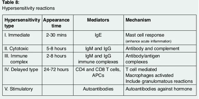
Hypersensitivity
The term hypersensitivity refers to the process whereby the adaptive immune response over-reacts to a variety of infectious and inert antigens resulting in damage to the host tissue. Five types of hypersensitivity reactions exist – all of which vary in their timing following contact with the antigen ((TTaabbllee 88)).
• Type I hypersensitivity: allergy Allergies may affect approximately 17% of the population45. The term atopic is used to describe those individuals who possess a genetic predisposition to allergy. Allergies may occur to otherwise innocuous antigens (known as aalllleerrggeennss) and infectious agents, e.g. worms. Type I hypersensitivity exists in two phases, the sensitisation and effector phases.
Firstly, a harmless allergen causes production of IgE antibody on first exposure. This IgE
February 8, 2002 OT
diffuses throughout the body until it comes into contact with mast cells and basophils. Both these cell types have receptors for IgE antibody. Although the patient experiences no symptoms after the initial binding, reintroduction of the antigen/allergen induces the production of more IgE and, furthermore, increase the likelihood of cross-linking with existing antibodies on the mast cell surface. Such cross-linking induces the mast cell to degranulate and release a host of inflammatory mediators such as histamine, prostaglandins and bradykinin.
Histamine characteristically causes the itchy symptoms experienced by patients and as a result of binding to H1 receptors in the eye, induces vasodilation and enhances mucous secretion by the goblet cells. Bradykinin augments vascular permeability, decreases blood pressure and contracts smooth muscle. Prostaglandins are also powerful inflammatory mediators.
Ocular correlates:
The following anterior segment conditions are due, if not only in part, to type I
hypersensitivity:
• Seasonal allergic conjunctivitis (type I) • Giant papillary conjunctivitis (types I and IV) • Vernal keratoconjunctivitis (VKC)
(type I and IV)
• Atopic keratoconjunctivitis (AKC) (types I and IV) ((FFiigguurree 77))
Seasonal allergic conjunctivitis can be easily recognised by chemosis and hyperaemia of the conjunctiva, lid swelling and excessive lacrimation. The cornea is not affected.
GPC is well known to optometrists as an allergic response to contact lenses, prosthetic lenses, protruding corneal sutures and scleral buckles. Histological examination of eyes suffering from GPC have revealed degranulated mast cells (type I hypersensitivity)46
and CD4+ T cells (type IV)47
, thus corroborating the theory that such a condition is mediated by both types of hypersensitivity.
Vernal conjunctivitis can present either in a palpebral form characteristically exhibiting
giant cobblestone papillae, or in a limbal form with gelatinous deposits known as Trantas’-dots, which represent degenerating epithelial cells and eosinophils. The first corneal change is a punctate epithelial keratitis, which if left unchecked develops into a macroerosion ((FFiigguurree 88)) and finally a corneal plaque develops ((FFiigguurree 99)). The conjunctiva itself is
characteristically oedematous and contains an array of immunological cells including lymphocytes and mast cells. Evidence of type I involvement includes the histological detection of, inter alia, degranulated mast cells, eosinophils and increased levels of IgE in affected eyes49. The detection of CD4+ T cells and macrophages is indicative of a delayed
inflammatory component49. Table 8:
Hypersensitivity reactions
Hypersensitivity Appearance Mediators Mechanism
type time
I. Immediate 2-30 mins IgE Mast cell response
(enhance acute inflammation)
II. Cytotoxic 5-8 hours IgM and IgG Antibody and complement III. Immune 2-8 hours IgM and IgG Antibody/antigen
complex immune complexes complexes
IV. Delayed type 24-72 hours CD4 and CD8 T cells, T cell mediated APCs Macrophages activated
Include granulomatous reactions V. Stimulatory Autoantibodies Autoantibodies against hormone
Figure 8:
Epithelial macroerosion in VKC
Figure 9:
Corneal plaque in VKC
Figure 7:
Sponsored by
a
Module 4 Part 2
Type II hypersensitivity: cytotoxic This classification of hypersensitivity involves either IgG or IgM antibodies, which may induce cellular lysis due to the involvement of the classical complement pathway (as seen in blood transfusion reactions) or recruit and activate inflammatory cells via complement. The components of complement include the C5a, which serves to attract inflammatory cells to the site of interest. The hypersensitivity reaction is a result of the excessive amount of extracellular mediators released by the inflammatory cells to antigens that are too big to be completely phagocytosed.
Antibodies to self-antigens, such as the acetylcholine receptor in myasthenia gravis, is not only another example of type II
hypersensitivity but also an example of autoimmune disease.
Ocular manifestations • Mooren’s ulcer • Cicatrical pemphigoid
Mooren’s ulcer is a rare peripheral ulcerative keratitis that exists either as a unilateral, non-progressive form which has a predilection for elderly patients or as a more severe, progressive form affecting both eyes of relatively young individuals. The signs range from a small patch of grey infiltrate near the margin to frank ulceration involving the entire corneal circumference and, in some cases, the central region as well. The healing process results in a thin, vascularised, opaque cornea. Investigators have identified a significant number of lymphocytes, neutrophils and plasma cells50in the cornea, thus providing unequivocal evidence to support the theory that this condition has an immunopathological aetiology.
Cicatrical pemphigoid is a chronic, blistering disease, which has a predilection for both the ocular and oral mucous membranes. Unlike its self-limiting counterpart, pemphigus vulgaris, cicatrical pemphigoid rarely affects the skin. Ocular cicatrical pemphigoid is a serious, bilateral condition that represents effective shrinkage of the conjunctiva. Although the initial presentation may be subacute and non-specific, it frequently progresses to symblepharon, entropion with secondary trichiasis, dry eye, ankyloblepharon and conjunctival fornix shortening. There is evidence to support the presence of IgG antibodies directed against self-antigen in the basement membrane of both the skin and eye51. Binding of the aforementioned antibodies may activate complement with subsequent recruitment of inflammatory cells into the area. The process of cicatrisation is achieved through the secretion of collagen via fibroblasts as a result of stimulation via cytokines released from the invading inflammatory cells.
Type III hypersensitivity: immune complex
Large pathogens with multiple antigenic sites have several antibodies bound to them forming
immune complexes. Normally, these complexes are removed by the mononuclear phagocytes in the liver and spleen with no adverse sequelae. However, persistence of immune complexes does occur in certain individuals leading to their deposition in tissue. As a consequence of the latter action, complement may be activated thus paving the way for inflammatory cells to enter the deposition site. Since blood vessels (which filter plasma at high pressure and exhibit a great deal of tortuosity) are more susceptible for immune complex deposition, the ciliary body is particularly vulnerable to this type of hypersensitivity reaction.
Ocular manifestations • Uveitis (Crohn’s disease)
• Peripheral corneal lesions associated with rheumatoid arthritis
• Stevens Johnson syndrome • Sjögren’s syndrome
The signs and symptoms of the above will be described in future articles in the series.
Type IV hypersensitivity: delayed-type hypersensitivity (DTH)
The term DTH has been used to describe such a reaction owing to its prolonged time-scale relative to the other hypersensitivity types. Although DTH can be transferred by T cells that have been previously sensitised by an antigen, it cannot be transferred in serum.
The sequence of events leading to DTH begins with initial presentation of the antigen peptide to T cells by APCs (e.g. Langerhans’ cells). The primed T cell migrates to the site of antigenic entry whereby it releases pro-inflammatory mediators such as TNF. The release of these cytokines facilitates blood flow and extravasation of plasma contents to the area. The activation of CD4 T helper and CD8 cells results in the release of IFN-γand, as a consequence, enhances macrophage activity in that area. Resolution of DTH is dependent on the efficacy with which such phagocytes can remove the offending antigen.
Recalcitrant infectious agents result in a chronic DTH that causes the chronically activated macrophages to fuse together and form multinucleated giant cells. In an attempt to contain the infectious agent, macrophages may undergo further inter connections to resemble an epithelial layer. Owing to the similarity to this layer they are referred to as epitheloid cells. Both epitheloid and multinucleated giant cells secrete factors that induce fibrosis resulting in granuloma formation. Thus granulomata are the hallmark of chronic inflammation. Damage and loss of function of the neighbouring tissues frequently ensues until the agent is removed either chemically or surgically.
Ocular manifestations • Ocular allergies (VKC, AKC GPC) • Idiopathic uveitis
• Sympathetic ophthalmia • Phlyctenulosis
Type V hypersensitivity
This relatively new category encompasses the concept of autoantibodies binding to hormone receptors that mimic the hormone itself. This results in stimulation of the target cells. Examples include thyrotoxicosis.
Autoimmunity
The ability to react against self-antigens is known as autoimmunity. However, a significant number of people exist who harbour auto-antibodies and yet remain asymptomatic. The corollary of this is that the presence of autoreactive cellsper seis not sufficient to trigger autoimmune disease. In fact,
autoimmune disease is a result of breakdown of one of the immunoregulatory mechanisms. Furthermore, autoimmune disease may be classified as either being organ specific (e.g. insulin dependent diabetes mellitus, Grave’s disease) or non-organ specific (e.g. Sjögren’s syndrome, ankylosing spondylitis).
It is important to realise that the causes of autoimmune diseases are multifactorial. The main predisposing factors are age, gender, infection and genetics. However, one of the most important factors of interest to immunologists and clinicians alike is the association between HLA and autoimmune disease. When determining the likelihood of contracting a disease both epidemiologists and clinicians refer to the relative risk. In the case of HLA antigen, the relative risk compares the chance of a person who has a particular HLA antigen acquiring a disease to those individuals who do not have such an antigen. The association is exemplified by the relative risk of suffering from ankylosing spondylitis (AS) in individuals who possess HLA-B27. In patients suffering from AS, the prevalence of HLA-B27 is 90% and this figure rises to 95% in patients who suffer both with the disease and acute iritis. Indeed, in the UK approximately 45% of patients who present with acute iritis will harbour HLA-B2752.
It is therefore important that patients who present with anterior uveitis are screened for HLA-B27 because although a positive result may not necessarily be diagnostic, its presence will certainly improve the sensitivity of further radiological tests.
TTaabbllee 99 compares the HLA associations with both ophthalmic disorders together with those systemic disorders relevant to the ophthalmic practitioner.
Conclusion
This article has only broached the fascinating subject of ocular immunology. A basic understanding of immunology is required if practitioners are to therapeutically manage their patients. Further articles in the series will help to reinforce the concepts of this challenging subject.
Acknowledgement
o
t
February 8, 2002 OT www.optometry.co.uk
34
Figures 1 to 6 reprinted from Immunology, Third Edition, Roitt, Brostoff and Male, 1993 by permission of the publisher Mosby.
About the author
Gregory Heath is an optometrist working part-time in private practice. He was recently awarded the diploma in clinical optometry at City University and is currently reading medicine at the Royal Free and University College, London, Medical School.References
References are available upon request. Please fax 01252-816176 or email [email protected].
Table 9:
HLA and ophthalmic disease
Disease HLA Relative risk 54 Ocular
association manifestation
Ankylosing spondylitis B27 90 Anterior uveitis
Reiter’s disease B27 33 Mucopurulent conjunctivitis
Anterior uveitis Keratitis
Rheumatoid DR4 7 Keratoconjunctivitis sicca
Keratitis Scleritis
Primary Sjögrens’ syndrome DR3, DR5 10 Keratoconjunctivitis sicca
Sarcoidosis DR3 Not known Anterior uveitis (acute and chronic)
Dacryoadentis Retinal vasculitis, neovascularisation, Optic nerve granulomata
Cicatrical pemphigoid DQw7 Not known Shrinkage of conjunctiva
Behcet’s disease B5 3 Anterior uveitis
Retinitis, periphlebitis, retinal oedema
Sympathetic DR4, A11, Not known Panuveitis
ophthalmia B40
Systemic lupus DR2, DR3 3 Punctate epithelial keratopathy
erythematosus Keratopathy
Necrotising scleritis
Retinal lesions (cotton wool spots) Autoimmune optic neuropathy
1
1.. WWhhiicchh oonnee ooff tthhee ffoolllloowwiinngg iiss nnoott ppaarrtt ooff tthhee iinnnnaattee iimmmmuunnee ssyysstteemm??
a. Mast cells b. Complement c. Phagocytes d. T cells
2
2.. WWhhiicchh oonnee ooff tthhee ffoolllloowwiinngg ssttaatteemmeennttss iiss ccoorrrreecctt rreeggaarrddiinngg tthhee iinnnnaattee iimmmmuunnee ssyysstteemm?? a. It is specific
b. It evokes a more potent response on secondary exposure
c. It represents the first line of defence d. It is able to memorise pathogens on
subsequent exposures
3
3.. WWhhiicchh oonnee ooff tthhee ffoolllloowwiinngg ssttaatteemmeennttss iiss ccoorrrreecctt rreeggaarrddiinngg tthhee cceellllss ooff tthhee iinnnnaattee ssyysstteemm??
a. Basophils are important phagocytes b. During phagocytosis the pathogen becomes
initially internalised as a phago-lysosome c. Eosinophils play an important role in
combating virally-infected cells d. Langerhans’ cells form a bridge between
innate and adaptive immunity
4
4.. WWhhiicchh oonnee ooff tthhee ffoolllloowwiinngg ssttaatteemmeennttss iiss ccoorrrreecctt rreeggaarrddiinngg tthhee aaddaappttiivvee iimmmmuunnee ssyysstteemm??
a. It consists of all types of lymphocytes b. T cells produce antibodies
c. T cells maturate in the thymus d. B cells are produced in the spleen
5
5.. WWhhiicchh oonnee ooff tthhee ffoolllloowwiinngg ssttaatteemmeennttss iiss ccoorrrreecctt rreeggaarrddiinngg TT cceellllss??
a. T cells can be subdivided into TH1and TH2 subtypes only
b. T cells alone can identify any type of antigen c. T cells express cell surface proteins denoted by
cluster determinant (CD) numbers d. All T cells are involved in initiating the
inflammatory response 6
6.. WWhhiicchh oonnee ooff tthhee ffoolllloowwiinngg ssttaatteemmeennttss iiss iinnccoorrrreecctt??
a. B cells have antibodies as their cell surface receptor
b. There are five types of antibody c. IgE is an important antibody in allergies d. All B cells differentiate into plasma cells 7
7.. WWhhiicchh oonnee ooff tthhee ffoolllloowwiinngg ssttaatteemmeennttss iiss ccoorrrreecctt rreeggaarrddiinngg ooccuullaarr iimmmmuunnee pprriivviilleeggee?? a. There is an absence of Langerhans’ cells in the
central cornea
b. Aqueous humour has no role c. Abundant vascular supply is vital d. All ocular cells express MHC class II
8
8.. WWhhiicchh oonnee ooff tthhee ffoolllloowwiinngg ssttaatteemmeennttss ccoonncceerrnniinngg ooccuullaarr iimmmmuunnoollooggyy iiss iinnccoorrrreecctt?? a. Levels of IgA and complement
increase when the eyes are closed b. IgA is the predominant antibody
in blood and tissue fluid
c. IgM, IgG and IgA antibody isotypes have been identified in the cornea
d. The sclera contains a smaller number of immune cells than the conjunctiva
9
9.. IInn aa ppaattiieenntt ssuuffffeerriinngg ffrroomm vveerrnnaall ccoonnjjuunnccttiivviittiiss,, w
whhiicchh oonnee ooff tthhee ffoolllloowwiinngg ssttaatteemmeennttss iiss ccoorrrreecctt??
a. Trantas’-dots represent infiltrating T cells b. Affected eyes have increased levels of IgD c. Histologically, mast cells, lymphocytes and
macrophages have been identified d. Is due to type III Hypersensitivity
1
100.. WWhhiicchh oonnee ooff tthhee ffoolllloowwiinngg ssttaatteemmeennttss iiss iinnccoorrrreecctt??
a. HLA-B27 is a risk factor for both anterior uveitis and ankylosing spondylitis
b. Granulomas are present in type IV hypersensitivity reactions
c. Histamine is an important vasoconstrictor d. IgE mediated hypersensitivity is of rapid onset
1
111.. WWhhaatt pprrooppoorrttiioonn ooff ppaattiieennttss wwiitthh aaccuuttee iirriittiiss wwiillll h
haarrbboouurr HHLLAA--BB2277?? a. 15%
b. 25% c. 45% d. 65%
1
122.. WWhhiicchh oonnee ooff tthhee ffoolllloowwiinngg ssttaatteemmeennttss iiss ccoorrrreecctt rreeggaarrddiinngg aa ttyyppee II hhyyppeerrsseennssiittiivviittyy rreeaaccttiioonn?? a. It always occurs in isolation
b. It is characterised by the presence of macrophages
c. It is associated with myasthenia gravis d. It involves the degranulation of mast cells
following the cross-linking of IgE bound to its cell surface