A subchronic toxicity test of ethyl acetate extract from endophytic fungus
Penicillium
sp. of kunyit putih (
Curcuma zedoaria
) against Swiss albino mice
Muharni
*, Heni Yohandini
Department of Chemistry, Faculty of Mathematics and Natural Sciences, University of Sriwijaya, Jl. Raya Palembang-Prabumulih Km 32,
Indralaya, Ogan Ilir, South Sumatra, 30662 Indonesia
Abstract: Ethyl acetate extract of endophytic fungus from Penicillium sp. of kunyit putih showed antibacterial activity in vivo but no acute toxicity. However, the extract may have toxic effects on major organs for long-term consumption. This study was carried out in order to test sub-chronic toxicity of the ethyl acetate from endophytic fungus Penicillium sp. of kunyit putih against mice (Mus musculus). A total of 50 male mice were divided into five groups. Groups I, II, III and IV were orally administered with ethyl acetate extracts of 250, 500, 1000 and 2000 mg/kg body weight (BW), respectively. Group V was used as a control without extract treatment. A toxic symptom was observed by analyzing several parameters, namely change in BW, hematologic and biochemical properties (SGOT & SGPT), macroscopic organs, and relative organ weight. The results showed that there was toxic symptom and statistically significant difference in the parameters of SGOT, SGPT and heart weight between treated and control groups. Based on analysis, we concluded that ethyl acetate extract from endophytic fungus Penicillium sp. of kunyit putih had subchronic toxicity effect.
Keywords: Subchronic toxicity; Endophytic fungi; Penicillium sp; Curcuma zedoaria
CLC number: R965 Document code: A Article ID: 1003–1057(2018)2–123–08
Received: 2017-11-13, Revised: 2017-12-20, Accepted: 2018-01-12. *Corresponding author. Tel.: +62-085381506355
E-mail: [email protected]
http://dx.doi.org/10.5246/jcps.2018.02.014
1. Introduction
Endophytic fungi are a type of microorganism found in almost all parts of the plant, which are able to produce certain secondary metabolites with their host plants[1,2]. For this reason, endophytic fungus is used as source to search new bioactive compounds[3]. Several authors have reported that endophytic fungi possess unique ability to produce secondary metabolite compounds that have high biological activity, which are even more active than compounds produced by their host plants[4]. In addition, active compounds also have a unique basic framework that is not related to the biosynthesis of its host compounds[5,6].
In our two previous works, two compounds have been successfully isolated from ethyl acetate extract of
endophytic Penicillium sp. Kunyit putih. The compounds are identified as 5(4′-etoxy-2′hydroxy-5′methyl-2′,3′ -dihydrofuran-3′-il (hydroxy) methyl-4-isopropyl-3 -methyl-2-piran-2-on and bis-ethylhexilphtalat[7]. Both compounds were tested for anti-bacterial activity against E. Coli (ATCC 25922), S. dysentriae, S. aureus (ATCC
25923) and B. subtilis (ATCC 6633) and displayed bacterial activity among all tested bacteria[8]. Active extract of
Penicillium sp. had also antibacterial activity in vivo by mice (Mus muscullus). The lab mice were induced by diarrhea bactery (E. coli and S. dysentriae) prior to the activity test. Result showed extract had active antidiarrhea in vivo with effective dose at 250 mg/kg·BW. This dose was equal to positive control of ciprolaxacin for S. dysentriae and chloramphenicol for S. typii at 10 mg/kg·BW. Acute toxicity test has indicated that the extract does not possess the acute toxic effects[9]. In this work, we studied subchronic toxicity test of ethyl acetate extract endophytic fungus Kunyit putih against mice (Mus muscullus).
ww
w.j
cps
.ac
2. Material and methods
2.1. Collection of test endophytic fungi
Endophytic fungi Penicillium sp was collected and prepared according to our previous study[7].
2.2. Preparation of ethyl acetate extract from endophytic fungus Penicillium sp.
Ethyl acetate extracts were prepared as previously reported[7,10,11].
2.3. Experiment animals
Male Swiss albino mice were used for the subchronic toxicity studies. The mice were obtained from animal house of Institut Teknologi Bandung, Indonesia. Test animals were acclimatized to laboratory conditions for 7 d before the experiments. During their acclimatization, mice were housed in cages and provided with pellet diet and clean water. The experiments were approved by the Ethics Committee Research Health of Sriwijaya University (No. 35/kepkrsmhfkunsri/2017) and carried out in accordance with current guidelines for the care of laboratory animals.
2.4. Subchronic toxicity test using mice (Mus muscullus)
Briefly, 50 male swiss albino mice (3-month-old) with BW of 20–25 g were evenly divided into five groups. Four groups were treated with the extracts of ethyl acetate from endophytic fungus Penicillium sp. of kunyit putih, and the last one was the control group without treatment. Groups I–IV were treated with ethyl acetate extract at a dose of 250 mg/kg·BW, 500 mg/kg·BW, 1000 mg/kg·BW and 2000 mg/kg·BW, respectively. The treatment was given by oral administration once a day for 90 d[12]. During the treatment, the mice were fed with normal food and drinking water. All procedures in the study were performed according to Ministry of
Health Guide for the Care and Use of Laboratory Animals[13].
Toxic symptoms were observed by means of body weight (BW) analysis, hematologic and biochemical parameters, and macroscopic organs. The behavior and motoric activity of the test animals were monitored every day, and their BW was measured every 15 days. After 30 and 90 d, three mice from each group were sacrificed. The mice were deprived of pellet food and only given free access to drinking water at 24 h prior to sacrifice. Blood samples were collected through cardiac puncture for haematological and biochemical analyses. The organs (liver, hearth and kidney) were excised, weighed and macroscopically examined. Parameters of hematologic and biochemical tests included levels of hemoglobin (Hb), serum glutamic oxaloacetic transaminase (SGOT) and serum glutamic-pyruvic transaminase (SGPT). The analyses of macroscopic organs were performed by comparing color and appearance of the vital organs of treated mice with control. Furthermore, the relative organ weights (ROWs) were also calculated. The weight of the vital organ of the treated mice was then compared with control group[14].
2.5. Statistical analysis
Data analysis was performed to evaluate differences among groups using one-way analysis of variance, followed by Duncan New Multiple Range Test (DNMRT). The significance level was set at 5% (P<0.05).
3. Results
3.1. Effect of the extract on motoric activity and BW
Subchronic toxicity test was used to evaluate effects of repeated or continuous administration of a test material on animals. Here, we first analyzed the motoric activity and BW of mice to observe treatment effects (Tables 1 and 2).
ww
w.j
cps
.ac
3.2. Effect of the extract on biochemical parameters
Table 3 presents the effect of subchronic administration of ethyl acetateextract from endophytic fungus Penicillium
sp. on biochemical parameters (SGOT and SGPT). Biochemical parameters (SGOT and SGPT) showed abnormal values for the treatment group.
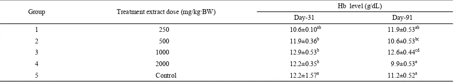
3.3. Effect of the extract on haematological parameters
Table 4 shows the effects of ethyl acetateextract on the hematological parameters (Hb) in subchronic study. Hb parameter showed no significant changes and remained within physiological range after the 90-day treatment period.
Group Treatment extract dose (mg/kg·BW) Number of rats Motoric activity
I 250 10 Normal
II 500 10 Normal
III 1,000 10 Normal
IV 2,000 10 Normal
V 0 (Control) 10 Normal
Group Treatment extract dose (mg/kg·BW) BW (g) days
0 15 30 45 60 75 90
I 250 22.5±1.25 22.8±2.80 22.0±2.00 25.2±1.70 24.3±2.50 24.0±1.50 23.5±0.75 II 500 23.5±1.75 21.8±1.50 22.9±1.75 24.1±1.50 24.9±2.00 24.0±1.50 23.5±1.50 III 1000 23.5±1.00 22.3±1.55 21.2±1.50 26.0±1.75 24.7±2.00 24.0±1.75 23.5±1.55 IV 2000 22.5±1.50 21.9±1.00 21.1±2.50 22.2±1.50 22.8±1.75 22.0±2.00 22.2±1.75 V Control 23.3±1.50 23.3±1.50 24.1±1.00 25.2±1.75 25.4±1.50 24.5±1.75 24.0±1.50
Group Treatment extract dose (mg/kg·BW) SGOT level (U/I) SGPT level (U/I) Day-31 Day-91 Day-31 Day-91
1 250 162±17.35b 211±11.53b 68±4.58b 89±5.57b
2 500 198±3.46c 351±11.00c 103±4.36c 136±4.00c
3 1000 251±2.65d 354±11.53c 156±4.00d 176±5.29d
4 2000 267±4.36e 374±5.29d 173±2.65e 189±3.61e
5 Control 69±2.65a 72±4.00a 46±1.73a 44±2.00a
Group Treatment extract dose (mg/kg·BW) Hb level (g/dL) Day-31 Day-91
1 250 10.6±0.10ab 11.9±0.53ab
2 500 11.9±0.36b 10.6±0.53bc
3 1000 12.9±0.53b 12.6±0.44cd
4 2000 12.2±0.35b 9.9±0.53e
5 Control 12.2±1.57a 11.2±0.52a
Table 1. The toxic symptoms of tested mice after extract injection.
BW: Body weight.
Table 2. Effect of ethyl acetate extract from endophitic fungus Penicillium sp. on BW changes in mice.
BW: Body weight.
Table 3. SGPT and SGOT levels for mice.
Note: Different superscript (a, b, c, d, e) in the same column indicate significant difference on Duncan test at level 5%.
Table 4. The results of hematologic evaluation.
Note: Different superscript (a, b, c, d, e) in the same column indicate significant difference on Duncan test at level 5%.
ww
w.j
cps
.ac
3.4. ROWs and macroscopic test
Macroscopic test was conducted on vital organ (liver, heart and kidney) to evaluate how extract toxicity changed the structure of test animal’s organ[15]. There was a significant difference in the weight of liver between the extract treated and control groups throughout the treatment period, and no significant difference was observed in the weights of heart and kidney (Table 5).
4. Discussion
After 1-h treatment of the ethyl acetate extract from endophytic fungus Penicillium sp. kunyit putih, mice showed no decrease in motoric activity, no signs of behavioral distress, no casualties and no observable toxicity symptoms. The experimented mice survived till the completion of the experimental duration at all levels of treatment. It indicated that the extract had no effect on their motoric activity. In addition to motoric activity, BW is important to initially describe the health of test animals. Change in BW is a highly sensitive indicator of health condition[16]. Table 2 shows that during 90-day treatment, the average BW of mices was relatively stable. From statistical analysis, no significant differences in BW were observed between control and treated groups during this period. The significance value was greater than 0.05, suggesting that the extract administration had no effects on BW and a negligible effect on the growth of animals.
Evaluation of blood parameters can be used to determine the levels of negative effect of foreign compounds[17–19]. Evaluation of hematologycal and biochemical parameters was conducted to observe liver and kidney functions. The damage in the liver can be used to indicate toxic substances. Two aminotransferase enzymes that are most commonly associated with liver cell damage are SGOT and SGPT. On day 31 and 91, three test animals from each group were sacrificed, and hematologic and biochemical parameter (Hb, SGOT and SGPT) as well as histopathological examination of liver, kidney and heart were evaluated. Table 3 shows the results of SGOT and SGPT levels.
SGOT and SGPT are two transaminases produced primarily by liver cells when hepatic cells are damaged. Measurement of the enzyme concentration in the blood can determine the levels of SGOT and SGPT, providing important information regarding the liver dysfunction[20]. Liver injury will also cause damage to the hepatocyte cell membrane, in which the permeability of hepatic cells will increase and the enzymes will be released into the blood circulation. One-way variance analysis showed that the SGOT level of treated groups (I–IV) was significantly different from the control group (V). However, the SGPT level was insignificantly different (Table 3). The treatment dose caused an increase in the SGOT value beyond the normal value for all given doses. Result of SGOT level on day 31 showed that the administration of extract to the test animals increased the SGOT level compared with the ROW of liver (g) ROW of heart (g) ROW of kidney (g) Dose of treatment (mg/kg·BW)
Day-31 Day-91 Day-31 Day-91 Day-31 Day-91
0 2.362±0.15a 2.615±0.25a 0.170±0.03a 0.235±0.05a 0.505±0.06a 0.634±0.08a 250 2.217±0.20ab 2.474±0.12b 0.187±0.02a 0.287±0.03a 0.594±0.11a 0.594±0.07a 500 2.032±0.09bc 2.274±0.06b 0.146±0.01a 0.180±0.01a 0.523±0.84a 0.656±0.03b 1000 1.796±0.18cd 2.171±0.15bc 0.132±0.01a 0.132±0.01b 0.413±0.08a 0.413±0.05b 2000 1.711±0.15d 1.796±0.22c 0.141±0.073a 0.141±0.01c 0.465±0.04a 0.465±0.04b
Table 5. ROWs of tested and control mice.
Note: Different superscript (a, b, c, d) in the same column indicate significant difference on Duncan test at level 5%.
ww
w.j
cps
.ac
control group (V). SGOT level of group I treated with 250 mg/kg·BW was 162±17.35 U/I, which was much higher than that of control group (69 U/I). Anova analysis suggested that extract administration affected the SGOT level. Duncan test on SGOT value at P<0.05 concluded that the higher extract dose administered, the higher SGOT level obtained. Highest level of SGOT was found from group IV, where the test animals were administered by 2000 mg/kg·BW extract, and the SGOT level was increased to 267±4.36 U/I. Statistical calculations proved that SGOT at this level was significantly different from all other test groups. Same conclusion was also obtained from group administered with 1000 mg/kg·BW. According to the above-mentioned findings, it can be concluded that extract administration even at dose 250 mg/kg·BW showed toxicity. After treatment for 91 d, it revealed that the SGOT level was significantly higher than that after 31 d.
Table 3 shows the SGPT measurement results for test animals after 31 and 91 d. The data exhibited that extract administration increased the SGPT level compare with control group. SGPT level of group I was (68±4.58) U/I, which was 47% higher than group IV (control) with an SGPT level of (46±1.73) U/I. Anova analysis followed by Duncan test concluded that the difference of SGPT level was significant. The highest SGPT level (173±2.65) U/I was observed in group administered with 2000 mg/kg·BW. The SGPT level in group administered with 1000 mg/kg·BW was proportionally higher than that of groups administered with 500 mg/kg·BW and 250 mg/kg·BW. By comparing duration of extract treatment, group I exhibited increased SGPT from day 31 to 91, from (68±58) to (89±5.57) U/I. The same pattern was observed in other doses. T-test calculation revealed, however, that the difference was not significant. At this point, we concluded that at dose of 250 mg/Kg, 31-day treatment showed symptom of toxicity. Increased levels of SGOT and SGPT indicated
toxicity of extract. Toxic effect caused liver necrosis, especially through its reactive metabolite. SGOT could also cause scuffle during treatment period, resulting in muscle trauma in the test animal. Another possible cause was disease or abnormalities of liver, kidney and heart. Statistical calculation using one-directional variance analysis at a 95% and P<0.05 showed that the SGOT change between five groups was significantly different but not for SGPT. Administration of extract from endophytic fungus Penicillium sp. increased measurabe SGOT and SGPT levels. The high levels of SGOT and SGPT were possibly due to toxic material intake, which triggered metabolism disturbance on hepatocyte, leading to morphological and functional damage in liver.
Hb level was also measured in order to assess toxic effect of extract administration (Table 4). Hb consists of mainly proteins in forms of protoporphyrin, globin and also iron (II). One gram Hb could raise oxygen dissolved 1.34 mL. Low level of Hb indicated low protein given (vitamin deficiency). Low level of Hb could also be caused by infection of test animal. Although low Hb indicated inappropriate condition, high Hb level could also indicate poor condition. Test animals were mostly under stressful condition if they showed high level of Hb. Results of Hb measurement showed that on day 31, the Hb level was within the normal range of 10–14 g/100 mL. This result designated that there was no significant difference in Hb parameter between treated group and control. According to Hb test result, we concluded that the extract did not cause adverse effect on hematology system of test animals. The duration of extract administration for all given doses based on T-test calculation also showed no significant difference (P>0.05).
Kidney and liver function analysis is highly useful in the toxicity screening of medicines and plant extracts as both are important for the survival of an organism[21,22]. The test was carried out by observing the physical
ww
w.j
cps
.ac
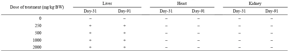
condition of organs, such as visual appearance, color and weight. Histopathology test was performed on day 31 and day 91. The result was depicted on Table 5. Observation on sample of mice liver histology (Mus musculus L) was conducted after mice were treated with ethyl acetate extract from endophytic fungus
Penicilium sp. of Kunyit putih for 31 d. The result showed that liver become darker, thicker and harder compare with control group. The same result also obtained from organs after 91-days treatment. These findings showed that damage occurred on hepatocyte in forms of pyknosis and necrosis. Hepatocytes suffering from pyknosis blackened although they still had cell membranes. Some cells seemed bigger than normal hepatocytes and some are smaller. Pyknosis or liver shrinkage is a cytoplasm homogenization[23]. Hepatocytes with pyknosis appeared darker than normal ones. Blackened nucleus and fragmentation indicated hepatocyte damage due to necrosis. The shrinkage of hepatocytes resulted in smaller size and irregular shape.
Chemical analysis of histopathology blood sample supported the above-mentioned conclusion. Liver damage increased the levels of SGOT and SGPT. Transaminase level increase in serum was due to necrosis of transaminase rich cells (Verma and Ahmed, 2009). This result indicated significant difference between treated group and control group. Variation on test extract concentration also gave significant impact on liver’s weight. Duncan test on liver weight confirmed that the lowest weight average was 1.711±0.15 U/I, which was detected in 2000 mg/kg·BW group. However, decrease in weight of an internal organ suggests toxicity resulting from exposure to toxic substances[24]. Table 5 shows the effect of duration time of extract treatment on liver’s weight. The weight of liver was increased as the treatment time was extended. T-test calculation proved that there was significant difference between treated group and control group.
Analysis of variance calculation on kidney’s weight at
5% significance level found P(0.000)>0.05. This result revealed that extract administration exerted no effect on the organ. Duncan test confirmed that there was no difference in kidney’s average weight between treated group and control group, suggesting no kidney damage in test animals. Visual observation on kidney with and without treatment showed the same condition, and no difference was observed. Treatment duration and dose given also showed no effect on kidney’s weight. T-test showed P>0.05, indicating no significant difference of treatment duration on weight of kidney. Histopathological observation on heart sample of test animal after 31-day treatment showed no damage, and it was relatively similar to the control group. Anova calculation showed
P(0.000)>0.05 on weight of heart, suggesting that extract treatment did not affect heart’s weight of test animals. Duncan test result supported the conclusion that the average weight of heart in tested groups was not significantly different from that in the control group. All statistical calculation approved that no damage occurred on heart due to extract administration. Treatment duration relatively exerted no effect on visual appearance or heart’s weight. T-test indicated no significant difference (P>0.05) for treatment duration, hence we concluded that extract administration duration did not affect weight of heart.
Physical condition was used to evaluate how extract administration affected liver, kidney and heart in addition to organ weight (Table 6). Table 6 shows a comparation between sample and control after extract administration started at an initial concentration of 250 mg/kg·BW. On day 31, with all concentrations, the liver became darker, thickened and harder compare with control group. Meanwhile, the heart and kidney exhibited similar physical condition compared with the control. Similar observation was found up to day 91. On day 91, extract concentration of 1000 mg/kg·BW made the liver smaller. These data confirmed the possibility of toxicity of the extract to the liver.
ww
w.j
cps
.ac
Based on data of hematology, blood chemistry and macroscopic test results, there was a significant difference in parameters of SGOT, SGPT and heart condition between treated mice and control. The difference appeared when 250 mg/kg·BW extract was given to the mice, therefore extract was considered to be toxic and could not be administered for long time.
Acknowledgment
We gratefully acknowledge the support Sriwijaya University, sponsored by “Penelitian Hibah Bersaing”, Ministry of Research Technology and Higher Education, Indonesia.
References
[1] Hung, P.Q.; Annapurna, K. Isolation and Characterization of Endophytic Bacterial in Soybean (Glycine sp.) Omonrice. 2004, 101, 92–101.
[2] Hundley, N.J. Struktur Elucidation of Bioactive Compounds Isolated from Endophytes of Alstonia Scholaris and Acmena Graveolens. Brigham Young University.2005. [3] Prihatiningtias. W.; Wahyuningsih, M.S.H. Prospek Mikroba
Endofit sebagai Sumber Senyawa Bioaktif. Fakultas Kedokteran Universitas Gadjah Mada. Yogyakarta. 2005.
[4] Tan, R.X.; Zou, W.X. A Rich Source of Functional Metabolites. Nat. Prod. Rep. 2001, 18, 448–459. [5] Radji, M. Peranan Bioteknologi dan Mikroba Endofit
dalam Pengembangan Obat Herbal. Majalah Ilmu Kefarmasian. 2005, 2, 113–126.
[6] Elfita, M.; Muharni, S. New pyran of an endophytic fungus Fusarium sp. Isolated from the leaves of brotowali (Tinaspora crispa). Indo. J. Chem. 2013, 13, 209–215. [7] Muharni, F.; Ruliza, M.O.; Susanti, D.A. Di-(2-ethylhexyl)
phthalate andPyranon Derivated from Endophytic fungi
Penicillium sp the Leave of Kunyit Putih (Curcuma zedoaria (Berg) Roscoe). Indones J. Chem.2014, 14, 290–296.
[8] Muharni, F.; Ruliza, M.O. Antibachterial and antioxidant activity testing of pyranon derivated compound from endophytic fungi Penicillium sp of kunyit putih (Curcuma Zedoaria (Berg) Roscoea). Tradit. Med. J. 2014, 19, 107–112.
[9] Muharni, Y.H.; Fitrya, R. Evaluation Acute toxicity and Antibacterial activity of Penicillium sp endophitic fungus extract of Kunyit Putih (Curcuma zedoaria) in mice (Mus musculus L.). J. Chem. Pharm. Res. 2015, 7, 147–155. [10] Barik, B.; Tayung, K.; Jagadev, P.; Dutta, S. Phylogenetic
placement of an endophytic fungus Fusarium oxysporum isolated from Acorus calamus rhizomes with antimicrobial activity. Eur. J. Biol. Sci. 2010, 2, 8–16.
[11] Aryantha, I.N.P.; Lestari, D.P.; Pangesti, N.P.D. Microbiol Indonesia. Potensi isolat bakteri penghasil IAA dalam peningkatan pertumbuhan kecambah kacang hijau pada kondisi hidroponik. Microbiol Indonesia. 2010, 9, 43–46. [12] Rhiouani, H.; Zh, I.; Lyoussi, B. Acute and sub-chronic
toxicity of an aqueous extract of the leaves of Herniaria glabra in rodents. J. Ethnopharmacol. 2008, 118, 1–2.
Table 6. The physical condition of organs.
Note : –: not affected +: affected.
Dose of treatment (mg/kg·BW) Liver Heart Kidney Day-31 Day-91 Day-31 Day-91 Day-31 Day-91
0 – – – – – –
250 + + – – – –
500 + + – – – –
1000 + + – – – –
2000 + + – – – –
ww
w.j
cps
.ac
[13] Departemen Kesehatan RI. 2000a. Pedoman pelaksanaan uji klinik obat tradisional. Direktorat Jenderal Pengawasan Obat dan Makanan. Direktorat Pengawasan Obat Tradisional. Jakarta.
[14] Majeed, M.; Nagabhushanam, K.; Natarajan, S.; Sarangbani, V.P.; Majeed, S.; Karri, S.K. Investigation of Acute, Sub-Acute, Chronic Oral Toxicity and Mutagenicity of Coleus forskohlii Briq. Hydroethanolic Extract, Standardized for 10% Forskolin in Experimental Animals. Int. J. Pharm. Pharm. Res. 2015, 5, 219–238.
[15] Diallo, A.; Eklu-gadegkeku, K.; Agbonon, A.; Aklikokou, K.; Creppy, E.E. Acute and Sub-chronic (28-day) Oral Toxicity Studies of Hydroalcohol Leaf Extract of Ageratum conyzoides L (Asteraceae). Trop. J. Pharm. Res. 2010,
9, 463–467.
[16] Hilaly, J.E.L.; Israili, Z.H.; Lyoussi, B. Acute and chronic toxicological studies of Ajuga iva in experimental animals. J. Ethnopharmacol. 2004, 91, 43–50.
[17] Agbaje, E.O.; Adeneye, A.A.; Daramola, A.O. Biochemical and Toxicological Studies of Aqueous Extract of
Syzigium Aromaticum (L.) Merr. & Perry (Myrtaceae) in Rodents. Afr. J. Tradit. Complement Altern. Med. 2009,
6, 241–254.
[18] Ibrahim, M.B.; Sowemimo, A.A.; Sofidiya, M.O.; Badmos, K.B.; Fageyinbo, M.S.; Abdulkareem, F.B.; Odukoya, O.A. Sub-acute and chronic toxicity profiles of Markhamia
tomentosa ethanolic leaf extract in rats. J. Ethnopharmacol. 2016, 193, 68–75.
[19] Roy, S.; Ukil, B.; Lyndem, L.M. Acute and sub-acute toxicity studies on the effect of Senna alata in Swiss Albino mice. Cogent Biol. Cogent. 2016, 10, 1–11. [20] Fitrya, M.; Fithry, N.A. A Subchronic toxicity test of
ethanol extract from tunjuk langit rhizome (Helminthostachys zeylanica), rattus noverticus (Wistar strain). Asian J. Pharm. Clin. Res. 2017, 10, 270–273.
[21] Olorunnisola, O.; Bradley, G.; Afolayan, A. Acute and sub-chronic toxicity studies of methanolic extract of Tulbaghia violacea rhizomes in Wistar rats. Afr. J. Biotechnol. 2002, 11, 14934–14940.
[22] Ping, K.Y.; Darah, I.; Chen, Y.; Sreeramanan, S.; Sasidharan, S. Acute and sub-chronic toxicity studies of methanolic extract of Tulbaghia violacea rhizomes in Wistar rats. Biomed. Res. Int. 2013, 1–14.
[23] Ngabekti, S.; Isnaeni, W. emanfaatan Kurkumin Untuk Mengeliminir Pengaruh Diazonin terhadap Kerusakan Hati Mencit(Mus musculus L). Manusia Dan Lingkung. 2000, VII, 1–2.
[24] Adeyemi, O.; Akindele, A.; Nwumeh, K. Acuteandsub-chronictox- icological assessmentof Byrsocarpus coccineus Schum. And Thonn. (Con-naraceae) aqueous leaf extract.
Int. J. Appl. Res. Nat. Prod. 2010, 3, 1–11.