Alamat Korespondensi email:
Akreditasi PB IDI–2 SKP
Clinical Manifestations of Ocular Tuberculosis
Elvira
Puskesmas Siulak Mukai, Kabupaten Kerinci, Jambi, Indonesia
ABSTRACT
Tuberculosis (TB) is chronic Mycobacterium tuberculosis (MTB) infection.1 This bacteria can affects the eye by direct invasion after haematogenous dissemination accompanied by local inflammation or via immunologic reaction related to delayed hypersensitivity reaction to the bacteria elsewhere in the body. Ocular TB can be a great mimicker of various uveitis depending on location, host response and the virulence of the organism.. Definitive diagnosis would require microbiological confirmation of Mycobacterium tuberculosis from ocular fluid/tissue. Tuberculin skin test and interferron-gamma release assays (IGRA) has been used to diagnose patient without systemic symptoms. Delayed diagnosis or treatment can result in vision loss. This review will focus on the clinical manifestation and diagnosis of ocular TB.
Keywords: Intraocular tuberculosis, ocular tuberculosis, tuberculosis.
ABSTRAK
Tuberkulosis (TB) adalah infeksi kronik oleh Mycobacterium tuberculosis.1 Bakteri ini dapat menginfeksi mata dengan cara invasi langsung setelah penyebaran hematogen yang sejalan dengan inflamasi lokal atau melalui reaksi hipersensitivitas tipe lambat. Manifestasi klinis TB okular dapat menyerupai berbagai bentuk uveitis, tergantung lokasi, respons inang, dan tingkat virulensi bakteri. Diagnosis definitif membutuhkan konfirmasi Mycobacterium tuberculosis dari jaringan atau cairan okular. Tes kulit tuberkulin dan interferron-gamma release assays (IGRA) dapat digunakan untuk diagnosis pasien tanpa manifestasi sistemik. Diagnosis dan terapi yang terlambat dapat mengakibatkan kebutaan. Artikel ini akan membahas tentang diagnosis dan terapi TB okular. Elvira. Manifestasi Klinis Tuberkulosis Okular.
Kata kunci: Tuberkulosis, tuberkulosis intraokular, tuberkulosis okular
INTRODUCTION
Tuberculosis (TB) is an airborne disease caused Mycobacterium tuberculosis.1 TB affects predominantly the lungs but may also affect the other organ (extra-pulmonary organ).1 Ocular TB is an extra-pulmonary form of the disease. The bacteria affect the eye either by a direct invasion after haematogenous dissemination accompanied by local inflammation or via immunologic reaction related to delayed hypersensitivity reaction to the bacteria elsewhere in the body.5
Ocular TB may affect all parts of the eye and present a complex clinical manifestations.5 Intraocular TB is a great mimicker of various uveitis and it can be considered as differential diagnosis of any type of intraocular inflammation. Diagnosis of ocular TB was challenging because lack of systemic or
pulmonary symptoms and some may have negative chest radiograph and negative response on tuberculin skin test.6 Systemic antituberculous therapy (ATT) with or without corticosteroid should be initiated immediately after diagnosis.7
EPIDEMIOLOGY
World Health Organization (WHO) has declared that tuberculosis to be a global emergency because still has high morbidity and mortality worldwide. In 2015, there were an estimated 10.4 million new TB cases worldwide, of which 5.9 million (56%) were among men, 3.5 million (34%) among woman and 1.0 million (10%) among children.2 Indonesia was in the second place of top 20 countries in absolute numbers and severity of TB burden.8 Intraocular manifestations are the most common and frequent presentation
of ocular TB. About 6.9-10.5% of uveitis cases were intraocular TB without a known of active systemic disease and 1.4-6.8% of patients with active pulmonary disease have concurrent ocular TB.3
MECHANISM OF INFECTION7
1. The most common mechanism of ocular involvement is from hematogenous spread. The uveal tract (iris, ciliary body and choroid) is the most frequently involved, presumably because of its high vascular content.
2. Unusual primary exogenous infection of the eye can occur in the lids or in the conjunctiva. Other less commonly infected include the cornea, sclera and lacrimal sac.
or by sputum contamination.
4. Some forms of ocular tuberculosis are hypersensitivity reaction.
CLINICAL MANIFESTATION
Mycobacterium tuberculosis is an obligate aerobic bacteria, usually found in oxyginated tissue. TB affects the lungs in 80% patients and in about 20% affects other organs including the eye. The most common ocular manisfestation are chorioretinitis and uveitis.5
Anterior Segment Ocular Tuberculosis
Primary infection of the conjunctiva has been reported, although it is unsual in developed countries.7 TB of the conjunctiva may give rise to conjunctival ulceration, conjunctival nodular lessions, hypertrophied papillary lession, or lead to scarring of the involved tissue.7,10 Ocular redness, discomfort, mucopurulent discharge, and lid edema can be found accompanying with lymphadenitis (more common in secondary form).7
Phlyctenular keratoconjunctivitis and interstitial keratitis are corneal manifestations of ocular TB.7 In phlyctenular keratoconunctivitis, a small pink nodule is noted at limbus and migrates centrally accompanied with superficial vessels.5 These lession can lead to epithelial defect and ulcer. Patient usually presents with gritty sensation, photophobia, foreign-body sensation, redness, and tearing. The severity depends on the degree of corneal involvement.7 Interstitial keratitis presents as peripheral stromal vascularised infiltrate in the superficial and middle of the cornea. Both conditions are described as a hypersensitivity reaction to tubercular proteins.5
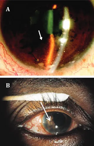
Figure 2. (A) The left eye shows a superior vascularized corneal scar with normal-appearing bulbar and tarsal conjuctiva.7 (B) Slit lamp picture of
the left cornea showing a peripheral corneal ulcer and vascularized nodule.7
Tuberculous scleritis should be considered in patients who are unresponsive to usual anti-inflammatory therapy.7 Anterior scleral involvement is more common; anterior
tuberculous scleritis may be localized or diffuse, most lesions are nodular and slighly elevated.10 Scleritis may be associated with cornea or iris leading to a condition of sclerokeratitis or uveitis.10
Granulomatous and non-granulomatous anterior uveitis can occur in TB with an insidious onset.7 Ocular manifestation of chronic granulomatous inflammation such as mutton-fat keratic precipitates on posterior aspect of cornea, nodule of the iris pupillary border (koeppe) or on iris surface (busacca). Localized nodule may lead to anterior synechiae.10 A nongranulomatous inflammation uveitis also can be occur in tuberculosis as small keratic precipitates and absence of iris nodules.7 Anterior uveitis can manifest as simple iritis or as iridocyclitis with involvement of ciliary body,7 inevitably
complicated as posterior synechiae and cataract.9 Clinical manifestations were predominantly non-granulomatous in HIV negative patients and granulomatous in HIV infected patients.6
Posterior Segment Ocular Tuberculosis
The most common clinical presentation of ocular TB is posterior uveitis (42%), followed by anterior uveitis (36%), panuveitis (11%), and intermediate uveitis (11%).11 The lesions predominantly present in the choroid as
Figure 3. Nodular scleritis in a patient with miliary TB7
Figure 4. (A) Anterior granulomatous uveitis with mutton fat keratic precipitates (black arrow) and iris nodule (white arrow).11 (B) right eye shows temporal
iris nodule.7
Figure 1. Classification the clinical presentation of ocular tuberculosis5
A
B
Ocular Tuberculosis
Anterior Segment
Conjungtiva, cornea, sklera, iris, cilliary body
Posterior Segment
Choroid, retina, optic nerve
Adnexa of Eye
serpiginous-like choroiditis, solitary or multiple choroidal nodules (tubercles), choroidal granuloma (tuberculoma), subretinal abscess, neuroretinitis, endophthalmitis, panophthalmitis, and retinal vasculitis.9
Serpiginous-like choroiditis (SLC) or multifocal serpiginoid choroiditis was a chronic, progressive, irregular geographic lesions in the fundus, midperiphery and periphery.12 SLC is seen at a young age, particularly from TB-endemic regions, may be unilateral or bilateral with vitreous inflammatory reaction.10 The higher prevalence of SLC in India and Turkey may be related to the higher incidence of latent TB compared to developed countries.12
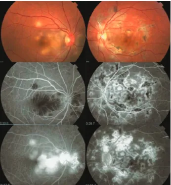
Bansal et al reported two distinct patterns of SLC : (1) multifocal lession of active choroiditis that were discrete and noncontiguous and then progressed to a diffuse, contiguous variety
and active serpiginous choroiditis or (2) diffuse plaque-like choroiditis showing amoeboid spread.13 On fluorescein angiography (FA), active lesions show initial hypofluorescence followed hyperfluorescence in late stage. On indocyanine green angiography (ICGA), the active lesions appear hypofluorescent during the early and late phases. SD-OCT reveals outer retinal disruption and increased outer retinal and choroidal reflectivity in the involved area.10
Choroidal tubercle are unilateral or bilateral, greyish-white to yellowish in color with indistinct margins, size varying from one half to several disc diameter, usually less than five in number.1 These lesions are mainly in posterior pole but can be seen in the mid-periphery.11 A choroidal tubercle may continues to grow as solitary mass, it known as tuberculoma.1 A solitary elevated mass-like lesion results from
a progressive, liquefied caseation necrotic with rapid multiplication of tubercular bacili and tissue destruction.14 Tuberculoma may be seen anywhere in the choroid-posterior pole, macula or juxta-papillary. They often mimicking a tumour, 4-14 mm in size and yellowish in color.1
Figure 6. (A) Fundus photograph of the right eye shows a tuberculoma at inferior macula.10 (B)
Multiple tubercles in the posterior fundus, some confluent.10
Optic nerve involvement may result from direct mycobacterial infection, by contiguous spread from the choroid or hematogenous dissemination or from hypersensitivity to the infectious agent. Papillitis, neuroretinitis and optic nerve tubercle were the most common clinical presentations.10 In a case report, the ocular manifestation of isolated optic disc TB was improved remarkably with antituberculosis treatment.15
Tuberculous retinal vasculitis occurs more frequently at veins than arteries.10 It presents with thick exudates, associated with retinal hemorrhages, active or healed focal choroiditis lesions, and moderate vitritis.10
Neovascularization and peripheral capillary occlusion have been described in retinal vasculitis.7 Macular edema may complicate tubercular retinal vasculitis. Optical coherence tomography (OCT) can be useful in evaluating macular edema or serous retinal detachment.10
Eales disease (ED) is a vaso-proliferative disorder of the retina characterized by peripheral retinal ischemia, neovascularization and recurrent vitreous hemorrhages.16 Several systemic diseases have been thought to be associated with its occurence but none have been proven. Positive polymerase chain reaction for Mycobacterium is found in around 50% patient with ED.17
Endophthalmitis and panophthalmitis were characterized with acute onset and rapid progression. The anterior chamber and vitreous reaction are typically severe10 Presentations include painless, progressive visual loss, decreased ocular motility, corneal cloudiness, sign of granulomatous ocular manifestation, and low intraocular pressure.7 Panophthalmitis may lead to scleral perforation.10
Adnexa of Eye
Lupus vulgaris is a chronic form of adnexa tuberculosis that affects eyelid skin and occurs in patient sensitive to tuberculin antigen. The lesions are solitary, small, reddish brown usually involving the head and neck region with gelatinous consistency (“apple jelly” color when pressure was applied).5,7 Non specific dacryoadenitis with or wtihout abscess formation is a usual presentation of lacrimal TB. Chronic dacryocystitis can present as sclerotic, painless, hard lobulated mass or as active caseous form (edematous lids with fluactuation and fistulization).5
DIAGNOSIS
Ocular TB may present with a wide spectrum of clinical manifestation and need uveal biopsy for culture or direct histopathological examination to provide definitive diagnosis. Most patient with ocular TB have no history of pulmonary or other systemic forms, such as in case of latent TB.9 Ocular TB should be in differential diagnosis in patient with uveitis of unknown etiology, recurrent or not responding to conventional therapy with findings suggestive of ocular TB.2 Intraocular TB can be assumed in the presence of cells in
anterior chamber or vitreous along with: broad posterior synechiae, retinal perivasculitis with or without discrete choroiditis, multifocal choroiditis, choroidal granuloma, optic disk granuloma, or optic neuropathy.18
Diagnosis and role of Anti-Tuberculous Therapy (ATT) in ocular TB management has been a challenge for ophthalmologist.19 In absence of gold standard tests to define the underlying pathogenic mechanisms, case definition has been assessed by expert opinion.20 There is no guidelines for intraocular TB (IOTB) diagnosis (Table).20
Microbiological confirmation of ocular TB can be made by performing acid-fast smear, microbacterial cultures, or polymerase chain reaction (PCR)-based assay on ocular fluids.19,20 Smear microscopy is cheap and easy to peform but has a highly false-negative result and cannot identify drug-resistance. Microbacterial culture on solid media is more sensitive than smear, but the average time to positive grow is 2-8 weeks and still needed to be tested biochemically for several weeks.21 In the last two decades, PCR has been used to detect MTB DNA from ocular fluid samples but with low sensitivity and lack of technique standardization.20 A commercially available and FDA approved NAAT, GeneXpert has been endorsed by WHO for detection of MTB. GeneXpert is a cartridge-based hemi-nested real-time automated PCR system which also can detect rifampicin resistance.20
Chest x-ray may provide evidence of active
or healed (primary or reactived) tuberculosis with consolidation, increased hilar densities, cavitation, fibrosis, and calcification or rarely lymph node enlargement. Lateral and PA view or additional top lordotic view should be ordered for a more sensitive visualization.22 High resolution CT scan can be used if chest x-ray is normal in clinically suspicious TB. If patient have no signs of active TB, immunological test (tuberculin skin test or interferon gamma test) should be performed in the same way as screening of latent tuberculosis.2
Management
Ocular TB is treated as extrapulmonary TB with the first-line combination regimen (antituberculosis therapy or ATT) comprising isoniazid 5 mg/kg/day (maximum 300 mg/ day), rifampicin 10 mg/kg (maximum 600 mg/day), pyrazinamide 20 to 25 mg/kg/day (maximum 1500 mg/day), and ethambutol 15 mg/kg/day (maximum 1000 mg/day).11 CDC guidelines recommend ATT for 6-9 months. Most studies prescribed ATT for at least 6 months, with maximum between 12-19 months.3 Patients on ATT should be monitored for any side effects or complications at every follow-up visit.2 Corticosteroids are used along with ATT to suppress inflammation caused by infection.3 Patients should be reviewed at the end of the initiation phase (2 months) and at the end of treatment (6-9 months).2,3 Treatment should not be stopped because of lack of response, unless other diagnosis has been made.2
Table. Proposed classification of intraocular tuberculosis20
Clinical Diagnostic Group Case Definition Criteria
Confirmed IOTB (both 1 and 2) At least one clinical sign suggestive of IOTB
Microbiological confirmation of Mycobacterium tuberculosis (MTB) from ocular fluids or tissues
Probable IOTB (1,2 and 3 together) At least one clinical sign suggestive of IOTB (other etiologies excluded) Evidence of chest x-ray consistent with TB infection or clinical evidence of extraocular TB or microbiological confirmation from sputum or extraocular sites.
At least one of the following: Documented exposure to TB Immunological evidence TB infection Possible IOTB (1,2 and 3 together) or (1 and
4) At least one clinical sign suggestive of IOTB (other etiologies excluded)Chest x-ray not consistent with TB infection and no clinical evidence of extraocular TB
At least one of the following: Documented exposure to TB Immunological evidence TB infection
Prognosis
Treatment efficacy has been difficult to define. Failure is defined as inability to taper oral corticosteroids to less than 10mg/day or topical oral corticosteroids to less than twice/ day, inability to stop oral immunosuppressive
agents as well as recurrence or persistence of inflammation within the first 6 months of ATT.2
CONCLUSION
Indonesia was in the second place of top 20 countries in absolute numbers and burden
severity of TB8 but no data on the incidence of ocular TB in Indonesia. Ocular TB may have wide spectrum of presentations, all parts of the eye maybe affected. The diagnosis should be kept in mind as differential diagnosis for ocular infection, especially in endemic area.
REFERENCES
1. Biswan J. Ocular tuberculosis- An update. Kerala J Ophthalmol. Chennai. 2009;XXI(4):351-7.
2. Figueira L, Fonseca S, Ladeira I, Duarte R. Ocular tuberculosis: Position paper on diagnosis and management. Rev Port Pneumol. 2016;10.004:1-8.
3. Kee AR, Gonzalez-Lopez JJ, Al-Hity A, Gupta B, Lee CS, Gunasekeran DV, et al. Anti-tubercular therapy for intraocular tuberculosis: A systematic review and meta-analysis survey of ophthalmology. Survey Ophthalmol 2016. doi: 10.1016/j.survophthal.2016.03.001.
4. World Health Organization. Global tuberculosis report 2016. Switzerland: WHO; 2016.
5. Goyal JL, Jain P, Arora R, Dokania P. Ocular manifestation of tuberculosis. Indian J Tuberculosis. 2015;4:4.
6. Nora RLD, Sitompul R, Susiyanti M, Edwar L, Soedarman S. Clinical characteristic and therapy of presumed ocular tuberculosis and their relation to HIV status. Med J Indones. 2012;21(4):214-9.
7. Albert DM, Raven ML. Ocular tuberculosis. Microbial Spectrum. 2016;4(6):1-17
8. World Health Organization. Use of high burden country lists for TB by WHO in the Post-2015 Era. Switzerland. 2016. 9. Shakarchi FI. Ocular tuberculosis: Current perspectives. Dove Medical Press Ltd. 2015; 9:2223-7.
10. Gupta V, Shoughy SS, Mahajan S, Khairallah M, Rosenbaum JT, Curi A, et al. Clinics of ocular tuberculosis. Informa Healthcare. USA. 2016;23(1):14-24. 11. Parchand S, Gupta V, Gupta A, Sharma A. Intraocular tuberculosis. J Postgrad Med Edu Res India. 2013;47(4):193-201.
12. Oray M, Zakiev Z, Cagatau T, Tugal-Tutkun I. Treatment result in serpiginous choroiditis and multifocal serpiginoid choroiditis associated with latent tuberculosis. Turk J Ophthalmol. 2017;47:89-93.
13. Bansal R, Gupta A, Gupta V, Dogra MR, Sharma A, Bambery P, et al. Tubercular serpiginous-like choroiditis presenting as multifocal serpiginoid choroiditis. Am Acad Ophthalmol. 2012;119:2234-42.
14. Gupta A, Gupta V. Tubercular posterior uveitis. Internat Ophthalmol Clinics. 2005.
15. Mansour AM, Tabbara KF, Tabbarah Z. Isolated optic disc tuberculosis. Karger. 2015;6:317-20.
16. Kumar V, Chandra P, Kumar A. Ultra-wide field angiography in the management of eales disease. Indian J Ophthalmol. 2016;64:504-7.
17. Mendonca MD, Guedes M, Matias G, Costa J, Viana-Baptista M. Steroid-responsive painful ophthalmoplegia: Tolosa-Hunt syndrome, eales disease, or both? International Headache Society. 2015.
18. Gupta A, Sharma A, Bansal R, Sharma K. Classification of intraocular tuberculosis. Ocul Immunol Inflamm. 2015;23:7-13. 19. Lee C, Agrawal R, Paveslo C. Ocular tuberculosis- A clinical conundrum. Ocul Immunol Inflamm. 2016;24(2):237-42. 20. Gupta A, Sharma A, Bansal R, Sharma K. Classification of intraocular tuberculosis. Ocul Immunol Inflamm. 2014;1-7.
21. Chang K, Lu W, Wang J, Zhang K, Jia S, Li F, et al. Rapid and effective diagnosis of tuberculosis and rifampicin resistance with Xpert MTB/RIF assay: A meta-analysis. J Infect. 2012; XX:1-9.