Ronald S. Duman, Jessica Malberg, Shin Nakagawa, and Carrol D’Sa
Studies at the basic and clinical levels demonstrate that neuronal atrophy and cell death occur in response to stress and in the brains of depressed patients. Although the mechanisms have yet to be fully elucidated, progress has been made in characterizing the signal transduction cas-cades that control neuronal atrophy and programmed cell death and that may be involved in the action of antide-pressant treatment. These pathways include the cyclic adenosine monophosphate and neurotrophic factor signal transduction cascades. It is notable that these same pathways have been demonstrated to play a pivotal role in cellular models of neural plasticity. This overlap of plasticity and cell survival pathways, together with studies demonstrating that neuronal activity enhances cell sur-vival, suggests that neuronal atrophy and death could result from a disruption of the mechanisms underlying neural plasticity. The role of these pathways and failure of neuronal plasticity in stress-related mood disorders are discussed. Biol Psychiatry 2000;48:732–739 © 2000 Society of Biological Psychiatry
Key Words: cAMP response element– binding protein,
brain-derived neurotrophic factor, apoptosis, neurogen-esis, mitogen-activated protein kinase
Introduction
R
ecent studies have resulted in considerable advance-ment of the monoamine hypothesis of mood disor-ders. Whereas earlier models focused on alterations in synaptic levels of monoamines, more recent studies dem-onstrate a major role for neural plasticity in the etiology and treatment of depression and stress-related illnesses. This neural plasticity includes adaptations of intracellular signal transduction pathways and gene expression. More-over, recent studies demonstrate that neural plasticity to stress and depression also involves alterations in neuronal morphology and cell survival. These findings suggest that neural plasticity may be necessary for the normal function and survival of neurons. Overlap of the signal transductionpathways that mediate neuronal plasticity and survival (e.g., the cyclic adenosine monophosphate [cAMP]– cAMP response element– binding protein [CREB] and neurotrophic factor–mitogen-activated protein [MAP] ki-nase cascades) also supports the notion of this relationship. The possibility that failure of neural plasticity could contribute to neurodegenerative disorders has recently been suggested (Finkbeiner 2000; Mesulam 1999). This review briefly discusses the background literature for a neuronal atrophy and cell death hypothesis of depression. Discussed in more detail are some of the intracellular cascades and target genes that control neuronal survival and plasticity and that could contribute to the actions of antidepressant agents.
Neuronal Atrophy and Death in Stress and
Depression
Recent basic and clinical studies have provided direct evidence of neuronal atrophy and loss in response to stress and depression. These studies are discussed in detail in other reviews found in this issue and will only be briefly mentioned here (also see Duman et al 1999). Basic research studies have demonstrated that stress can result in atrophy and death of CA3 pyramidal neurons in the hippocampus (Table 1; see McEwen 1999; Sapolsky 1996). In addition, stress decreases the neurogenesis of dentate gyrus granule neurons in the hippocampus of adult animals (Gould et al 1997, 1998). These damaging effects of stress could contribute to the reduction of hippocampal volume reported in patients with depression or posttrau-matic stress disorder (Bremner et al 1995; Drevets et al 1997; Sheline et al 1996); however, there are no reports to date that directly demonstrate a reduction in the number of neurons in hippocampus. This point needs to be addressed with additional postmortem analysis of cell number using unbiased stereological procedures.
In contrast, recent postmortem studies have demon-strated that the number of neurons and glia are reduced in the cerebral cortical regions, including prefrontal cortex, of depressed patients (Table 1; Ongur et al 1998; Raj-kowska et al 1999). In addition, one of these studies also found that the size of neurons in cortical areas was reduced (Rajkowska et al 1999). These studies demonstrate that cell loss and damage in depression are not restricted to the From the Laboratory of Molecular Psychiatry, Departments of Psychiatry and
Pharmacology, Yale University School of Medicine, Connecticut Mental Health Center, New Haven, Connecticut.
Address reprint requests to Ronald S. Duman, Ph.D., Connecticut Mental Health Center, 34 Park St., New Haven CT 06508.
Received March 15, 2000; revised May 5, 2000; accepted May 9, 2000.
© 2000 Society of Biological Psychiatry 0006-3223/00/$20.00
hippocampus. In addition, a reduction in the number of glia indicates that the loss of neurons is probably not the result of an inflammatory response, which would be expected to result in the opposite effect on the number of glia. These results suggest that neuronal loss may have occurred long before the time of death, possibly during the developmental period. Alternatively, this type of neuronal loss could result from programmed cell death or apoptosis. Additional basic research studies of the prefrontal cortex are required to determine if stress could produce similar effects and to investigate the cellular mechanisms respon-sible for cell loss.
Cellular Actions of Stress
There are many possible mechanisms that could contribute to the atrophy and loss of neurons observed in response to stress and depression. Although there is still a great deal that is not known, significant progress has been made in our understanding of how stress and adrenal glucocorti-coids damage certain populations of stress-vulnerable neurons (McEwen 1999; Sapolsky 1996). These studies demonstrate a role for glutamate excitoxicity and a reduc-tion in metabolic capacity, the latter resulting from a reduction in glucose uptake. Prolonged exposure to stress or glucocorticoids could be sufficient to produce cell damage via these mechanisms. Another possibility is that these effects could produce a state of neuroendangerment whereby cells become more vulnerable to other types of insult, such as hypoxia–ischemia, hypoglycemia, or viral infection. The mechanisms by which stress or glucocorti-coids decrease the neurogenesis of granule cells in the dentate gyrus have not been determined.
In addition to these effects, stress is also reported to decrease the expression of brain-derived neurotrophic factor (BDNF) in the hippocampus (Smith et al 1995). Brain-derived neurotrophic factor, a major neurotrophic
factor in the brain, is critical for the survival and guidance of neurons during development, but is also required for the survival and function of neurons in the adult brain (McAl-lister et al 1999; Thoenen 1995). For example, BDNF has been demonstrated to play a critical role in long-term potentiation, a cellular model of learning and memory, demonstrating that this neurotrophic factor can influence plasticity (Figurov et al 1996; Korte et al 1995). Decreased levels of BDNF in response to stress could lead to a loss of normal plasticity and eventually damage and loss of neurons. Reduction in BDNF expression, which is activity dependent, also suggests that neuronal activity may be decreased by stress, although there is no evidence for this to date. Assuming that this is the case, it is notable that a reduction in neuronal activity could contribute to the damaging effects of stress by reducing the survival of neurons (see below).
Neurotrophic Factors and Cell Survival
Neurotrophic factors are known to be critical for the survival of neurons during development in vivo and in culture (for complete references see reviews by Goldberg and Barres 2000; McAllister et al 1999; Thoenen 1995). In addition, neurotrophic factor support increases the survival of injured neurons in the adult brain. During development, neuronal survival is dependent on the presence of target-derived neurotrophic factor expression. In the absence of neurotrophic factor the neurons undergo a process of programmed cell death or apoptosis; however, this neuro-trophic hypothesis of neuronal survival may be an over-simplification (Goldberg and Barres 2000). The survival of a neuron may also be dependent on its synaptic connections with the target cell, as well as other cells (Goldberg and Barres 2000). Evidence for this possibility is supported by studies in cultured cells demonstrating that a combination of depolarization or activation of the cAMP pathway with exposure to a neurotrophic factor increases the survival and growth of neurons (Ghosh et al 1994; Goldberg and Barres 2000; McAllister et al 1996).
Activity-induced survival and growth of neurons could occur by several possible mechanisms (for complete ref-erences see Goldberg and Barres 2000). First, an increase in the number of synaptic connections and neuronal activity could elevate the expression of neurotrophic factor in the postsynaptic cells. This type of activity-dependent expression has been reported in many systems and for several different neurotrophic factors, including BDNF. Second, an increase in the number of synaptic connections could result in increased supply of neurotrophic factor from the presynaptic terminal. For example, BDNF is transported in an anterograde manner from dentate gyrus granule neurons to CA3 pyramidal neurons. Third, depo-Table 1. Stress and Depression Result in Neuronal Atrophy
and Cell Death in the Cerebral Cortex and Hippocampus
Prefrontal cortex
Decreased number and size of neurons in depressed patients Decreased number of glia in depressed patients
Decreased volume of the subgenual prefrontal cortex Influence of stress has not been determined Hippocampus
Decreased number and size of rodent CA3 neurons caused by exposure to stress
Decreased neurogenesis of rodent granule cells caused by exposure to stress
Decreased volume of the hippocampus in depressed patients Stereological analysis of cell number in the hippocampus of
depressed patients has not been determined
larization or activation of the cAMP cascade in postsyn-aptic neurons could increase the neuronal responsiveness to neurotrophic factor stimulation. Activity-dependent ex-pression of BDNF and other neurotrophic factors could have relevance to the atrophy and loss of neurons in depressed patients (see below).
CREB and Neurotrophic Factor–Mediated
Neuronal Survival
The process of programmed cell death includes shrinkage of neurons, condensation of chromatin, and finally complete cell disintegration (Nijhawan et al 2000). Although it was originally thought that neurotrophic factors increased cell survival by providing necessary trophic support, it is now clear that their survival-promoting effects are mediated by inhibition of the cell death pathway. Programmed cell death is controlled by factors that either promote or inhibit the cell death pathway. Proteins that are required for programmed cell death are the caspases, a family of cysteine proteases. Caspases are activated by proteolytic cleavage, resulting in degradation of cellular proteins that are necessary for neuro-nal survival. Conversely, other factors, the Bcl-2–like pro-teins, inhibit the cell death pathway, and thereby increase neuronal survival. Bcl-2 prevents cell death by inhibiting pathways that activate caspases; however, another member of
this group, Bad, blocks the actions of Bcl-2 and thereby promotes cell death.
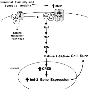
Recent studies have elucidated the mechanisms that underlie neurotrophic factor inhibition of cell death (Fig-ure 1; Finkbeiner 2000). These mechanisms include reg-ulation of both Bcl-2 and Bad. One of the intracellular pathways activated by neurotrophic factor coupled trk receptors is the MAP kinase cascade (Russell 1995). Interestingly, recent studies demonstrate that one of the targets of this pathway is CREB (Finkbeiner 2000). Activation of CREB via the MAP kinase cascade is mediated by ribosomal S6 kinase (Rsk). Thus, in addition to activation by cAMP-dependent protein kinase, as well as other second messenger– dependent protein kinases (protein kinase C and calcium/calmodulin-dependent pro-tein kinase), CREB can be phosphorylated and activated by Rsk.
The question as to how Rsk-2 and CREB mediate the survival or anti-apoptotic actions of neurotrophic factors has been addressed by two elegant studies (Figure 1; Bonni et al 1999; Riccio et al 1999). In one of these studies (Bonni et al 1999) BDNF-induced survival of cerebellar granule cells was found to be dependent on activation of Rsk and two important actions of this kinase. First, activated Rsk increases the phosphorylation of Bad, and thereby inactivates this cell death factor. Second, Rsk
increases the phosphorylation of CREB, resulting in up-regulation of the expression of Bcl-2, which inhibits cell death pathways. Riccio and colleagues (1999) also dem-onstrated a requirement for CREB and upregulation of Bcl-2 expression in neurotrophic factor–mediated survival of sympathetic neurons or cerebral cortical neurons. The promoter of the Bcl-2 gene contains a CRE that confers responsiveness to CREB, although additional transcription factors are thought to be required for induction of Bcl-2 expression (Finkbeiner 2000).
It is conceivable that the cell loss observed in depres-sion could result from alterations in the factors that control programmed cell death (e.g., increased Bad or decreased Bcl-2–like proteins) and the pathways that control the function and expression of these proteins (e.g., decreased Rsk and CREB). One study has already reported that levels of CREB are decreased in the cerebral cortex of depressed patients (Dowlatshahi et al 1998). Additional
studies are needed to confirm this finding and to examine other proteins involved in cell survival and plasticity.
Antidepressant Treatment Upregulates the
cAMP-CREB Cascade and Expression of
BDNF
Recent studies demonstrate that antidepressant treatment upregulates the cAMP–CREB cascade and expression of BDNF (Figure 2; Duman et al 1999). Regulation of the cAMP–CREB cascade includes increased coupling of the stimulatory guanosine triphosphate (GTP)– binding pro-tein (Gs) to adenylyl cyclase, increased particulate levels
of cAMP-dependent protein kinase, and increased expres-sion of CREB. A recent study has also demonstrated that antidepressant treatment increases CREB phosphorylation and CRE-mediated gene expression in limbic brain re-gions (Thome et al 2000). Upregulation of CREB and
Figure 2. Influence of antidepressant treatment on the cyclic adenosine monophosphate (cAMP)– cAMP response element– binding protein (CREB) cascade. Antidepressant treatment increases synaptic levels of norepinephrine (NE) and serotonin (5-HT) via blocking the reuptake or breakdown of these monoamines. This results in activation of intracellular signal transduction cascades, one of which is the cAMP–CREB cascade. Chronic antidepressant treatment increases Gscoupling to adenylyl cyclase (AC), particulate levels of
cAMP-dependent protein kinase (PKA), and CREB. CREB can also be phosphorylated by Ca21
-dependent protein kinases, which can be activated by the phosphatidylinositol pathway (not shown) or by glutamate ionotropic receptors (e.g., N-methyl-D-aspartate [NMDA]). Glutamate receptors and Ca21
BDNF occurs in response to several different classes of antidepressant treatments, including norepinephrine (NE) and serotonin selective reuptake inhibitors and electrocon-vulsive seizure, indicating that the cAMP–CREB cascade and BDNF are common postreceptor targets of these therapeutic agents (Nibuya et al 1995, 1996). In addition, upregulation of CREB and BDNF is dependent on chronic treatment, consistent with the therapeutic action of antide-pressants. A role for the cAMP–CREB cascade and BDNF in the actions of antidepressant treatment is also supported by studies demonstrating that upregulation of these path-ways increases performance in behavioral models of depression (Duman et al 1999).
The mechanisms underlying the upregulation of CREB and BDNF are currently being studied. The promoter of the BDNF gene contains a CRE and has been shown to be induced by CREB (Shieh et al 1998; Tao et al 1998). The mechanisms underlying the regulation of CREB are less clear, but one model we have proposed is that antidepres-sant treatment increases the phosphorylation and expres-sion of CREB via activation of cAMP-dependent protein kinase and Ca21-dependent kinases. This could occur via NE and serotonin receptors and the corresponding second messenger pathways, the cAMP and phosphatidylinositol cascades, which stimulate these kinases. Activation of Ca21-dependent kinases via glutamate receptors could also contribute to the regulation of CREB phosphorylation (Figure 2). Evidence for antidepressant regulation of N-methyl-D-aspartate– glutamate receptors has been re-ported (Paul et al 1994). It is also possible that antidepres-sant- and/or monoamine-induced upregulation of CREB could occur via activation of the MAP kinase pathway and Rsk. Recent studies demonstrate that activation of G protein– coupled receptors, including b-adrenergic and serotonin1A receptors, can also activate the MAP kinase
cascade (Luttrell et al 1999; Mendez et al 1999). These findings provide a novel pathway by which the NE and serotonin neurotransmitter systems, and psychotropic drugs that act on NE and serotonin, could influence CREB and other intracellular targets.
Upregulation of CREB and BDNF raises the possibility that antidepressant treatment could oppose the cell death pathway. This could occur via increased expression of Bcl-2. Studies are needed to determine if antidepressant treatment increases Bcl-2 expression. Lithium treatment has been reported to increase the expression of Bcl-2 in brain and cultured cells, and inhibits apoptosis of cultured cerebellar granule neurons (Chen et al 1999; Chen and Chuang 1999; Nonaka et al 1998). These findings suggest that regulation of the cell death pathways could also contribute to the actions of agents used for the treatment of bipolar disorder. It is also possible that antidepressant treatment could influence the function of Rsk, another
hypothesis that warrants future investigation. Direct stud-ies of antidepressant inhibition of cell death are more complicated and would involve conditions where cell death occurs naturally or is induced. For example, does antidepressant treatment influence the cell death that occurs during development or is induced by other types of stimuli? Another possibility is to study the influence of antidepressants on cell death in genetic models where a gene mutation increases cell death (e.g., Bcl-2 null mutant mice).
Antidepressant Treatment Increases
Hippocampal Neurogenesis
In addition to a potential role in cell survival, it is possible that antidepressant treatment also regulates other pro-cesses, such as neurogenesis, that could influence the number of neurons. Recent studies support this hypothesis and demonstrate that chronic, but not acute, antidepressant treatment increases the neurogenesis of dentate gyrus granule cells (Malberg et al 1999). These studies demon-strate that chronic administration of different classes of antidepressant treatment, including NE and serotonin se-lective reuptake inhibitors and electroconvulsive seizure, increases the proliferation and survival of new neurons. In contrast, increased neurogenesis is not observed in re-sponse to chronic administration of nonantidepressant psychotropic drugs.
located in the vicinity of the progenitor cells that are activated by antidepressant treatment and that release a proliferation factor onto the progenitor cells. Studies are currently underway to identify proliferation factors that could be regulated by CREB and could mediate the action of antidepressants.
Upregulation of granule cell neurogenesis could be one mechanism by which antidepressant treatment increases neuronal number in hippocampus. Increased granule cell neurogenesis could also influence the survival and func-tion of the CA3 neurons that receive input from the granule cells. This could occur via increased synaptic input and depolarization-induced survival of CA3 neurons, and increased levels of BDNF that is supplied by antero-grade transport from granule cells. A recent study demon-strating that neurogenesis also occurs in the cerebral cortex (Gould et al 1999b) raises the possibility that the cell loss in the cerebral cortex of depressed patients could result from decreased neurogenesis. Studies to determine the influence of stress and antidepressants on cortical neurogenesis are needed to test this hypothesis.
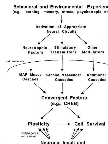
Neural Plasticity and Cell Survival
It is notable that CREB and BDNF have also been demonstrated to play a critical role in neural plasticity, particularly in cellular and behavioral models of learning and memory (Figurov et al 1996; Korte et al 1995; Silva et al 1998). Taken together, these studies demonstrate that there is considerable overlap between the intracellular pathways that mediate neuronal depolarization and neural plasticity and those that control cell survival (Figure 3). In addition to BDNF and CREB, it is likely that there are other convergent factors that mediate the actions of neural plasticity and cell death. This has resulted in the hypoth-esis that cellular activity and plasticity is a necessary requirement for neuronal survival. As mentioned above, increased synaptic activity could promote cell survival via a number of mechanisms, including increased amounts of, and responsiveness to, neurotrophic factors.
The possibility that failure of the mechanisms underly-ing neural plasticity could contribute to neurodegenerative disorders has been discussed (Mesulam 1999). We would like to extend this model and propose a hypothesis that stress-related affective illnesses, as well as other psychi-atric disorders, result in part from a loss of neuronal plasticity. For example, it is possible that the atrophy and loss of neurons in depressed patients may result from a reduction in the normal level of synaptic activity. This could be combined with stress-induced loss of neurotro-phic factors, or other factors that compromise neuronal function and activity (e.g., hypoxia–ischemia, hypoglyce-mia, viral infections). Another possibility is that there are
genetic factors, including mutations of the genes encoding intracellular signaling proteins, that influence neural plasticity.
Support for this hypothesis is provided by brain imaging studies. Reduced blood flow, which suggests decreased neuronal activity, has been reported in prefrontal regions when sadness is induced in healthy individuals (Mayberg 1997). In addition, relapse of depressive symptoms is reported to decrease metabolism in the dorsolateral pre-frontal cortex (Bremner et al 1997). These findings sug-gest that sadness and depression induce a reduction in neuronal activity in the prefrontal cortex. It is possible that
if this type of hypofunction is sustained for a long period of time it could result in a loss of activity-induced neurotrophic factors in this brain region. This could contribute to the reduction in volume and cell size and number that has been reported in depressed patients (Drevets et al 1997; Ongur et al 1998; Rajkowska et al 1999).
Development of Novel Therapeutic Agents
Studies demonstrating that an upregulation of the cAMP– CREB cascade is involved in the action of antidepressant treatment provide one set of novel targets for development of therapeutic agents. This includes the agents that act on the receptors that are directly coupled to the cAMP cascade (e.g., the serotonin7 receptor) or that could
di-rectly increase levels of cAMP (e.g., selective inhibitors of cAMP phosphodiesterase [PDE4] isozymes). These pos-sibilities are discussed in detail in previous reviews (Du-man et al 1997, 1999). Other targets of interest are the various components of the MAP kinase and cell death pathways. It is possible that agents that activate the MAP kinase pathway or Rsk could promote cell survival and have antidepressant efficacy. Further studies are needed to validate these targets and to utilize available inhibitors and activators of the MAP kinase cascade to test this possibility.
Summary and Conclusions
The studies discussed in this review raise the possibility that atrophy and cell death are intimately related to synaptic interactions between neurons and normal pro-cesses of neuronal plasticity. Failure of these propro-cesses and loss of synaptic interactions could contribute to a loss of neurotrophic factor input to neurons that could eventu-ally lead to further atrophy and death of neurons. Failure of neuronal plasticity could result from disruption of signaling processes at a number of levels, including the cAMP–CREB and neurotrophic factor–MAP kinase cas-cades; however, this is only a partial list of the complex array of intracellular signaling systems in the brain, and future studies are required to further elucidate the role of these pathways in depression. In addition, disruption of these processes could occur via environmental stimuli that compromise a cell’s ability to undergo neuronal plasticity. These findings lend support to the notion that affective disorders, as well as other psychiatric disorders, could result from a failure of plasticity. This hypothesis could be tested by brain imaging studies that characterize the brain regions where a reduction in neuronal activity occurs in response to thought processes related to depression or other disorders. In addition, this model could be further tested by a combination of behavioral and pharmacologic
interventions that correct or produce normal levels of neuronal activity in the affected brain regions. This hy-pothesis also supports the possibility that other types of neuronal insult (e.g., those resulting from cardiovascular disease) could explain their high rate of depression asso-ciated with these illnesses (Duman et al 1997).
The authors acknowledge the support of the U.S. Public Health Service (Grants Nos. MH45481, MH53199, and 2 PO1 MH25642), the Veterans Administration National Center Grant for PTSD, VA Medical Center, and the National Alliance for Research on Schizophrenia and Depression. Aspects of this work were presented at the conference “Depression in the Twenty-First Century: New Insights into Drug Development and Neurobiology,” February 21–22, 2000, Dana Point, California. The conference was sponsored by the Society of Biological Psychiatry through an unrestricted educational grant provided jointly by Pharmacia & Upjohn and Janssen Pharmaceutica.
References
Bonni A, Brunet A, West AE, Datta SR, Takasu MA, Greenberg ME (1999): Cell survival promoted by the Ras-MAPK signaling pathway by transcription-dependent and -indepen-dent mechanisms. Science 286(5443):1358 –1362.
Bremner JD, Innis RB, Salomon RM, Staib LH, Ng CK, Miller HL, et al (1997): Positron emission tomography measurement of cerebral metabolic correlates of tryptophan depletion-induced depressive relapse. Arch Gen Psychiatr 54:364 –374. Bremner JD, Randall P, Scott TM, Bronen RA, Seibyl JP, Southwick SM, et al (1995): MRI-based measurement of hippocampal volume in patients with combat-related post-traumatic stress disorder. Am J Psychiatry 152:973–981. Chen G, Zeng WZ, Yuan PX, Huang LD, Jiang YM, Zhao ZH,
Manji HK (1999): The mood stabilizing agents lithium and valproate robustly increase the levels of the neuroprotective protein bcl-2 in the CNS. J Neurochem 72:879 – 882. Chen RW, Chuang DM (1999): Long term lithium treatment
suppresses p53 and Bax expression but increases Bcl-2 expression. A prominent role in neuroprotection against excitotoxicity. J Biol Chem 274:6039 – 6042.
Dowlatshahi D, MacQueen GM, Wang JF, Young LT (1998): Increased temporal cortex CREB concentrations and antide-pressant treatment in major depression. Lancet 352:1754 – 1755.
Drevets WC, Price JL, Simpson JR, Todd RD, Reich T, Vannier M, Raichle ME (1997): Subgenual prefrontal cortex abnor-malities in mood disorders. Nature 386:824 – 827.
Duman RS, Heninger GR, Nestler EJ (1997): A molecular and cellular theory of depression. Arch Gen Psychiatry 54:597– 606.
Duman RS, Malberg J, Thome J (1999): Neural plasticity to stress and antidepressant treatment. Biol Psychiatry 46:1181– 1191.
Finkbeiner S (2000): CREB couples neurotrophin signals to survival messages. Neuron 25:11–14.
Ghosh A, Carnahan J, Greenberg ME (1994): Requirement for BDNF in activity-dependent survival of cortical neurons.
Science 263:1618 –1623.
Goldberg JL, Barres BA (2000): The relationshop between neuronal survival and regeneration. Annu Rev Neurosci 23: 579 – 612.
Gould E, Beylin A, Tanapat P, Reeves A, Shors TJ (1999a): Learning enhances adult neurogenesis in the hippocampal formation. Nat Neurosci 2:260 –265.
Gould E, McEwen BS, Tanapat P, Galea LAM, Fuchs E (1997): Neurogenesis in the dentate gyrus of the adult tree shrew is regulated by psychosocial stress and NMDA receptor activa-tion. J Neurosci 17:2492–2498.
Gould E, Reeves AJ, Graziano MSA, Gross CG (1999b): Neurogenesis in the neocortex of adult primates. Science 286:548 –552.
Gould E, Tanapat P, McEwen BS, Flugge G, Fuchs E (1998): Proliferation of granule cell precursors in the dentate gyrus of adult monkeys is diminished by stress. Proc Natl Acad Sci
U S A 95:3168 –3171.
Kempermann G, Kuhn HG, Gage FH (1997): More hippocampal neurons in adult mice living in an enriched environment.
Nature 386:493– 495.
Korte M, Carroll P, Wolf E, Brem G, Thoenen H, Bonhoeffer T (1995): Hippocampal long-term potentiation is impaired in mice lacking brain-derived neurotrophic factor. Proc Natl
Acad Sci U S A 92:8856 – 8860.
Luttrell LM, Ferguson SSG, Daaka Y, Miller WE, Maudsley S, Della Rocca GJ, et al (1999):b-Arrestin-dependent formation of b2 adrenergic receptor-Src protein kinase complexes.
Science 283:655– 661.
Malberg JE, Eisch AJ, Nestler EJ, Duman RS (1999): Chronic antidepressant administration increases granule cell neuro-genesis in the hippocampus of the adult male rat. Soc
Neurosci Abstr 25:1029.
Mayberg HS (1997): Limbic-cortical dysregulation: A proposed model of depression. J Neuropsychiatry 9:471– 481. McAllister AK, Katz LC, Lo DC (1996): Neurotrophin
regula-tion of cortical dendritic growth requires activity. Neuron 17:1057–1064.
McAllister AK, Katz LC, Lo DC (1999): Neurotrophins and synaptic plasticity. Annu Rev Neurosci 22:295–318. McEwen BS (1999): Stress and hippocampal plasticity. Annu
Rev Neurosci 22:105–122.
Mendez J, Kadia TM, Somayazula RK, El-Badawi KI, Cowen DS (1999): Differential coupling of serotonin 5-HT1A and 5-HT1B receptors to activation of ERK2 and inhibition of adenylyl cyclase in transfected CHO cells. J Neurochem 73:162–168.
Mesulam M-M (1999): Neuroplasticity failure in Alzheimer’s Disease: Bridging the gap between plaques and tangles.
Neuron 24:521–529.
Nibuya M, Morinobu S, Duman RS (1995): Regulation of BDNF and trkB mRNA in rat brain by chronic electroconvulsive seizure and antidepressant drug treatments. J Neurosci 15: 7539 –7547.
Nibuya M, Nestler EJ, Duman RS (1996): Chronic antidepres-sant administration increases the expression of cAMP re-sponse element binding protein (CREB) in rat hippocampus.
J Neurosci 16:2365–2372.
Nijhawan D, Honarpour N, Wang X (2000): Apoptosis in neural development and disease. Annu Rev Neurosci 23:73– 87. Nonaka S, Hough CJ, Chuang DM (1998): Chronic lithium
treatment robustly protects neurons in the central nervous system against excitotoxicity by inhibiting N-methyl-D -aspar-tate receptor-mediated calcium influx. Proc Natl Acad Sci
U S A 95:2642–2647.
Ongur D, Drevets WC, Price JL (1998): Glial reduction in the subgenual prefrontal cortex in mood disorders. Proc Natl
Acad Sci U S A 95:13290 –13295.
Palmer TD, Takahashi J, Gage FH (1997): The adult rat hippocampus contains primordial neural stem cells. Mol Cell
Neurosci 8:389 – 404.
Paul IA, Nowak G, Layer RT, Popik P, Skolnick P (1994): Adaptation of N-methyl-D-aspartate receptor complex fol-lowing chronic antidepressant treatments. J Pharmacol Exp
Ther 269:95–102.
Rajkowska G, Miguel-Hidalgo JJ, Wei J, Dilley G, Pittman SD, Meltzer HY, et al (1999): Morphometric evidence for neuro-nal and glial prefrontal cell pathology in major depression.
Biol Psychiatry 45:1085–1098.
Riccio A, Ahn S, Davenport CM, Blendy JA, Ginty DD (1999): Mediation by a CREB family transcription factor of NGF-dependent survival of sympathetic neurons. Science
286(5448):2358 –2361.
Russell DS (1995): Neurotrophins: Mechanisms of action.
Neu-roscientist 1:3– 6.
Sapolsky RM (1996): Stress, glucocorticoids, and damage to the nervous system: The current state of confusion. Stress 1:1–19. Sheline YI, Wany P, Gado MH, Csernansky JG, Vannier MW (1996): Hippocampal atrophy in recurrent major depression.
Proc Natl Acad Sci U S A 93:3908 –3913.
Shieh PB, Hu S-C, Bobb K, Timmusk T, Ghosh A (1998): Identification of a signaling pathway involved in calcium regulation of BDNF expression. Neuron 20:727–740. Silva AJ, Kogan JH, Franklind PW, Kida S (1998): CREB and
memory. Annu Rev Neurosci 21:127–148.
Smith MA, Makino S, Kvetnansky R, Post RM (1995): Stress alters the expression of brain-derived neurotrophic factor and neurotrophin-3 mRNAs in the hippocampus. J Neurosci 15:1768 –1777.
Tao X, Finkbeiner S, Arnold DB, Shaywitz AJ, Greenberg ME (1998): Ca21
influx regulates BDNF transcription by a CREB family transcription factor-dependent mechanism. Neuron 20:709 –726.
Thoenen H (1995): Neurotrophins and neuronal plasticity.
Sci-ence 270:593–598.
Thome J, Sakai N, Shin KH, Steffen C, Zhang Y-J, Impey S, et al (2000): cAMP-response-element mediated gene transcrip-tion is up-regulated by chronic antidepressant. J Neurosci 20:4030 – 4036.