ANESTHETIC MANAGEMENT OF A PATIENT WITH
CENTRAL AIRWAY COMPRESSION DUE TO POSTERIOR
MEDIASTINAL MASS
Nina Sulen1, Barbara Petani1, Ivan Bačić2 and Domagoj Morović2
1Department of Anesthesiology and Intensive Care Medicine, 2Department of Surgery, Zadar General Hospital, Zadar, Croatia
SUMMARY – Patients with mediastinal masses present unique challenge to anesthesiologists. Patients with anterior mediastinal masses have well documented cases of respiratory or cardiovas-cular collapse during anesthesia and in postoperative period. Masses in the posterior mediastinum have been traditionally regarded to carry a significantly lower risk of anesthesia related complicati-ons but cases of near fatal cardiorespiratory complicaticomplicati-ons have been reported. We describe anesthe-tic management of a patient with posterior mediastinal mass compressing the trachea and the left main bronchus presented for left thoracotomy and tumor excision. The patient experienced pain and cough, and exhibited positional dyspnea. Airway was successfully secured with awake nasotracheal intubation and placement of single lumen endobronchial tube.
Key Words: Mediastinal Neoplasms – Complications; Airway Obstruction; Airway Management; Bronchoscopy; Case Reports
Correspondence to: Nina Sulen, MD, Zadar General Hospital, Bože Peričića 5, HR-23000 Zadar, Croatia
E-mail: nina.sulen@zd.t-com.hr
Received March 1, 2015, accepted September 13, 2015
Introduction
The literature is focused mostly on anesthetic management of patients with anterior mediastinal masses, which have well documented cases of respi-ratory or cardiovascular collapse during anesthesia and in postoperative period1. Masses in the posterior mediastinum have been regarded to carry a signifi-cantly lower risk. However, there are reports of near fatal cardiorespiratory complications during manage-ment of patients with masses in the posterior medi-astinum1-3. We describe anesthetic management of a patient with posterior mediastinal mass compressing the lower trachea and the left main bronchus pre-sented for left thoracotomy and tumor excision. The patient experienced pain and cough, and exhibited positional dyspnea. Airway was successfully managed
with awake nasotracheal intubation and placement of single lumen endobronchial tube (SLEBT).
Case Report
A 36-year-old woman with posterior mediastinal mass presented for left thoracotomy and excision of tumor. The patient had good nutritional status and was previously healthy. For the last 2 months, she had dry cough and felt pain under the right costal arch that irradiated towards the right scapula. She was un-able to sleep in supine position due to dyspnea. Her breath sounds were normal and her blood pressure was 130/80 mm Hg. In supine position, she experi-enced dyspnea that she could tolerate for a short pe-riod. Her symptoms were similar in the left and right lateral position. Laboratory findings were within the normal limits.
Contrast enhanced computed tomography (CT) scan showed a homogeneous, well defined mass, 8.1 cm x 7.5 cm x 5.3 cm in size, in the posterior
medi-astinum that shifted the trachea anteriorly and to the right and exerted compression on the posterior tra-cheal wall narrowing tratra-cheal diameter to 50% at the point of maximal compression (Fig. 1). The left main bronchus was also compressed and bronchial diameter reduced to 35% of normal (Fig. 2).
Fiberoptic bronchoscopy revealed significant ex-traluminal compression of the trachea (Fig. 3) and the
left main bronchus (Fig. 4). Spirometry performed in upright position was within the normal limits. Since our patient experienced positional dyspnea with sig-nificant compression of the trachea and the left main bronchus, our plan was to perform awake nasotrache-al fiberoptic intubation.
On the morning of surgery, the patient was trans-ferred to the operating room in sitting position. Stan-dard monitors were applied and the patient was pre-medicated with 1 mg of midazolam. Epidural catheter
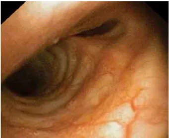
Fig. 4. Bronchoscopy image at the level of tracheal carina showing patent right main bronchus and almost complete obstruction of the left main bronchus.
Fig. 1. Transverse section of computed tomography scan showing posterior mediastinal mass compressing and dis-placing the trachea anteriorly and to the right.
Fig. 2. Coronal section of computed tomography scan showing posterior mediastinal mass compressing the tra-chea and the left main bronchus.
Fig. 3. Bronchoscopy image showing extraluminal com-pression on the posterior tracheal wall.
was placed on Th5/6 level for intra- and postopera-tive analgesia without difficulties. Right radial artery was cannulated for invasive blood pressure monitor-ing. Rigid bronchoscope and thoracic surgeon were available in the operating room. Both nostrils were prepared for nasal intubation with ephedrine solution and 2% lidocaine gel. Additional 2% lidocaine solu-tion was applied through a narrow catheter posisolu-tioned in the nasopharynx. Right-sided single lumen endo-bronchial tube, 6.5 mm internal diameter, 45 cm in length (Rush, Teleflex Medical, Wayne, Pennsylva-nia, USA) was coupled with fiberoptic bronchoscope LF-GP with outer diameter 4.1 mm (Olympus, To-kyo, Japan). Endobronchial tube was positioned at a depth of 13 cm in the right nostril and bronchoscope was passed through the tube. Additional 2% lidocaine solution was applied on the aditus. On advancing the bronchoscope, the compressed segment of the trachea was visualized. The right bronchus was patent but there was almost complete occlusion of the left main bronchus. Endobronchial tube was passed over the bronchoscope beyond tracheal narrowing at the level just above the carina without difficulties. Propofol and fentanyl were given and sevoflurane was started. The patient was breathing spontaneously. She was turned in supine position and with increasing depth of an-esthesia manually assisted ventilation was applied. Bronchoscopy was performed again to position the tube in the right main bronchus. After turning the patient in lateral decubitus position, the tube was re-checked and tracheal cuff was briefly released to allow collapse of the lung on the operated side. Anesthesia was maintained with sevoflurane and fentanyl. Dur-ing preparation for surgery, arterial pressure dropped to 90/60 mm Hg and the patient received 4 mg of etilefrine. Fifteen minutes after beginning of the surgery, pressure controlled ventilation was initiated without difficulties and a bolus dose of rocuronium was administered. Sevoflurane was reduced and the patient remained hemodynamically stable thereafter. The patient was extubated in the operating room and transferred to the Intensive Care Unit. Histopatho-logic diagnosis was bronchogenic cyst.
Discussion
Literature reports are focused mostly on manage-ment of patients with anterior mediastinal masses
while masses in the posterior mediastinum have been regarded to carry a significantly lower risk. How-ever, there are reports of near fatal cardiorespiratory complications during management of patients with masses in the posterior mediastinum. Since mediasti-nal compartments are not bounded with firm bound-aries, sufficient tumor growth anywhere within the mediastinum can cause significant compression of vital structures. Masses in the posterior mediastinum cause compression of the trachea, main bronchi, left atrium and left ventricle. Posterior tracheal wall due to its membranous structure can be especially prone to external compression1. Respiratory and cardiovascular decompensation requiring urgent cardiopulmonary bypass due to posterior mediastinal mass has been re-ported2. Patients with mediastinal masses need very meticulous preoperative assessment and planning. Dyspnea, cough and syncope, especially when chang-ing position from upright to supine, are very signifi-cant symptoms. Position that is most comfortable for the patient must be determined preoperatively. Seri-ous complications are reported in patients with un-remarkable history and clinical examination, so care-ful evaluation of CT scan is necessary, supplemented with fiberoptic bronchoscopy and echocardiography in selected cases1,4. Patients with compression of major airways are at an increased risk of respiratory compli-cations during postoperative period as well3,6.
Since our patient had positional dyspnea and sig-nificant compression of the trachea with associated bronchial compression, she was at an increased risk of perioperative airway decompensation1. For patients with central airway obstruction, the safest option is securing distal airway with awake fiberoptic intuba-tion and placement of endotracheal tube distal to tra-cheal obstruction.
If general anesthesia is indicated, volatile or intra-venous induction is performed with preservation of spontaneous breathing. The use of muscle relaxants should be avoided but if positive pressure ventila-tion is possible without significant increase in airway pressures, they can be given. If respiratory decom-pensation ensues intraoperatively, rigid bronchos-copy may be necessary for ventilation. In the event of respiratory or cardiovascular collapse, changing patient position to the most comfortable one as de-termined preoperatively can lead to improvement4.
Airway collapse despite spontaneous ventilation has been reported in patients with anterior mediastinal mass with airway compression beyond the tip of the endotracheal tube7.
Besides securing airway beyond the compressed tracheal segment, lung isolation was required for planned thoracotomy and excision of tumor. Rec-ommended technique for lung isolation in patients with predicted difficult airway is awake orotracheal or nasotracheal intubation followed by placing a bronchial blocker (BB) or replacing single lumen tracheal tube for double lumen tracheal tube over the airway exchange catheter after induction of gen-eral anesthesia. Lung isolation with endobronchial placement of single lumen tracheal tube is rarely performed8,9. Nasotracheal intubation was selected for our patient because it usually enables easier glot-tic visualization and smoother tube passage into the trachea with less breath holding, which is es-pecially undesirable in patients with central airway obstruction. A tube long enough to enable nasotra-cheal intubation and positioning of the tip distally to the compressed tracheal segment was required, so SLEBT was selected.
Single lumen endobronchial tubes are double cuffed single lumen tubes adjusted for left- or right-sided bronchial intubation. They were used in the past for lung separation but they were substituted with double lumen tubes and bronchial blockers in mod-ern thoracic anesthesia. However, SLEBT can have role in particular clinical settings for airway manage-ment in the surgery of distal trachea and main bronchi and for lung separation in obese patients10. Modified SLEBT was successfully used for airway management in severe tracheobronchial compression11. Also, when dealing with difficult airway in emergency, SLEBT could be easier to place than double lumen tube and protect the lung more safely than BB because it is less prone to dislodgement12. For our patient, advantage of SLEBT compared with double lumen tube was small-er outsmall-er diametsmall-er that allowed smooth awake nasal intubation and passage through the significantly nar-rowed trachea. Compared to single lumen tube with BB, the advantage of SLEBT was sufficient length that allowed nasotracheal intubation with reliable positioning of the tip of the tube distally to tracheal obstruction.
Conclusion
For symptomatic patients with central airway ob-struction due to posterior mediastinal mass, the safest option for airway management is awake fiberoptic in-tubation and placement of endotracheal tube distally to tracheal obstruction. Single lumen endobronchial tube can be used in a particular clinical setting for managing lung separation in patients with difficult airway.
References
1. Blank RS, de Souza DG. Airway management of patients with an anterior mediastinal mass: continuing professional development. Can J Anesth. 2011;58:853-67. doi: 10.1007/ s12630-011-9539-x
2. Anderson DM, Dimitrova GT, Awad H. Patient with pos-terior mediastinal mass requiring urgent cardiopulmonary bypass. Anesthesiology. 2011;114:1488-93. doi: 10.1097/ ALN.0b013e31821a8af1
3. Lalwani P, Chwala R, Kumar M, Tomar AS, Raman P. Pos-terior mediastinal mass: do we need to worry much? Ann Card Anaesth. 2013;16:289-92. doi: 10.4103/0971-9784.119183 4. Erdos G, Tzanova I. Perioperative anaesthestic
manage-ment of mediastinal mass in adults. Eur J Anaesthesiol. 2009;26:627-32. doi: 10.1097/EJA.0b013e328324b7f8 5. Bechard P, Letourneau L, Lacasse Y, Cota D, Bussieres JS.
Perioperative cardiorespiratory complications in adults with mediastinal mass: incidence and risk factors. Anesthesiology. 2004;100:826-34.
6. Slinger P. Management of the patient with a central air-way obstruction. Saudi J Anaesth. 2011;5:241-3. doi: 10.4103/1658-354X.84094
7. Gardner JC, Royster RL. Airway collapse with an ante-rior mediastinal mass despite spontaneous ventilation in an adult. Anesth Analg. 2011;113:239-42. doi: 10.1213/ ANE.0b013e31821f9c95
8. Brodsky JB. Lung separation and the difficult airway. Br J Anaesth. 2009;103(Suppl 1):i66-i77. doi:10.1093/bja/aep262 9. Globokar MD, Novak Janković V. Difficult airway and one
lung ventilation. Acta Clin Croat. 2012;51(3):477-82. 10. Conacher ID, Velasquez H, Morrice DJ. Endobronchial
tubes – a case for re-evaluation. Anaesthesia. 2006;61:587-90. doi:10.1111/j.1365-2044.2006.04644.x
11. Harte BH, Jaklitsch MZ, McKenna SS, Body SC. Use of a modified single-lumen endobronchial tube in severe tracheo-bronchial compression. Anesthesiology. 2002;96:510-1. 12. Merli G, Guarino A, Della Rocca G, Frova G, Petrini F,
Sorbello M, et al. Recommendations for airway control and difficult airway management in thoracic anesthesia and lung separation procedures. Minerva Anestesiol. 2009;75:59-96.
Sažetak
ANESTEZIOLOŠKI POSTUPAK KOD BOLESNIKA S KOMPRESIJOM SREDIŠNJEGA DIŠNOG PUTA ZBOG MASE U STRAŽNJEM MEDIJASTINUMU
N. Sulen, B. Petani, I. Bačić i D. Morović
Bolesnici s masama u medijastinumu predstavljaju jedinstven izazov za anesteziologe. U literaturi se uglavnom govori o anesteziološkim postupcima kod bolesnika s masama u prednjem medijastinumu za koje postoje brojni dobro dokumenti-rani slučajevi respiracijskog i kardiovaskularnog kolapsa tijekom anestezije i u poslijeoperacijskom razdoblju. Smatra se da mase u stražnjem medijastinumu imaju značajno manji rizik vezan za anesteziju, ali su i kod ovih bolesnika opisani slučaje-vi teških perioperacijskih kardiorespiracijskih komplikacija. Prikazan je anesteziološki postupak kod bolesnice s tumorom u stražnjem medijastinumu koji je uzrokovao kompresiju traheje i lijevog glavnog bronha, predviđene za torakotomiju i eksciziju tumora. Bolesnica je imala simptome kašlja, bolova i dispneje u ležećem položaju. Dišni put je uspješno osiguran nazotrahealnom intubacijom budne bolesnice i postavljanjem jednoluminalnog endobronhalnog tubusa.
Ključne riječi: Medijastinalni tumori - komplikacije; Dšini put, opstrukcija; Dišni put, zbrinjavanje; Bronhoskopija; Prikazi slučaja