ER
S &
M
C
D
O
WE
LL
: S
N
AKE
S AN
D
H
EM
IP
EN
ES
A
M
N
H
BU
LLE
TIN
3
85
20
14
BULLETIN OF THE AMERICAN MUSEUM OF NATURAL HISTORY
N
EW
T
A X A AND
C
RYPTIC
S
PECIES OF
N
EOTROPICAL
S
NAKES (
X
ENODONTINAE),
WITH
C
OMMENTARY ON
H
EMIPENES AS
G
ENERIC AND
S
PECIFIC
C
HAR ACTERS
CH A RLES W. MYERS
A N D SA MU EL B. M
C
D OW ELL
American Museum Novitates
Bulletin of the American Museum of Natural History
Anthropological Papers of the American Museum of Natural History
Publications Committee Robert S. Voss, Chair Board of Editors
Jin Meng, Paleontology
Lorenzo Prendini, Invertebrate Zoology Robert S. Voss, Vertebrate Zoology Peter M. Whiteley, Anthropology Managing Editor
Mary Knight
Submission procedures can be found at http://research.amnh.org/scipubs
O n t h e c o v e r :
Juvenile and adult colorations of
Xenodon
“
rabdocephalus
,” a geographically widespread
complex of cryptic species and fer-de-lance mimics.
Photos by C.W. Myers.
All issues of Novitates and Bulletin are available on the web from http://digitallibrary.amnh.org/dspace
Order printed copies from http://www.amnhshop.com or via standard mail from: American Museum of Natural History—Scientific Publications
Central Park West at 79th Street New York, NY 10024
NEOTROPICAL SNAKES (XENODONTINAE), WITH
COMMENTARY ON HEMIPENES AS GENERIC
AND SPECIFIC CHARACTERS
CHARLES W. MYERS
SAMUEL B. M
CDOWELL
Division of Vertebrate Zoology
Department of Herpetology
American Museum of Natural History
New York, New York
BULLETIN OF THE AMERICAN MUSEUM OF NATURAL HISTORY
Number 385, 112 pp., 40 figures, 1 map Issued March 6, 2014
Abstract . . . 3
Introduction . . . 4
Higher-level taxonomy. . . 5
Descriptions of new and reassigned taxa. . . 6
Eutrachelophiini, new tribe . . . 6
Eutrachelophis, new genus . . . 6
Eutrachelophis bassleri, new species . . . 8
Eutrachelophis, undescribed species . . . 14
Eutrachelophis steinbachi(Boulenger), new combination . . . 14
Global comparisons . . . 20
Viscera . . . 20
Head glands . . . 21
Head muscles . . . 22
Skull and dentition . . . 26
Color pattern . . . 37
Hemipenes . . . 39
Summary of global comparisons. . . 42
Discussion . . . 43
Hypothesis for hemipenial transformation inEutrachelophis. . . 45
On the apical disc . . . 47
Loss of a ‘‘generic character’’—the apical disc . . . 49
Commentary on hemipenes as generic and specific characters . . . 52
Hemipenial terminology for snakes . . . 55
Are hemipenes ‘‘conservative’’ characters? . . . 59
Examples of intraspecific variation in snake hemipenes and cloacae. . . 61
Evolutionary plasticity and extreme divergence of snake hemipenes . . . 67
Theoretical bases for hemipenial evolution . . . 72
Eberhard’s thesis. . . 72
Hoxgene expression . . . 73
Taxonomic use of hemipenial data . . . 74
Conclusions and brief summation . . . 75
Museum abbreviations . . . 76
Acknowledgments . . . 77
Appendix 1: Nomenclatural considerations, by C.W. Myers . . . 77
Notes on the tribe Xenodontini Bonaparte . . . 77
Color pattern variation in Xenodon rabdocephalus, sensu lato, with X. suspectus Cope removed from synonymy . . . 79
Lectotype designation forXenodon rabdocephalus(Wied) . . . 83
Xenodon angustirostrisW. Peters removed from synonymy . . . 89
Named genera based on loss of the apical disc. . . 90
Postscript . . . 92
Appendix 2: The Vidian canal and venous foramina in the prootic of alethinophidian snakes, by S.B. McDowell . . . 92
‘‘The Vidian canal’’ . . . 92
Venous drainage and foramina. . . 102
References . . . 104
Eutrachelophis, new genus is established to accommodate E. bassleri, new species, and E. steinbachi(Boulenger), new combination; a third species close to E. bassleri awaits naming. These taxa are placed in the Eutrachelophiini, new tribe, to express hypothesized relationship with the Xenodontini, which are defined by presence of hemipenial apical discs (a character lost in several species). The acalyculate spiny hemipenis ofEutrachelophis bassleriis unique among ‘‘xenodontines’’ in having a noncapitate, well-formed capitulum in the form of a nude dome; bifurcation is lacking even in the insertion of the major retractor muscle; the sulcus spermaticus is centrolineal in the retracted organ but becomes centrifugal during eversion.
The hemipenis ofEutrachelophis steinbachiis strikingly different in being deeply divided, with long spiny lobes tipped with tufts of sender spines, but it resembles those of some other colubrids (e.g., South American Xenodon suspectus; African Mehelya poensis). Based on hemipenial comparisons, E. bassleri and E. steinbachi seem unlikely congeners. Nonetheless, global comparisons of viscera, head glands, head muscles, color pattern, skull, and dentition indicate that they are congeneric despite hemipenial differences. NeitherE. basslerinor E. steinbachi
shows sufficient resemblance to any other ‘‘xenodontine’’ that would suggest an alternative phylogeny. Overall resemblance in so many details, especially of the skull, is not reasonably explained by convergence.
Therefore, contrary to dogma, the hemipenes in this case provide no clues to generic affinity. An explanatory hypothesis hasEutrachelophis bassleriandE. steinbachiderived from common stock, but with hemipenial lobes in the bassleri lineage suppressed during embryonic development. It further suggests that the unusual broad, hemispherical nude apex inE. bassleri
is homologous with the interlobular smooth, expandable terminal basin inE. steinbachi. The hemipenial differences in Eutrachelophis are not inconsistent with growing awareness that evolution of male genitalia may outpace changes in other characters without predictable limits to complexity. Fine-scale Hoxgene expression might account for the novel hemipenis of E. bassleri.
Although it is well established that snake hemipenes generally give at least a hint of relationship, a widely held belief that they are taxonomically stable and relatively free of selection pressures must be abandoned. Hemipenes (and probably female cloacae) are not ‘‘neutral’’ or ‘‘uncorrelated’’ characters but are subject to intense selection pressure requiring successful copulation, hence successful reproduction. The belief that one description or illustration suffices to typify a species (or genus) has no merit without proper sampling. Intraspecific variation is commonplace in geographically widespread species—sometimes, not always, signaling the presence of unnamed cryptic species.
Examples are given of intraspecific variation in different kinds of hemipenial features. Also provided are examples of evolutionary plasticity and extreme divergence in snake hemipenes, with a few references to female cloacae, about which much less is known. The hemipenes of two apparent sister species,Enulius flavitorquesand ‘‘Enuliophis’’sclateri, might represent a case of hemipenial divergence as extreme as seen between Eutrachelophis bassleri and E. steinbachi. Attention is called to examples of extraordinary folding and coiling of retractor muscles and even a folding hemipenis, all of which enable long hemipenes to fit within short tails in both Scolecophidia (Typhlina) and Alethinophidia (Prosymna). Folding of the hemipenis and retractor muscle is illustrated for the AfricanProsymna ambigua bocagii,which has hemipenes longer than the tail. Axial architecture and limblessness of snakes have been attributed toHox
gene expression, which we suggest may also be the mechanistic basis for the appearance of hemipenial novelties without gradual change.
Myers (1986) and the late Garth Underwood (2001, letters published herein), working with biometrician Clive Moncrieff, had independently concluded that the apical disc—the defining character of tribe Xenodontini—has been lost in some populations of the geographically widespreadXenodon ‘‘rabdocephalus’’ (Wied). That finding is here extended also toXenodon suspectus Cope, which is resurrected from the synonymy ofX. ‘‘rabdocephalus’’ based on a variable but cohesive color pattern and a hemipenis tipped with slender apical spines. A
lectotype is designated for X. rabdocephalus,sensu stricto, which has been said to lack apical discs; the lectotype, however, has small apical discs on long slender lobes.
Underwood and Moncrieff did not find the apical disc on hemipenes in Central American or in most South American populations ofXenodon‘‘rabdocephalus.’’ Its absence is confirmed for populations in Central America and northwestern Colombia, for which the name Xenodon angustirostrisW. Peters, 1864 (type locality Veragua) is tentatively resurrected. But sample sizes are small and hemipenial characteristics need to be elucidated for more populations. In addition to unrecognized species, South American ‘‘rabdocephalus’’ overall includesX. angustirostris,X. suspectus, andX. rabdocephalussensu stricto.
Xenodon rabdocephalussensu lato is a complex of an unknown number of cryptic or ‘‘hidden’’ species, in which speciation events appear to be signaled by hemipenial changes. However, hemipenial data are too sparse at this time to allow separation of taxa because color patterns maintain a degree of consistency throughout an enormous geographic range (Mexico to Bolivia). These fer–de-lance mimics presumably are under strong selection pressure to maintain aBothrops -like color pattern. A comprehensive study is needed, with molecular input to the extent possible. Loss of the apical disc among species ofXenodon may have been confined to species like ‘‘rabdocephalus’’ that have long-lobed hemipenes primitively tipped with small discs. Such loss characters can helpfully define species, but do not suffice for genera. Junior synonyms of
XenodonincludeAcanthophallusCope, 1894 (type species Xenodon colubrinusGu¨ nther, 1858); and ThalesiusYuki, 1993 (type speciesXenodon werneriEiselt, 1963), both based on absence (loss) of the apical disc. Waglerophis Romano and Hoge, 1972, with apical discs on long hemipenial lobes, also is a synonym. (These interpretations ofXenodonsynonymy agree with Zaher, 1999, and Zaher et al., 2009.)
Terminology applicable to snake hemipenes is reviewed, particularly for a few characters needing clarification. Illustrations show the difference between capitation and noncapitation of hemipenes or hemipenial lobes with distinct capitula (‘‘heads’’). A commonly overlooked character—the smooth terminal basin of some bilobed hemipenes is described. The simple tripartite system for describing the orientation of sulci spermatici is updated. Complementary comparisons of retracted and everted hemipenes are encouraged, to provide better understanding and sometimes to increase the number of taxonomically useful characters.
A discussion is appended of the Vidian canal and venous foramina in the prootic of Alethinophidian snakes. The Vidian canal in Pythonidae and the fossilDinilysiashows the most lizardlike, presumably primitive, pattern. Anilioids show departures from this pattern, as do Acrochordoidea and Colubroidea. Boidae differ from Pythonidae in the loss of a true basipterygoid process (replaced, at least embryonically, by a cartilaginous nodule). Among the variable colubroid conditions, the Vidian canal in Eutrachelophis bassleriandE. steinbachiis similar to that ofLiophisin being short and entirely enclosed in the sphenoid, with its anterior orifice set well in from the sphenoid-parietal suture.
INTRODUCTION
This is a study in which we originally set out to discover the affinities of two South American snakes. One was an unnamed species from the upper Amazon drainage of Peru and Ecuador; the other wasRhadinaea steinbachi Boulenger (1905) of east-central Bolivia—a snake not assignable to any currently defined genus (fide Myers, 1974: 22). These are small slender colubrids that share a low number (15) of dorsal scale rows
and similar nuchal color patterns, by which they are readily distinguished from nearly all other snakes. Externally they easily appeared congeneric, but differences in the male genitalia would seem to place them in different tribes or even in different genera. The hemipenis ofR. steinbachiis long, deeply divided, and spinose to the tips, whereas that of the new species is short, undivided, proximally spinose, and distally nude.
The systematic problem is an old one: Do the resemblances reflect affinities or do
striking differences in a taxonomically im-portant structure indicate lack of kinship? We initially thought the latter and suspected convergence in external features, but after looking to many additional characters we concluded that there actually is a close relationship. We think that the affinities between the two species are best expressed by placing them in a single new genus, to which a third, more recently discovered species is added. At the same time, we erect a new tribe in the subfamily Xenodontinae.
Although the hemipenial differences be-tween the first two species are striking, they are not inconsistent with growing awareness that evolution of male genitalia may proceed in advance of changes in other characters, without predictable limits to complexity (Eber-hard, 1985, 2004). The taxonomic decision is based on morphology; tissue samples have not been collected, although we hope that molec-ular data will eventually provide additional insight.
This study also led to broad comparisons on use of the hemipenis for systematic purposes—leading to conclusions that it
sometimesis worthless as a generic character, but that, on the other hand, it often is indispensable in defining species, particularly those in which hemipenial evolution has outpaced change in other characters. The first situation applies to cases of extreme hemipenial diversity, as illustrated in this paper. The second is applicable to the cryptic species of Xenodon ‘‘rabdocephalus,’’ a com-plex of fer-de-lance mimics distributed from Mexico to Bolivia; these ‘‘hidden’’ species are presumably under heavy selection pressure to maintain theirBothrops-like coloration.
First, however, we provide a brief historical framework and the rationale for our practical approach to the higher-level taxonomy of ‘‘colubrid’’ and ‘‘xenodontine’’ snakes.
HIGHER-LEVELTAXONOMY
The vast majority of Neotropical colubrid snakes have long been considered to belong to the poorly characterized subfamily Xeno-dontinae. Two large assemblages have been informally called Central American and South American xenodontines, although there is broad geographic superposition.
These two groups received suggestive support as clades from early microcomplement fixa-tion studies of serum albumins (Cadle, 1984a, 1984b, 1984c, 1985). Some hemipenial differ-ences were pointed out by Myers and Cadle (1994: 27–28), who thought that the hemi-penes of the South American clade were relatively primitive compared with the Cen-tral American clade, which is characterized by several derived hemipenial features.
Zaher (1999) recognized the mainly South American clade as Xenodontinae, sensu stricto, based on two hemipenial synapomor-phies (enlarged calyces on the asulcate sides of the lobes, and enlarged lateral spines), which are seen either individually or together in most species. Zaher (1999) recognized the mainly Central American clade as subfamily Dipsadinae, based on several hemipenial synapomorphies suggested by Myers and Cadle (1994: 27).
Zaher et al. (2009) subsequently reworked the classification of caenophidian snakes based on a new molecular phylogeny. The family Colubridae was greatly restricted from ‘‘its long-standing use [for] all caenophidians that were not acrochordids, elapids, or viper-ids’’ (Zaher et al., 2009: 132). In addition to confirming removal of a number of Old World groups whose relationships had been ques-tioned, the name Colubridae in its common sense was most significantly affected by raising its main subfamilies (colubrines, na-tricines, dipsadines) to familial level. The subfamily Xenodoninae was returned to its earlier concept of a single group of New World ‘‘xenodontines’’ by submersing it within family Dipsadidae. The Xenodoninae were then judged to have no known synapo-morphies because those presented by Zaher (1999) were moved to the Dipsadidae (see Zaher et al., 2009: 140). The Xenodoninae were nonetheless retained as a subfamily in order to avoid ‘‘changing the well-established taxonomic hierarchy for this group’’ (Zaher et al., 2009: 141–142).
Myers (2011: 11ff.) responded narrowly to Zaher et al. (2009) by arguing that the aforesaid familial changes were unnecessary. He believed that ‘‘discussion is hampered and becomes confused when new taxonomies are generated from new (uncorroborated) phylo-genetic hypotheses, especially when familiar
groups are renamed and redefined in major
ways’’ (Myers, 2011: 12). As a practical reason for retaining Colubridae, sensu lato, Myers (2011: 13) noted that he could not unambiguously assign the new genus Amnes-teophisto any of four subfamilies to which he had compared it.
We agree with Zaher and others that sound classification should reflect phylogeny, but we disagree fundamentally as to whether taxonomy needs to be changed almost automatically with new phylogenetic insight. We value any insights provided by the molecular phylogeny generated by Zaher et al. (2009), but for practicalreasons (sensu Myers, loc. cit.) we continue to recognize the Dipsadinae, Xenodontinae, and Colubrinae as subfamilies of Colubridae. We continue to use ‘‘colubrid’’ as a sort of common name for snakes in or closely related to members of these subfamilies. We follow Zaher (1999: 3) in using, for pragmatic reasons, the term ‘‘xenodontines’’ in quotes for many dipsadine and xenodontine genera of uncertain assign-ment, including some colubrids used herein for anatomical comparisons (e.g., Diadophis,
Rhadinophanes,Taeniophallus).
It must not be thought that our few critical comments are directed solely at Zaher et al. (2009); we are suspicious of all phylogenies that are published rapidly in order to yield ‘‘provisional’’ taxonomies (e.g., Kelly et al., 2011; see fn. 17 herein). Although new genera and new species must be dealt with in a timely fashion, ‘‘provisional’’ higher-level classifica-tions can and should compete among them-selves before new taxonomies are accepted by biologists who actually use them. Nonsystem-atists need at least ‘‘provisional’’ stability; we find it admirable when authors of molecular phylogenies (e.g., Vidal et al., 2008) decline to make taxonomic decisions straightaway when-ever data are insufficient for resolving lineages adequately. Snake taxonomy remains unstable and more than ever it is in a state of change, and so should be approached as flexibly and as conservatively as possible.
The new taxa are named and described forthwith. Additional global comparisons rel-evant to the taxonomy and intensive discus-sion, including resurrection of a few previously named cryptic species, are given in sections following.
DESCRIPTIONS OF NEW AND REASSIGNED TAXA
Colubridae Oppel, 1811: 50 (as ‘‘Colubrini’’)
Xenodontinae Bonaparte, 1845a: 377; 1845b: 4 (as ‘‘Xenodontina’’)
Eutrachelophiini,new tribe
TYPEGENUS: Eutrachelophis, new genus. CONTENT: One genus with three species from western and middle Amazonia (map 1). DEFINITION ANDDIAGNOSIS: Distinguished from all other snakes by the generic defini-tion below. Tribal status is conferred primarily to express hypothesized relation-ship with the Xenodontini, which are defined mainly by the presence of hemi-penial apical discs (lost in a few species as demonstrated herein). The Eutrachelophiini and Xenodontini have hemipenes (divided except in one species) with noncapitate capitula ornamented solely with spines and spinules and with the apices either nude or spiny; the sulcus spermaticus is divided proximally, with branches centrifugal or becoming so. Most ‘‘xenodontine’’ genera are characterized by hemipenes that are distally calyculate or flounced, frequently with some form of capitation. Few other genera of ‘‘xenodontines’’ are characterized by acalyculate spiny hemipenes.
REMARKS: The new tribe Eutrachelophiini is assigned to the Xenodontinae primarily on the basis of hemipenial comparisons with a cluster of genera (tribe Xenodontini) that includesLiophisand the type genusXenodon. Relationships among the various genera remain to be clarified.
Eutrachelophis, new genus
TYPESPECIES:Eutrachelophis bassleri, new species.
ETYMOLOGY: The intended meaning of the generic name is ‘‘beautiful-necked snake.’’ It is compounded from the prefixeu- (beautiful)
+trachelos(neck)+ophis(a serpent), all from the Greek. Gender masculine.
CONTENT: Two named species, as de-scribed or redede-scribed below: Eutrachelophis bassleri,new species,E. steinbachi(Boulenger,
1905), new combination. A new species related toE. bassleriawaits description.
DEFINITION ANDDIAGNOSIS: Small terres-trial colubrids lacking hypapophyses (hemal
keel present) on posterior trunk vertebrae. High number (about 25–30) of prediastemal maxillary teeth followed by diastema and two enlarged, ungrooved teeth (the last offset
laterad1); differentiated rear maxillary teeth not accompanied by correspondingly con-spicuous differentiation of Duvernoy’s gland. Spiny hemipenis either divided, with spines to apices of lobes, or single with distal nude area; hemipenis lacking calyces, flounces, or apical discs; sulcus spermaticus forked proximally, with branches centrifu-gal (at least when organ is everted). Eye large, with round pupil. Habitus slender, with smooth dorsal scales in 15-15-15 rows; single scale pits present or absent; no anal ridges. Normal complement of colubrid head plates; , 150 ventrals; anal plate divided, , 90 subcaudals, paired. Color pattern with black-rimmed pale ocelli or elongated spots on head or on head and neck; dorsum with dark stripes or spots anteriorly, becoming nearly uniform posteriorly.
The above combination of traits is unique. Externally, the species of Eutrachelophis are readily differentiated from most other New World snakes by the combination of 15 dorsal scale rows and the presence of conspicuous ocellar markings on head or on head and neck. There may be a vague resemblance in pattern with some specimens of the variable Taeniophallus occipitalis, which also has 15 scale rows, but occipitalis
differs in having a white canthal line (fig. 15B), more ventrals (. 160), fewer maxillary teeth (about 13–17 + 2), and a calyculate hemipenis.
DISTRIBUTION: Lowland rain forest, in western Amazonia—in Ecuador, Peru, Boli-via—and middle Amazonia in Brazil (map 1).
Eutrachelophis bassleri, new species Figures 1–3, 10
Leimadophissp., Dixon and Soini, 1977: 54. cf.Liophissp., Dixon and Soini, 1986: 114–115.
HOLOTYPE: AMNH R-52926, an adult male from Pisqui Hills, [upper] Rı´o Pisqui, Province of Loreto, Peru, obtained by Harvey Bassler on January 15, 1927. The type locality is situated west of the Rı´o Ucayali in the region of 8u00– 8u229 S, 75u30–75u509 W (see Remarks). This specimen (fig. 1A) is in good condition except that the maxillae and mandibles have been dissected out (possibly by Bassler), although still associated with the specimen. Total length 345 mm, tail length 101 mm; 2 preventrals (gulars wider than long), 133 ventrals + half ventral at anal plate, 67 pairs of subcaudals not counting terminal spine.
PARATYPES (11): ECUADOR: Pastaza
Province: mouth Rı´o Pucayacu, between Sarayacu and Montalvo, USNM 232826 (R. Olalla, Aug. 1948); Sarayacu, Rı´o Bobonaza, USNM 232825 (R. Olalla, Nov. 1962). PERU: Hua´nuco Province: [Rı´o] Pachitea, AMNH R-52682 (H. Bassler, date?); Serranı´a de Sira, ridge above Rı´o Llullapichis, 510 m (9u299S, 74u499W), NMW 31795 (M. Henzl and B. Wallno¨ ver, May 20, 1988). Loreto
Province: Mishana, TCWC 40555, 41424,
41425 (P. Soini; collected over an eight year period fide Dixon and Soini, 1986: 114); Pampa Hermosa, Rı´o Cushabatay, AMNH R-55786 (H. Bassler, Sept. 1927);2Pebas, Rı´o Ampiyacu, 250 ft., AMNH R-25193 (collec-tor?); Rı´o Tapiche, AMNH R-52441 (H. Bassler, Jan. 1928); upper Rı´o Utuquinia, AMNH R-53473 (H. Bassler, Feb. 1928).
ETYMOLOGY: The species is named in memory of Harvey Bassler (1883–1950), a former Research Associate in the American Museum’s Department of Herpetology. Bass-ler accumulated five of the 12 known specimens of this rare species during a decade devoted to petroleum exploration and zoo-1That is, offset laterad from a straight line through
the posterior several teethoroffset laterad from a line from the last ‘‘prediastemal’’ tooth to the first enlarged tooth (Myers, 1974: 28; 2011: 9–10, 27–28). Posteriorly offset teeth, whether grooved or not, characterize most ‘‘xenodontines,’’ although there is great variability in maxillary shape and the first enlarged tooth (‘‘fang’’) sometimes may better be described as offset mediad to the general line of the tooth row. Presence or absence of a diastema often is diagnostic, but in occasional ‘‘xenodontines’’ (e.g., Rhadinaea decorata) it also seems to be correlated with the total number of maxillary teeth. The offset of the ultimate enlarged tooth (‘‘fang’’) is relatively slight inEutrachelophis.
2
A catalog later compiled by Bassler at the American Museum bears an unexplained note indi-cating that this and three other snakes ‘‘may’’ have been obtained on the upper Rı´o Maran˜ o´n in September 1924. The Pampa Hermosa record is more in keeping with Bassler’s otherEutrachelophisrecords (all in Rı´o Ucayali drainage) for this species, however, and it is the one plotted on the distribution map.
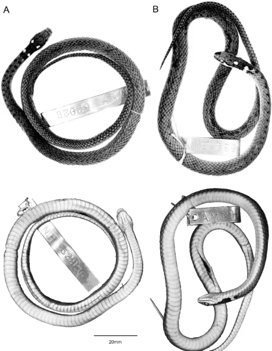
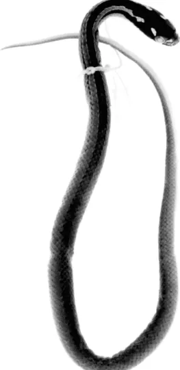
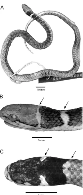
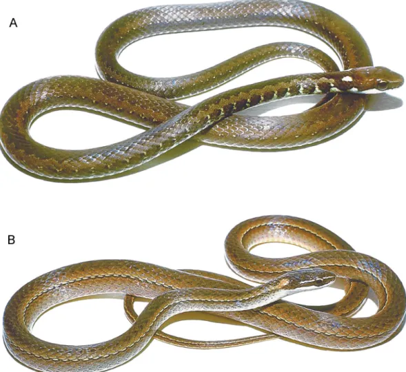
Fig. 1. Eutrachelophis bassleri, new species. Dorsal and ventral views of adult specimens:A.AMNH R-52926 (male holotype), from upper Rı´o Pisqui, Peru.B.AMNH R-53473 (female paratype), from upper Rı´o Utuquinia, Peru, near border with Brazil. The conspicuous nuchal markings are ‘‘cream’’ or ‘‘yellow’’ in life. Specimens shown life size.
logical and ethnographic collecting in eastern Peru.3
DIAGNOSIS: Eutrachelophis bassleri is a small snake (,400 mm total length) that resembles the somewhat larger E. steinbachi
(to 558 mm) in having 15 dorsal scale rows and conspicuous paired ocelli on the nape. It differs fromsteinbachiin having a postocular wedge of pale color extending dorsad from the lip, and in having a pale broken, posterior collar (the rounded upper ends of which often resemble a second pair of ocelli when viewed from above), and in lacking oblique pale dorsolateral markings touching the eye.
E. basslerifurther differs in having dorsolat-eral lines of vague dark spots (or fused crossbars) rather than dark anterior stripes; a lateral line of pale dashes or dots lies on scale row 4 inbassleri, on row 6 insteinbachi.
Eutrachelophis bassleri differs radically from
steinbachi in having an undivided hemipenis with a nude, dome-shaped apex.
DISTRIBUTION: Lowland rain forest in the western part of the Amazonian basin (map 1). Known localities for Eutrachelophis basslerirange from Pebas, Peru, on the upper Amazon River, northwestward to east-cen-tral Ecuador in the Rı´o Bobonaza drainage, and southward in Peru through the Rı´o Ucayali drainage to nearly10u S. The species probably occurs also in extreme western Brazil and possibly in southern Colombia. There is an unnamed, clearly related species in central Amazonia, Brazil (see comments and illustration under Eutrache-lophisspecies following Remarks).
DESCRIPTION OFTYPE SPECIMENS
Eutrachelophis bassleri is a small slender snake that probably is sexually mature by
300 mm total length, with a maximum known length of 377 mm. Following is a combined description of the 12 specimens in the type series.
PROPORTIONS AND SCUTELLATION: Head wider than high and wider than neck; eye large, its diameter greater than distance from its anterior edge to nostril in adults (relatively larger in juveniles, which have eye diameter.
distance to snout tip). Body slender, slightly higher than wide, with rounded ventrolateral edges; tail slenderly tapering. Less than 400 mm total length, with tail comprising 27%–30%of the total. Five adult males 320–377 mm (x¯5
350.8 mm) total length, tail/total length 0.281– 0.296 (x¯50.2894); two adult females 339 and 369 mm total length, tail/total 0.268, 0.271. Smallest specimen with complete tail, 160 mm total length (42 mm tail), a juvenile male with threadlike vasa deferentia and unenlarged kidney tubules. Two subadults approaching maturity at 245 mm (
U
) and 259 mm (-
) total length, as evidenced by enlarging ova in the female and enlarging vasa deferentia and kidney tubules in the male.Dorsal scales smooth, in 15-15-15 rows; anal ridges lacking; a few specimens with single apical pits (situated either medially or off center) discernible on some scales, espe-cially on neck. Ventral plates 128–139 (8 males 128–138, x¯5133.3; 4 females 134–139, x¯5136.8). Anal plate divided. Subcaudals in 62–70 pairs (7 males 65–70, x¯ 5 67.9; 3 females 62–66, x¯563.7).
Rostral plate wider than high, tipped forward, readily visible from above. Interna-sal and prefrontal plates paired, each pre-frontal laterally in contact with nasal, loreal, and preocular. Supraocular large, about as long as frontal and more than half as wide. Frontal pentagonal or slightly hexagonal, about two times longer than wide, longer than distance to snout. Interparietal suture shorter than length of frontal. Loreal higher than wide, variable in shape (e.g., tipped forward and rhomboidal, or vertical and rectangular). One high, narrow preocular, rarely two preoculars (on one side only in two specimens); two postoculars, the lower one varying in size, distinctly smaller than or nearly equal to the upper. Temporals 1+2 in eight specimens; two other individuals with this basic pattern but having the primary and 3
Harvey Bassler was a petroleum geologist who spent a decade (1921–1931) exploring the upper tributaries of the Amazon, all the while making a magnificent herpetological collection, including 4200 snakes. His base of operations for upriver expeditions was Iquitos, where specimens were also obtained. He brought his collection to the American Museum in 1934, where he worked on his snakes up to World War II, when he was called away by the U.S. Government to work on the urgent need to increase rubber production in the Amazon Basin, Bassler left no scientific publications, but his collections have been repeatedly mined over the years and continue to provide new insight. (Bassler, ms.; Myers, 2000: 139–141).
upper secondary temporals fused into one long scale; one specimen with 1+1+1/1+1 (left/right) temporals. Eight supralabials (8/7 in one), with labials 2 (usually) or 2–3 touching loreal and 3–5 bordering eye. Eight, nine (usually), or rarely 10 (one with 10/9) infralabials, of which 1–4 (if only 8 infra-labials) or 1–5 touch anterior genials and 4–5 or 5–6 touch posterior genials. First infra-labials in contact behind mental except in one juvenile, where widely separated. Anterior and posterior genials long and narrow, subequal. Tiny, inconspicuous tubercles (presumed sen-sory organs) on head plates and chin.
COLOR AND PATTERN IN PRESERVATIVE: Brown or gray-brown above—gray after loss of stratum corneum—with head and neck usually darker than body for length of 9–11 scales behind parietal plates. A pair of conspicuous, black-rimmed white ocelli atop
nape, occupying parts of several dorsal scales (usually not including a complete scale) just behind each parietal plate. A second pair of often conspicuous albeit incomplete ocelli situated farther back, dorsolaterally at about level of ventrals 4–5; in lateral view these markings are seen to be the expanded, rounded dorsal ends of an incomplete white collar that is medially broken by a space of several dorsal scales (fig. 2B).4 Most of rostral plate and supralabials immaculate white, this color extending dorsad as a triangular wedge just behind each eye (a rectangular wedge in one juvenile); a black streak across tops of anterior supralabials, extending thinly under and up behind eye, then becoming wider and dropping obliquely along the white postocular wedge to lower side of neck (fig. 2B). A few specimens with a vertical white preocular bar (fig. 2B), but this region dark in most. One juvenile with an ill-defined whitish blotch on frontal plate and a pair of vague whitish parietal blotches (fig. 2C).
A pair of dorsolateral lines of irregular and often vague black spots, spaced about every other scale, mainly on scale rows 6 and/or 7 on each side (fig. 1). Aforesaid dark spots not always discernible throughout body and virtually absent on some specimens (possibly due to method of preservation); often largest behind the broken collar cum ocelli on neck, where spots may fuse into vague dark crossbars that, in some juveniles, may sepa-rate vague dorsolateral pale spots (fig. 2C). A lateral line of white dashes or dots on scale row 4, sometimes weakly indicated along side of neck but more often nearly confined to rear half of body, where the pale dashes are accentuated by black pigmentation along their lower edges. Inconspicuous dorsolateral dark spots (when present) and lateral line of pale dashes extending onto tail for most of its length. Dark body color encroaching slightly onto sides of ventrals and subcaudals. Ventral surfaces immaculate pale yellow or white.
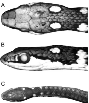
Fig. 2. Heads ofEutrachelophis bassleri, new species. A, B. AMNH R-53473, adult female (369 mm total length), in dorsal and lateral view,
33.9. C. TCWC 41425, juvenile male (160 mm total length), 32.5. In addition to the white or yellow nuchal markings, the postocular extension of white or yellow labial color (B) is diagnostic of
E. bassleri and its unnamed sister species (cf. fig. 4); the white preocular bar is present or absent inE. bassleri.
4This feature may be geographically variable. The dorsal ends of the broken collar are narrow in the two paratypes from Ecuador—not conspicuously rounded as in the Peruvian specimens.
COLOR IN LIFE: Based on a few color notes quoted below, this is a prettily colored snake in life, with black head and nape bearing conspicuous white or yellow ocellar markings; white or yellow lips and a trian-gular postocular marking; ventral surfaces cream or changing from white to yellowish. Body and tail in at least one individual sea green anteriorly, then reddish brown, and grayish brown posteriorly.
Dixon and Soini (1977: 54; 1986: 115) published color notes on one or more of the three paratypes from Mishana, Peru.
Dorsum light brown with dorsolateral pair of small black spots from posterior body to tail tip; an irregular yellowish, dotted lateral line on 4th scale row, bordered below by black; venter and subcaudals cream; extreme edge of ventrals with grayish black flecks; top of head black, lips bright yellow; yellow triangular spot behind eye; pair of yellow spots behind parietals; incom-plete yellow nuchal collar.
Eye color was not given, but some preserved specimens retain a pattern of a pale upper sector that is distinct from the dark lower part (fig. 2B); see also below.
Martin Henzl (letter, March 17, 1990) kindly provided the following color notes on a Peruvian specimen (NMW 31795) collected on a ridge above the Rı´o Llullapi-chis, in the Serranı´a de Sira.
Head and nape black, upper lips white, small white triangle laterally behind eyes, pair of dorsolateral cream spots on occiput, another pair of similar spots on the nape contacting the white ventral color in a small channel. Anterior third of body sea-green, second third of body reddish brown, posterior third and tail grayish brown. Indistinct dark paravertebral blotches that soon join in a zigzag stripe becoming more and more indistinct posteriorly; thin light lateral stripe from midbody onto tail. Chin and anterior third of venter white, posterior two-thirds and underside of tail yolky yellow. Iris dark brown with bronze blotch in upper part; oral cavity greyish, tongue dark with pink tips.
HEMIPENIS
RETRACTEDORGAN OFEUTRACHELOPHIS BASSLERI: The following description is based on the uneverted left hemipenis of AMNH
R-55786, which was opened by midventral incision and then removed and pinned flat for illustration (fig. 3A). Data also were taken from the uneverted right organ of TCWC 41424, which was examined in situ.
Major retractor muscle originating at level of subcaudals 27 or 28, and anteriorly inserting without division on end of single (unbifurcated) hemipenis at level of subcaudals 10 or 12. The distal fourth of the uneverted organ (fig. 3A) is a nude area of folded tissue—the proximal part of this area being somewhat puckered and the whole region apparently capable of consider-able expansion. The midsection (about 40%of length of organ) is densely spinulate and less folded than the distal fourth, although the area between the branches of the sulcus spermaticus appears capable of greater expansion than the rest of the midsection. There is a densely spiculate section around the proximal part of the otherwise distal nude section. There are roughly 30 large to very small spines from the basal region distad into the densely spiculate area.5The extreme base of the organ (proximal 10%) is nearly nude, with only a few spinules. There are no basal spines obviously enlarged compared with the others and no basal naked pocket (sensu Myers, 1974: 32). The deeply incised sulcus spermaticus divides low on the organ and its branches terminate abruptly about two-thirds of the way onto the distal nude section. The sulcus lies mostly on the lateral wall of the uneverted organ (fig. 3A).
EVERTED ORGAN OF EUTRACHELOPHIS BASSLERI: The right hemipenis of AMNH R-25193 (paratype) was manually everted by Myers in the late 1970s, using the ‘‘Waite Gibson Technique’’ later described in Myers and Cadle (2003: 299). The distal nude section (fig. 3A) everted as a domelike head or capitulum that lacks a free overhanging edge (fig. 3B–D). The dome is nude, except for branches of the sulcus spermaticus and except proximally, where it is densely spicu-late as seen in the retracted organ (fig. 3A). Owing to differential tissue expansion, the
5Tiny, usually mineralized, hemipenial spines are commonly called ‘‘spinules,’’ butspiculesandspiculate seem to be more appropriate terms for these even tinier structures that also are mineralized (they can be ‘‘felt’’ with a very fine teasing needle and transiently stained with Lugol’s iodine solution).
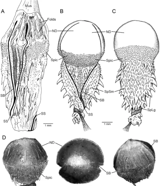
Fig. 3. Hemipenes ofEutrachelophis bassleri, new species. The distal nude section is greatly distensible, becoming hemispherical on eversion. Because of differential tissue expansion, the close-lying branches of the sulcus spermaticus diverge and acquire a centrifugal orientation, entering onto the nude dome from nearly opposite sides of the everted organ.A.Uneverted left hemipenis of AMNH R-55786 (paratype), opened by midventral incision.B.Everted right hemipenis in sulcate view, AMNH R-25193 (paratype; depiction of the manually everted organ).C.Same organ as in B, in asulcate view.D.Distal half of everted hemipenis showing appearance of the maximally inflated, mostly nude dome, from left to right: asulcate side, apex, and sulcate side. Left organ of TCWC 41424, paratype. (This organ previously had been manually turned inside out, but, because the base had been badly damaged, only the distal half was tied off and injected with petroleum jelly in an attempt to determine maximum inflation). Abbreviations:Folds, expansion folds;Mus,major retractor muscle;ND,nude dome;SB,a branch of the sulcus spermaticus;SS, sulcus spermaticus (proximal to bifurcation);Spic,densely spiculate area below and around base of nude dome;SpLg,large lateral and basal spines;SpSm,small spines on sulcate and asulcate faces.
sulcus branches acquired a centrifugal orien-tation upon hemipenial eversion, entering onto the bulbous dome nearly from opposite sides of the organ (see figs. 3B, D). There are no greatly enlarged spines, only relatively large spines that are positioned laterally and basally, with numerous small spines or spinules on the sulcate and asulcate sides of the hemipenial body (fig. 3B,C).
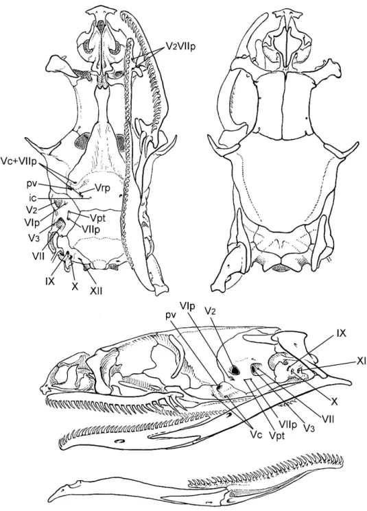
The left organ of TCWC 41424 had been manually turned inside out prior to our examination, and, although the base had been torn in the process, it was possible to tie off and temporarily inflate the distal half (see views in fig. 3D). This partly everted hemi-penis appears to have the apical nude dome fully expanded, although manually everted organs often do not retain their original elasticity (Myers and Cadle, 2003: 295, 300). SKULL AND DENTITION: The skull was removed from AMNH R-55786. See fig-ure 10 and text under Global Comparisons. Ten maxillae in 10 specimens bear 26–29 (x¯5
27.3) small, subequal teeth, followed by a distinct diastema and two ungrooved fangs, the last fang slightly offset laterad. The maxillary fangs are about twice as large as the prediastemal teeth and are further differ-entiated by having knifelike rear edges. AMNH R-55786 has 19 palatine teeth, fol-lowed by about 30–31 pterygoid teeth; 34 dentary teeth. The holotype has 32 (right) or 34 (left) dentary teeth on the previously excised mandibles.
VERTEBRAE, HEAD MUSCLES, GLANDS, ANDVISCERA: See under Global Comparisons.
REMARKS
The general location of Bassler’s ‘‘Pisqui Hills’’—the type locality of Eutrachelophis bassleri—can be determined from an unpub-lished report (Bassler, MS.). Bassler first explored and mapped the Rı´o Pisqui in 1923, traveling 186 km by river from its mouth (149 m elev.) to the head of canoe navigation (259 m), followed by an addition-al 18 km on foot beyond the first cataracts— a straight-line distance of 93 km by Bassler’s careful reckoning. He determined latitude and longitude as 7u429 S, 75u009 W at the mouth of the Rı´o Pisqui and as 8u229 S, 75u309W at the end of his traverse; it may be
noted that the first set of coordinates agrees within a few minutes to recent maps and gazetteers. Bassler described the first 45 km (by river) as ‘‘a flood plain subject for the greater part to a period of inundation each year and the channel here is not permanent for lateral erosion is active.’’ Upriver, ‘‘this recent flood plain merges into a plain determined by firmer though still practically unconsolidated sediments and here the chan-nel is deeper and appears to change very little even over long periods of time.’’ Somewhere on this upper plain Bassler wrote that,
hills 200 ft. high were observed but between these and the mountains [to westward] the hills are usually under 100 ft in altitude above the general level of the plain. Beyond the plain the mountains [evidently the northern end of Cordillera Azul, an Andean front range] rise abruptly with stupendous cliffs and a very rugged skyline, with relief of 3000 ft. or more.
The 200 ft-high (60 m) hills on the higher flood plain of the upper Rı´o Pisqui must certainly be what Bassler gave as ‘‘Pisqui Hills’’ for specimens collected on this and subsequent trips, and the locality can with confidence be placed within the area bounded by parallels 8u009–8u229S and meridians 75u309–75u509W.
Eutrachelophis, undescribed species Figure 4
In 1993, the late Paulo E. Vanzolini sent to Myers what he recognized as a new species of snake from Cabeceira do Rio Urucu, Ama-zonas, Brazil. It clearly is closely related to
Eutrachelophis bassleri and seemed likely to be a different species. But it could not be so identified with assurance because (1) ocellar head and neck patterns in Eutrachelophis
and other genera are somewhat variable, and (2) because it is a female collected at about 5uS, 65uW—far to the east of knownbassleri
localities (see map 1). Ana Lucia C. Prudente fortunately has obtained additional material that she will describe separately.
Eutrachelophis steinbachi (Boulenger), new combination
Figures 5–9, 11–12
Rhadinaea Steinbachi Boulenger, 1905: 454 (two syntypes, female and young, from ‘‘the Province
Fig. 4. Eutrachelophisspecies (MZUSP 10530, head in dorsal and lateral view; a female from Cabeceira do Rio Urucu, central Amazonian Brazil). This unnamed snake appears to be the sister species of E. bassleri.See Remarks and compare with figure 2. Formalin preservation caused the integument of this specimen to become slightly translucent, giving visibility to a large ‘‘supralabial’’ gland (SG); this large gland (similarly positioned in all three species of Eutrachelophis) is adherent to the medial side of the supralabial integument. The mucous and serous (Duvernoy’s) parts of the gland cannot be distinguished in
Eutrachelophiswithout histological examination.
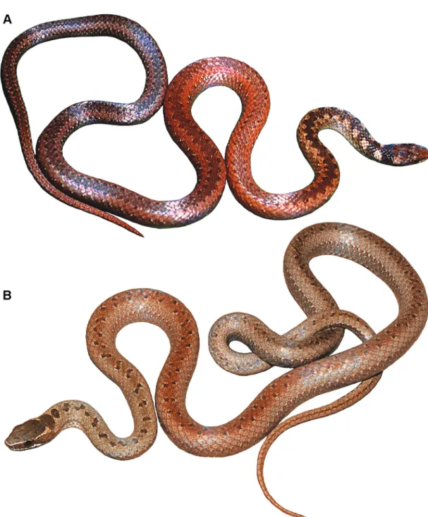
Fig. 5. Eutrachelophis steinbachi (Boulenger). Adult female shown approximately life size (BMNH 1946.1.21.62, lectotype by present designation).
Sara, Department Santa Cruz de la Sierra, collected by Hr. J. Steinbach’’).
Aporophis melanocephalus Griffin, 1916: 171–172 (holotype CM R-18, ‘‘a female, taken at Las Juntas, Bolivia, 250 M. above sea-level, by Jose´ Steinbach in December, 1913’’).
Rhadinea steinbachi Boulenger: Dunn, 1922: 220 (Aporophis melanocephalusplaced in synonymy).
Liophis steinbachi (Boulenger): Amaral, ‘‘1929b’’ [1930]: 174. Peters and Orejas-Miranda, 1970: 179. Myers, 1974: 22 (comment on unsatisfacto-ry generic assignment); Myers and Cadle, 1994: 2. Dixon, 1980: 15, 20 (listed as incertae sedis).
Rhadinea steinbocki(misspelling): Clark, 1945: 428 (mention of hemipenis).
LECTOTYPE: The lectotype (fig. 5) by pres-ent designation is BMNH 1946.1.21.62, one of the two original syntypes. It is an adult female 558 mm total length (150 mm tail length), with one preventral, 140 ventrals, 67 subcaudals. The second syntype (paralectotype) is BMNH 1946.1.21.63, a juvenile male (fig. 6).
DIAGNOSIS: Eutrachelophis steinbachi is likeE. bassleriin having 15 dorsal scale rows and a usually conspicuous pair of ocellar markings6 on the nape. It is a larger snake (to 558 mm total length) than bassleri
(, 400 mm) and is readily differentiated by details of color pattern. E. steinbachi has a pair of oblique pale markings touching the upper anterior and upper posterior edges of the eye (both lacking inbassleri), and the dark head color extends onto the neck as unbroken dorsal and lateral stripes.E. steinbachilacks the broken, ocellarlike nuchal collar (present inbassleri). A lateral line of pale dashes, where present, lies on scale row 6 in steinbachi, on row 4 in
bassleri. E. steinbachi differs absolutely in having a divided hemipenis with spinose tips.
DISTRIBUTION:Eutrachelophis steinbachiis known only from a small section of central Bolivia near the eastern base of the Cordillera Oriental, at elevations of perhaps 250–500 m (map 1). Its habitat has not been recorded, but presumably it is a forest species.
SPECIMENS EXAMINED: All 12 specimens seen by us were obtained by the Bolivian collectors Jose´ Steinbach and his son Fran-cisco Steinbach over a span of years (circa 1904–1928); these were sold to several muse-ums. Museum data for these and one additional specimen (13 total) follow (see Remarks for commentary): BOLIVIA: no data, FMNH 35662 (F. Steinbach).
Depart-ment Santa Cruz: no other data, UMMZ
69550; Buena Vista, no elevation, FMNH 35641 (F. Steinbach, April–May, 1928); Buena Vista, 450 m, AMNH R-125695, UMMZ 60736 (J. Steinbach, Nov. 1923 and Jan. 1924); Buena Vista, 500 m, UMMZ 60734–60735 (J. Steinbach, May and Sept. 1923); Las Yuntas [5Las Juntas?], 50 m, CM
Fig. 6. Eutrachelophis steinbachi (Boulenger). The paralectotype (BMNH 1946.1.21.63), a juve-nile male shown 1.63life size.
6
In the variational repertory ofE. steinbachi, the postparietal (nape) ocelli may lack dark edges posteriorly and be confluent with (but usually paler than) the light ground color between dorsal and lateral stripes (e.g., fig. 6).
R-18 (holotype ofAporophis melanocephalus, J. Steinbach, Nov.–Dec. 1913). Province Sara, BMNH 1946.1.21.62
U
, 1946.1.21.63 -(U
lectotype and juvenile-
paralectotype ofRhadinaea steinbachi, J. Steinbach, no date, received at BMNH Oct.17, 1904, fide A.F. Stimson, in litt.); Province Sara, Rı´o Sirutu [5Rı´o Surutu´ ?], UMMZ 63216 (J. Steinbach, Jan. 1925).
ADDITIONALSPECIMEN: ‘‘Syntypus: NMW 23106 (
-
) Bolivia; gekauft von ROSEN-BERG (?)’’ fide Tiedemann and Ha¨ upl (1980: 61). Only two BMNH specimens (see above) were mentioned in the original de-scription, so this specimen certainly is not a syntype as stated; there seems also a question as to whether it was purchased from Rosen-berg. The specimen is correctly identified, however, based on scale counts and photo-graphs kindly supplied by J.R. Dixon (see fig. 7); some of Dixon’s data on NMW 23106 are incorporated in the following description.DESCRIPTION
PROPORTIONS ANDSCUTELLATION: Eutra-chelophis steinbachi is a slender snake that attains a maximum known length of 558 mm. Head wider than neck; eye large, its diameter greater than distance from its anterior edge
to nostril, going about 1.3–1.5 times into length of snout. Body higher than wide, with rounded ventrolateral edges; tail slenderly tapering. The only adult male having a complete tail is 454 mm in total length, with a tail/total length ratio of 0.313; the snout-vent length of this specimen is 312 mm, which is exceeded by a broken-tail male of 345 mm SVL. Two adult females are 531 and 558 mm total length, with tail/total ratios of 0.273 and 0.269, respectively.
Juveniles have relatively shorter tails: Two females are each 185 mm total length, with identical tail/total ratios of 0.254. Two males of 211 and 222 mm total length have tail ratios of 0.270 and 0.284, respectively. A larger male 251 mm SLV and 363 mm total length (tail/total50.308) is still immature, as judged from soft hemipenial spines and unenlarged kidney tubules and vasa defer-entia—suggesting that sexual maturity in this species is not attained before roughly 400 mm total length.
Dorsal scales smooth, in 15-15-15 rows; anal ridges lacking; apical pits absent on several specimens carefully examined. Ven-tral plates 134–140 (6 males 134–136, x¯ 5
134.8; 6 females 137–140, x¯ 5 137.7). Anal plate divided. Subcaudals in 66–81 pairs (4 males, 70–81, x¯576.0; 5 females 66–73, x¯
568.8).
Rostral plate wider than high, visible from above. Internasal and prefrontal plates paired, each prefrontal laterally in contact with nasal, loreal, and preocular. Supraocular large, about as long as frontal and more than half as wide. Frontal pentagonal or slightly hex-agonal, over 1.5 times longer than wide, equal or slightly longer than distance to snout. Interparietal suture varying from conspicu-ously shorter than to nearly equal length of frontal. Loreal higher than wide, tending in shape toward a slanting rhomboid. One high, narrow preocular, rarely two (on one side only in one specimen); two postoculars, the lower being smaller. Temporals 1+2 (one specimen with 2 + 2 + 3 on one side of head). Eight supralabials, with labial 2 touching loreal and labials 3–5 bordering eye. Infralabials vari-able, range 9–11, but counts differing on left and right sides in six of seven specimens; first pair in contact behind mental. Tiny incon-spicuous tubercles on head plates and chin.
Fig. 7. Eutrachelophis steinbachi (Boulenger), handheld preserved specimens.Upper.A specimen (NMW 23106) cited as ‘‘syntypus’’ by Tiedemann and Ha¨ upl (1980: 61) but not mentioned in Boulenger’s (1905: 454) original description. Low-er.The syntype (BMNH 1946.1.21.63) designated lectotype in this paper (photograph courtesy of James R. Dixon).
COLOR AND PATTERN IN PRESERVATIVE: In alcohol, Eutrachelophis steinbachi is gray-ish brown (gray where stratum corneum has fallen away). Top and sides of head darker brown, with the dark head coloring extending posteriorly as a middorsal and pair of lateral stripes for a distance of about a fourth to a third of the body length before fading out. There are three pairs of conspicuous, black-edged white spots atop the head and nape (figs. 5–8), as follow: (1) An elongated white spot slants anterodorsally in front of the upper edge of each eye (from top of preocular onto side of prefrontal). (2) A similarly elongated white spot slants posterodorsally behind the upper edge of each eye (from upper postocular onto parietal). The oblique postocular marking may be better defined and more vivid than its preocular counter-part. (3) The third pair of black-rimmed white markings are on the nape and may appear as rounded ocelli (fig. 8) or elongated spots (fig. 5), situated posterolaterally about one scale-length behind each parietal. The pale nape ocelli, however, are not always discrete, but are often fused posteriorly with the light dorsolateral ground color adjacent to the dorsal stripe (fig. 6); this fusion occurs in eight of 12 specimens, either on one side only (3 specimens) or on both sides (5 specimens), partly determined by the undu-latory courses of the dark neck stripes. The lower parts of the supralabials and underside of the head are white.
The middorsal dark stripe undergoes one to several undulations anteriorly on the neck, where it varies in width from about four to seven scale rows and sometimes fuses briefly with the lateral stripe. As the middorsal stripe straightens out it becomes edged by a line of
white dashes along the middle of the sixth scale row; posteriorly the middorsal stripe starts to fade first in the center, and in some specimens may be represented all the way to the tail by a double line of black dots (sixth scale row on each side) marking its former edges. At its start, the lateral dark stripe occupies scale row 3 and adjacent halves of rows 2 and 4, but it soon narrows to a line confined to row 3 before breaking up into a series of dark dots, which extend (inconspic-uously) far posteriad in a few specimens. In some individuals the lower sides posteriorly are somewhat darker than the dorsum. The body color extends onto the edges of the ventral and subcaudal plates; ventral surface otherwise immaculate white. No information is available on coloration in life.
HEMIPENIS
The following description is entirely based on retracted hemipenes (fig. 9). The un-everted left organs of AMNH R-125695 and FMNH 35662 had been opened midven-trally; they were removed and pinned flat for detailed study and illustration. Supplementa-ry notes were provided by examination of in situ organs in UMMZ 60734 and 63216.
Major retractor muscle originating at level of subcaudal 37 for the right hemipenis of FMNH 35662, anteriorly dividing at the levels of subcaudals 20 (1 specimen), 19 (2), or 15 (1), and inserting on the ends of the hemipenial lobes at subcaudals 17 (1), 15 (2), or 14 (1). The hemipenis therefore is relatively long, spanning 14–17 subcaudals. The two lobes are narrow and long, comprising nearly two-thirds the total length of the hemipenis, which bifurcates at the level of subcaudal 6 in three specimens and at subcaudal 7 in another.
The extreme base of the organ is virtually nude except for a sparse distribution of spinules; a relatively deep basal groove on the dorsal wall might persist on the everted hemipenis as a basal naked pocket (sensu Myers, 1974: 32), but this is uncertain. Two large and two medium-sized spines arise across the middle of the undivided base of the organ; these spines are nearly straight, and the tips of the two largest extend nearly to the base of the hemipenis, on either side of the sulcus (fig. 9). Above the big spines are numerous small,
Fig. 8. Head of Eutrachelophis steinbachi
(Boulenger) (UMMZ 60736), showing details of color pattern. The oblique alignment of the pale ocelli differentiates E. steinbachi from E. bassleri
straight to slightly recurved spines, those close to the sulcus being very small. The proximal 40%–50%of each hemipenial lobe is profusely covered by short, thick spinules. The distal 50%–60% of a lobe is densely covered with straight, relatively long thin spines, and the sulcus branch ends abruptly in this region, at a length about 80%up the lobe.
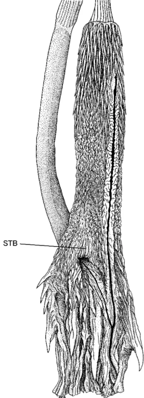
There is at the base of the lobes an in-terlobular nude space—the STB, or ‘‘smooth terminal basin’’ (see Hemipenial Terminology for Snakes)—which is closely edged by small, short spines (fig. 9). This ‘‘basin’’ has apparent expansion folds and therefore lacks the sulcus-like smoothness seen in some other taxa (e.g., compare with the STB shown in fig. 18). The expansion folds suggest that the basin will be considerably enlarged after eversion.7
Outside the STB, the long hemipenial lobes are completely spiny as described above. The sulcus proximally lies on the lateral wall (both on left and right organs) and divides halfway up the base, at the level of subcaudal 3. The branches of the sulcus are deeply incised and have a centrifugal orientation, lying on the ventral wall of the ventral lobe and on the dorsal wall of the dorsal lobe; each sulcus branch terminates well short of the apex.
SKULL AND DENTITION: The skull was removed from one specimen of
Eutrachelo-phis steinbachi (AMNH R-125695). See
figure 11 and text under Global Compari-sons. Eight maxillae in as many specimens bear 25–28 (x¯525.6) small, subequal teeth, followed by a diastema and two offset, ungrooved fangs (fig. 12); maxillary fangs about twice as large as the prediastemal teeth, with knifelike rear edges. AMNH R-125695 has 19/20 palatine teeth, followed by about 34/36 pterygoid teeth; dentary teeth 35/33.
VERTEBRAE, HEADMUSCLES, GLANDS, AND VISCERA: See under Global Comparisons.
REMARKS
As indicated above, Eutrachelophis stein-bachi (Boulenger) is known to us from a dozen specimens. Excluding one specimen of
Fig. 9. Uneverted hemipenis ofEutrachelophis steinbachi (Boulenger); right organ of AMNH R-125695, opened by midventral incision, 35. Abbreviation:STB,smooth terminal basin.
7
The AMNH specimen ofEutrachelophis steinbachi was obtained by exchange. Both hemipenes had been opened, so it was not possible to obtain a manual eversion as was done for E. bassleri. The organ depicted in figure 9 suffered the same fate as that of another important specimen (see appendix 1: Hemi-penis of Lectotype), but in each case there is a contralateral organ to save the day. A check of the left in situ hemipenis of AMNH R-125695 verifies that the STB was accurately drawn in figure 9. Hussam Zaher (personal commun.) manually everted the hemipenis of a MZUSP specimen ofE. steinbachi; a description or illustration showing the STB is awaited with interest.
questionable provenance, all originated over a period of time (circa 1903–1928) from the Bolivian collectors Jose´ Steinbach and his son Francisco Steinbach. Their locality des-ignations are of a somewhat general nature, but the several Steinbach localities for this species seem to lie at the eastern base of the Andes in the department of Santa Cruz.
Buenavista (5 Buena Vista of modern maps and gazetteers) and Provincia Sara are the only localities forEutrachelophis steinbachi
shown on map 1, although the old ‘‘Province Sara’’ [Provincia Gutie´rrez]—the type locali-ty—is a region rather than a single collecting site.8 Jose´ Steinbach lived in Buena Vista (17u279S, 63u409W), the most frequently men-tioned locality. His locality Rı´o ‘‘Sirutu’’ for a UMMZ specimen may be one of several spelling variants of Rı´o Surutu´, a known Steinbach locality (Paynter, 1992: 142–143). According to Sydney Anderson (personal commun.), ‘‘The most probable collecting areas near the Rı´o Surutu´ would have been reasonably near the town of Buenavista … probably to the west or southwest of that village.’’
The locality for one specimen sent to the Carnegie Museum was given by Steinbach as Las Yuntas, 250 m., Dpto. Santa Cruz de la Sierra (fide C.J. McCoy, in litt.);9this became the type of
Aporophis melanocephalus, described by Grif-fin (1916), who failed to publish the depart-ment and who changed ‘‘Las Yuntas’’ to ‘‘Las Juntas.’’ The last is in fact the modern Bolivian name for at least one town previously with the former spelling, but there are at least two places with the name ‘‘Las Juntas’’ in the western part of the department of Santa Cruz,10and we are
uncertain which (if either) of them is the type locality ofA. melanocephalus. The holo-type of melanocephalus is a juvenile female only 185 mm in total length (tail5 47 mm) rather than ‘‘291 mm’’ (tail 47 mm) as given by Griffin; the right maxilla has been subse-quently removed and shows 25+2 teeth; there are about 137 ventrals and 69 subcaudals.
GLOBAL COMPARISONS
Despite striking differences in their hemi-penes, the very considerable external similarity betweenEutrachelophis bassleri, new species, andE. steinbachi(Boulenger) is accompanied by great similarity in skull and dentition, head muscles and glands, and general visceral anatomy. The vertebrae are also similar, with a ventral keel but no hypapophysis on the posterior vertebrae; neither species shows any suggestion of winglike or shelflike expansions on the zygapophyses or any expansion of the distal edge of the moderately high neural spine. These vertebral similarities are not so impressive, however, because they are shared with the majority of colubrid snakes.11
VISCERA
In both species the tongue is long and extends back nearly (E. steinbachi) or quite (E. bassleri) to the heart. There is no left lung in the two specimens dissected and the trachea ends opposite the apex of the heart in both specimens.12 In each species the pulmonary (right) lung has the usual reticulation of raised alveolar rims on its lining (rather than the essentially smooth lining surface seen in a few colubrids such as Amastridium, Compsophis, andPsammodynastes); this alveolar reticula-tion is continued forward on the membranous dorsal wall of the trachea to fade into pitting and then into quite smooth membrane 8
This subdivision of Depto. Santa Cruz has not had stable borders. The Province of Sara was larger in the early part of the 20th century than at present. It included the more recent provinces of Sara, Santiesteban, and Ichilo, which were shown on a map published in 1980 (S. Anderson, personal commun.). The name Provincia Gutie´rrez appears to be a more recent replacement for Provincia Sara (Paynter, 1992: 59, 138).
9The Steinbachs’ ‘‘Depto. Santa Cruz de la Sierra’’ apparently is a descriptive phrase specifying the western part of the large Department of Santa Cruz—not to be confused with ‘‘Santa Cruz de la Sierra’’ as applied to the city in the same region.
10One Las Juntas is about 120 km SW Buena Vista, and another is about 140 km SSE Buena Vista on the Rı´o Grande (from Mapa de la Repu´ blica de Bolivia, 1:1,500,000, R. Zumelzu y Cia., La Paz, 1947).
11
An unfortunate consequence of this is that it is unlikely that the fossil record, which is almost entirely of vertebrae for snakes, will ever give any useful information aboutEutrachelophisand the many other genera whose vertebrae are too nondescript to be surely recognizable.
12Data on viscera, head glands and muscles, skull, and vertebrae were obtained by dissection of AMNH R-55786 (E. bassleri) and AMNH R-125695 (E. steinbachi); both adult males.
anteriorly, but in neither does this forward extension of alveolar reticulation bulge out beyond the dorsal tips of the cartilaginous tracheal semirings to form a conspicuous ‘‘tracheal lung.’’ In E. bassleri the forward extension of alveolar reticulation reaches within a head length of the head, but in E. steinbachiit reaches only about a heart length anterior to the heart. Thus, both species of
Eutrachelophisseem to fall in the intermediate range, between Neotropical ‘‘xenodontines’’ that unequivocally have a tracheal lung (e.g.,
Coniophanes, Conophis, Darlingtonia, Hy-drops, Rhadinaea decorata, R. laureata, Ur-otheca godmani) and those that unequivocally lack a tracheal lung (e.g.,Atractus major,Ialtris,
Taeniophallus brevirostris,T. occipitalis); pro-bably the majority of ‘‘xenodontines’’ fall into the intermediate range with Eutrachelophis. The liver is separated from the heart by a moderate interval (eight ventrals in both specimens examined), as in most ‘‘xenodon-tines.’’ (But in a broad range of dipsadines, including Amastridium, Arrhyton vittatum,
Darlingtonia, Rhadinaea calligaster, and Ur-otheca godmani, for example, the liver reaches forward nearly or quite to the level of the apex of the heart. At the opposite extreme, in Pseu-doeryx plicatilis[AMNH R-52229] the liver is separated from the heart by 30 ventrals.)
As in other colubrids, with the exception of a few Old World genera (e.g., Boaedon,
Bothrophthalmus, Liophidium, Pareas, Pseu-doxyrhopus), the rectal caecum is absent in
Eutrachelophis bassleriandE. steinbachi. The general evidence from the viscera is consistent with a close affinity between Eu-trachelophis bassleri and E. steinbachi, but cannot be considered convincing evidence for close relationship because the two species are not at all unusual in visceral features and the resemblances between them are shared with many other ‘‘xenodontines.’’ It is the head structure that shows a sufficient number of shared unusual characters to make a special common ancestry the most likely explanation for the resemblances betweenEutrachelophis bassleriandE. steinbachi.
HEADGLANDS
Both species show an unusually large temporal extension of the Harderian gland,
exposed behind the orbit but posteriorly inserted deep to the muscle adductor man-dibulae externus superficialis and superficial to the adductor externus profundus (sensu Zaher, 1994). An equally large (and similarly placed) temporal extension of the Harderian gland also occurs inRhadinophanes monticola
(UTA R-4176) andContia tenuis(AMNH R-69062). In all these snakes, the deep insertion of the gland between adductores externus superficialis and profundus seems to form a functional complex, with the two muscles acting to compress and evacuate the gland; the profundus retains its usual function as a powerful adductor of the lower jaw, but the superficialis is reduced to a thin layer of fibers across the lateral surface of the rear portion of the gland and probably functions mostly—or entirely—as a compressor of the gland. Secretions from the Harderian gland discharge into the subbrillar space to lubri-cate the eye; the secretions also pass through the lachrymal duct into the vomeronasal (Jacobson’s) organ, for a function still speculative (e.g., Bellairs and Boyd, 1947, 1950; Minucci et al., 1992; Rehorek et al., 2003: 358 [the last authors do not identify a method for lubricating the eyeball under the brille, and only observe that ‘‘there is no nictitating membrane in the orbit of either snake species’’]).
In neither Eutrachelophis bassleri nor E. steinbachi could a rictal gland (sensu Mc-Dowell, 1986) be found, nor could any infolding of the oral mucosa just medial to quadrato-maxillary ligament that might rep-resent a rictal gland; the quadrato-maxillary ligament ends anteriorly on the skin of the last supralabial, and so does not reach forward to the region where a rictal gland might be expected. McDowell (1986) argued that the ‘‘rictal gland’’ is the homolog of the ‘‘anterior temporal gland’’ and of at least the sheath of the venom gland of various other snakes, and that these are, in turn, homolo-gous to the Mundplatte, or rictal fold, of lizards. Furthermore, he stated that since the lizard rictal fold is normally a long and deep invagination of oral mucosa deep to the quadrato-maxillary ligament, the absence of the rictal gland (and even of the portion of the ligament that should accompany it) in the two species of Eutrachelophis would be less
lizardlike (and presumably, more specialized) than the presence of the gland; this absence would represent an unusual, but not unique, shared specialization. The gland is a minute vestige or absent inTaeniophallus brevirostris, and it could not be found inConophis vittatus
(AMNH R-65108), Contia tenuis (AMNH R-73392), Farancia abacura (AMNH R-110941), Helicops angulatus (AMNH R-52746), or Thamnodynastes pallidus(AMNH R-4446). In Urotheca multilineata (AMNH R-98288), at the opposite extreme, the rictal gland has become greatly expanded as a floccular, thin-walled glandular structure covering most of the temporal region just beneath the skin; although large, it is a solid mass of glandular tissue rather than a hollow pocket (thus, it is quite different in appear-ance from the lizard rictal fold) and may represent a secondary enlargement of the small to very small pocket seen in most ‘‘xenodontines.’’
All three species ofEutrachelophis have a well-differentiated ‘‘supralabial gland,’’ the outline of which in some preserved specimens can easily be seen through the postorbital supralabial integument, as in figure 4 (un-named species). The gland is similarly posi-tioned in E. bassleri and E. steinbachi, in which it is adherent to the medial side of the supralabial integument. The serous (Duver-noy’s) portion of this gland is not (to gross examination, at least) clearly differentiated from the mucous part of the gland in either species. Enlargement of the rear maxillary teeth is not accompanied by a correspond-ingly conspicuous differentiation of the gland, which tapers posteriorly rather than showing enlargement behind the level of the eye. Unfortunately, the serous and mucous contributions to this gland in Eutrachelophis
and many other ‘‘xenodontines’’ are not distinguishable without histological prepara-tion (Taub, 1966, 1967).
In both named species of Eutrachelophis
the lateral nasal gland is well defined and lies in a clearly defined aditus conchae of the nasal capsule—that is, in an invagination of the lateral wall of the cartilaginous capsule that forms a vertically oriented protrusion (the concha) into the lateral wall of the nasal passage; the vertical edge of this protrusion defines a lateral diverticulum (the paracapsular
recess, or sakter) of the nasal cavity, housed in the prefrontal bone, with the facial wing of the prefrontal forming a partial lateral wall for the paracapsular recess, the in-traorbital wing of the prefrontal forming a posterior wall for the recess, and the roof of the lachrymal canal of the prefrontal forming the floor of the recess. Two bony processes define the rim of the aditus conchae: the conchal process of the septo-maxilla rises along the anteromedial rim of the aditus; and the conchal process of the prefrontal, rising from dorsomedial rim of the anterior orifice of the lachrymal canal of the prefrontal, lies along the posterolateral rim of the aditus. The above conditions are as in the great majority of ‘‘xenodontines,’’ and Colubroidea in general. But some ‘‘xenodontines’’ have reduced the nasal conchae and its aditus, so that the bulk of the lateral nasal gland lies more superficial-ly: in Tretanorhinus nigroluteus (AMNH R-70222) and in Coniophanes quinquevittatus
(AMNH R-74493) the concha is present, but with a reduced aditus, so that much of the gland lies in a shallow depression on the side of the nasal capsule; inApostolepis, Carpho-phis,Farancia,Hydrops, andPseudoeryx, the concha (and its aditus) is only vaguely defined and the conchal process of the prefrontal is a blunt vestige or absent.
HEADMUSCLES
Both Eutrachelophis bassleri and E. stein-bachi have unusually weak jaw adductors, with the adductor externus medialis essen-tially confined to the lateral surface of the braincase, leaving a broad exposure of the parietal and supraoccipital between the left and right muscles.
The adductor externus superficialis (sensu McDowell, 1986 5 adductor externus pro-fundus of most authors) and the adductor posterior are also weak muscles, so that the bony crests of the compound mandibular bone lateral (for the adductor externus superficialis) and medial (for the adductor posterior) to the mandibular adductor fossa are both low. These two mandibular adduc-tors arise from the quadrate to insert on the mandible and therefore have the same mechanical force whether the quadrate is in