BIOASSAY TECHNIQUES FOR DRUG
BIOASSAY TECHNIQUES FOR
DRUG DEVELOPMENT
Atta-ur-Rahman
and
M.Iqbal Choudhary
H.E.J.Research Institute of Chemistry
University of Karachi, Pakistan
and
William J.Thomson
Arena Pharmaceuticals
San Diego, USA
harwood academic publishers
Australia • Canada • France • Germany • India • Japan •
Luxembourg Malaysia • The Netherlands • Russia • Singapore •
“To purchase your own copy copy of this or any of taylor & Francis or Routledge's collection of thousands of ebooks please go to
www.eBookstore.tandf.co.uk.”
Copyright © 2001 OPA (Overseas Publishers Association) N.V. Published by license under the Harwood Academic Publishers
imprint, part of The Gordon and Breach Publishing Group. All rights reserved.
No part of this book may be reproduced or utilized in any form or by any means, electronic or mechanical, including photocopying
and recording, or by any information storage or retrieval system, without permission in writing from the publisher. Printed in
Singapore. Amsteldijk 166
1st Floor 1079 LH Amsterdam
The Netherlands
British Library Cataloguing in Publication Data
A catalogue record for this book is available from the British Library.
ISBN 0-203-30453-5 Master e-book ISBN
CONTENTS
PREFACE ix
1.0 GENERAL INTRODUCTION 1
PART A: BENCH-TOP AND PRIMARY BIOASSAY SCREENING
1.1 Toxicity Assays 8
1.2 Antimicrobial Assays 13
1.3 Antiviral and Anticancer Assays 23
1.4 Antimitotic Assay 35
1.5 Genotoxicity Assays 37
1.6 Assays for Control of Tropical Diseases 44
1.7 Assays for Agrochemicals 59
1.8 Hepatotoxicity Assays 68
1.9 Hypoglycemic/Antidiabetic Activity Assays 75
1.10 Diuretic Activity Assay 80
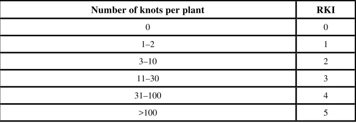
1.11 Anthelmintic Activity Assays 81
1.12 Antifertility/Anti-implantation Assays 84
1.13 In Vitro Assay for Platelet Aggregation 86
1.14 Anti-inflammatory Assay 88
1.15 Immunomodulating Assay 89
1.16 Antiepileptic (Anticonvulsant) Assay 91
1.17 Analgesic Assays 94
1.18 Gastroprotective/Antiulcer Assays 95
1.19 Radiolabelling Bioassays 98
1.20 Anti-emetic Assay 100
PART B: HIGH-THROUGHPUT SCREENING
2.1 Introduction 103
2.2 Enzyme Assays 108
2.3 Cell-based Receptor Functional Assays 142
2.5 References 189
PREFACE
The so-called “green wave”, triggered by a growing ecological awareness, has resulted in an increased interest in herbal formulations throughout the world, particularly in the last decade. The consumption of medicinal plants has almost doubled in the West during that period. The efficacy of a number of herbal formulations has been tested by valid phytopharmaceutical techniques and the number of plant-based drugs or health foods has increased steadily to meet the growing demand. Over the years a new relationship between phytochemists and pharmacologists has accordingly developed which, in many cases, has proved to be very productive.
Unfortunately, despite recent advances in chromatographic and spectroscopic techniques and the rich tradition of the use of herbal medicines, the majority of natural product chemists in developing countries are involved in empirical phytochemical practices and very little effort has been directed towards isolating the bioactive chemical constituents from the natural sources. This is due to the lack of expertise and infra-structure for biological screening and the often long waiting times required for such screening if samples are sent to other pharmacology laboratories. It is therefore highly desirable to establish in-house bioassays in phytochemistry laboratories which are inexpensive, rapid and do not require a specialized knowledge of biochemistry, biology or pharmacology. A number of phytochemical laboratories in the West have therefore established simple “bench-top” bioassays which can be carried out by non-specialists. The results obtained from such processes have strongly justified such a multidisciplinary approach. It is hoped that this manual of bioassay techniques will fulfill the need for a comprehensive text and prove useful to a large number of natural products chemists from around the world.
Bioassays can be divided into various broad groups based on the target life forms on which they are carried out. These could be:
1. Whole animals
2. Isolated organs of vertebrates
3. Lower organisms e.g. fungi, bacteria, insects, molluscs, lower plants, etc. 4. Cultured cells (such as cancer cells) and tissues of human or animal origin 5. Isolated subcellular systems, such as enzymes, receptors, etc.
The purpose of these bioassays is to rapidly screen for interesting biological activities, which can then be followed by more detailed mechanism-based studies of a multidisciplinary nature.
We therefore felt that there was a strong need of a book which would present the more important “bench top” bioassays. These can be integrated into the research programs by natural product chemists and thereby their efforts could be targeted to new drug discovery. A few specialized bioassays have also been included in the latter part of the book (e.g. anticancer screening using human cancer cell lines) for those phytochemistry laboratories having the desire, expertise and funds to implement these relatively more demanding techniques.
The second part of the book includes a number of enzyme-based assays, cell-based functional bioassays and receptor radioligand binding assays along with detailed descriptions of each type. The style of presentation of this section is deliberately more detailed as it constitutes rapidly developing technology and we feel that the material would be of considerable interest to the readers. However a major emphasis of the book is to present those bioassays which can be readily set up in any phytochemistry laboratory with limited funds, facilities or technical know-how. The majority of these bioassays have been presented in a step-wise format, the object being to make the procedures as simple as possible so that they can be implemented in chemistry laboratories by technical personnel with little background of microbiology, biochemistry or pharmacology.
1.0
GENERAL INTRODUCTION
Extracts from natural product sources have served as a valuable source of molecular diversity in many drug discovery programs, and several important drugs have been isolated from natural products. In any natural product isolation program in which the end-product is to be a drug or a lead compound, some type of bioassay screening or pharmacological evaluation must necessarily be used to guide the isolation process towards the pure bioactive component.
The pharmacological evaluation of extracts of organisms and pure isolates is an essential aspect of the drug discovery process and developments in the area of in vitro techniques have substantially transformed this facet of natural product chemistry. While it previously took weeks or even months to test a sample for some assays, it can now take only a few hours.
One should also distinguish between the “primary bioassay screens” from “secondary screens”. Primary bioassays are assays which can be rapidly applied to a large number of samples to determine if any bioactivity of the desired type is present. They should therefore have high capacity, low cost and provide the results quickly. They need not be quantitative. “Secondary testing” procedures involve more detailed testing of lead compounds on a number of model systems in order to select compounds for clinical trials. They are usually low capacity, slow and costly assays.
The main requirements which a primary bioassay screen should meet are the following:
(1) The bioassay results should predict some type of therapeutic potential, either directly or by analogy with clinically effective drugs which have also been screened by the same procedure.
(2) Potentially useful pharmacological activity should not go undetected even though the activity may be either unexpected or unique.
(3) The probable nature of the activity should be indicated so that subsequent research can be organized intelligently.
(4) The primary bioassay screening test should be tolerant of the many impurities present in a crude extract and yet it should be sensitive enough to reveal presence the
potentially interesting substances present in low concentrations (levels of about 0.0001% of an active compound in an extract, based on the dried weight of the extracted organism, should be detectable).
(5) The bioassay procedure should be unbiased and it should allow for the coding of all samples, including both “known” reference materials (standards) and “unknown” test samples.
(6) The results obtained should be reproducible.
procedure can be used to direct the extraction, isolation and purification work of the natural product chemist.
(8) Completion of a single bioassay screen should not require more than 1.0–2.0 g of the crude dry natural material (plant or animal extract).
(9) The primary bioassay screen should have a high throughput, even if the information content is low, with the results becoming available quickly.
(10) The procedure should not require expensive equipment or a sophisticated laboratory environment so that the primary level screening experiment can be conducted
synchronously with the fractionation process.
(11) The procedure should be compatible with the use of dimethyl sulfoxide (DMSO) since DMSO is commonly employed to solubilize extracts or pure polar compounds for screening.
(12) The procedure should be simple enough to be taught easily to laboratory technicians so that highly trained and qualified researchers are not required for the routine operation of the bioassay program.
(13) The test animals (if required for the bioassay) should be easily obtainable, easily handled, easily bred and resistant to infections.
(13) Finally the bioassay should be economical to conduct over extended time periods.
A hit rate of 1% or less is generally considered a reasonable and one then proceeds from primary screening to secondary screening for profile selectivity and in order to establish biological activity.
The bioassay-guided natural product drug discovery research can be broadly divided into four approaches:
(1) the use of a single bioassay technique in order to search for a specific type of pharmacological activity (such as antidiabetic, cardiotonic or antiinflammatory activity);
(2) the use of a battery of specific bioassay techniques with each procedure directed to discover a different type of useful activity;
(3) the use of a single bioassay technique designed to detect multiple activities (non-specific bioassay). For example cytotoxicity bioassays can be employed to predict a variety of biological activities such as antitumor, insecticidal and antimicrobial activities. Another example of this approach is the study of drug-induced symptomatology (CNS depressant, tranquilizing, psychotropic, skeletal muscle relaxant, sympathetic stimulant, diuretic, metabolic poison, vasodilatory etc.) after a single intraperitoneal injection of an extract to an intact anaesthetized rat;
(4) the use of a combination of a variety of bioassays in order to detect specific activities as well as to detect multiple activities.
The choice of the screening approach to be adopted generally depends on the target disease as well as on the available information about the target organism (plant, marine animal, etc.) to be studied. For example if a plant has a ethanopharmacological history of use against a particular disease, then one would logically use a specific bioassay technique (single goal screening) which can predict the reputed therapeutic activity in order to isolate the lead which is responsible for that biological activity. Similarly, based
on the chemotaxonomic knowledge of related species, one can select one or more bioassay screens based on the reputed or reported bioactivity (or use) of related species. Bio-rational selection is based on the knowledge of plants and animals and their behavior in certain circumstances. For example some primates may eat certain types of grasses in cases of indigestion. The zoo-pharmacognosic knowledge can hence help in the selection of specific bioassays. Similarly certain plants many exhibit resistance against insect attack. They can therefore be screened for insecticidal compounds by using pesticidal bioassays. In the case of random or blind collection of common, unusual or uninvestigated organisms, it is better to use a battery of bioassay screens at the extract level and follow the most prominent activity subsequently through a specific bioassay technique (Scheme-1). A typical flow diagram of a bioassay-guided isolation of bioactive isolates from natural sources (plants, microorganisms, marine animals, etc.) is presented in (Scheme-2).
Scheme-2: Bioactivity-directed isolation of natural products.
PART-A
1.1
TOXICITY ASSAYS
1.1.1Brine-Shrimp Lethality Assay
Bioactive compounds are often toxic to shrimp larvae. Hence, in vivolethality to shrimp larvae can be used as a rapid and simple preliminary monitor for bioactive compounds during the isolation of natural products. The eggs of the brine shrimp Artemia salina (Leach) are readily available as fish food in pet shops. When placed in artificial sea water, the eggs hatch within 48 hours, providing large numbers of larvae. These tiny shrimp larvae have been extensively used as a tool to monitor the cytotoxicity of samples under study. This is a rapid, inexpensive, in-house, general bioassay which has been developed for screening, fractionation and monitoring of physiologically active natural products (Meyer et al., 1982).
Materials
1.Artemia salina Leach (brine shrimp eggs)* 2. Sea salt+
3. Small tank with perforated dividing dam and cover to grow shrimps; lamp to attract shrimps
4. Syringes; 5.0 ml, 0.5 ml, 100 µl and 10 µl 5. 2 dram vials (9 per sample+1 control) 6. Magnifying glass
7. Organic solvents (methanol, dichloromethane, chloroform, DMSO etc.) 8. Distd. water
9. Pasteur pipettes 10. Aluminium foil
11. Test sample (crude extract of organism, pure natural product or synthetic compound)
The steps involved in the Brine-shrimp lethality assay are as follows:
1. Artificial “sea water” is prepared by dissolving ca. 3.8 g sea salt per liter of water and filtered.
2. “Sea water” is placed in a small unequally divided tank and shrimp eggs added to the larger compartment of the tank which is darkened by covering it with aluminium foil. The illuminated compartment attracts shrimp larvae (nauplii) through perforations in the dam.
3. Allow 2 days at room temperature (22–29°C) for the shrimps to hatch and mature. 4. Prepare vials for testing; for each fraction, test initially at 1000, 100, and 10 µg/ml; prepare 3 replicates for each concentration making a total of 9 vials; weigh 20 mg of sample and add 2 ml of organic solvent (20 mg/2 ml); from this solution transfer 500, 50, or 5 µl to vials corresponding to 1000, 100, or 10 µg/ml, respectively. Evaporate solvent under nitrogen and then place under high vacuum for about 30 min; the volatile organic solvents will evaporate overnight. Alternatively, polar insoluble materials may be dissolved in DMSO, and upto 50 µl may be added per 5 ml of “sea water” before DMSO toxicity affects the results.
5. After 2 days (when the brine shrimp larvae have matured), add 5 ml “sea water” to each vial and add 10 shrimps per vial with the help of Pasteur pipette (30 shrimps per dilution). The vials are maintained under illumination.
6. After 24 hours have elapsed, count and record the number of surviving shrimps, with the aid of a 3×magnifying glass.
7. Analyze data with a Finney computer program (Probit analysis) to determine LC50 values and 95% confidence intervals.**
8. Additional dilutions of less than 10 µg/ml may be needed for potent materials. Intermediate concentrations can be prepared and tested to narrow the confidence intervals.
1.1.2Brine-Shrimp Microwell Cytotoxicity Assay
A new microplate assay for cytotoxicity or lethality determination using brine-shrimp (Artemia salina) has been developed which gives results comparable to the vial method described under the heading of brine-shrimp lethality assay (Section 1.1.1). The assay reliably correlates with KB cell toxicity assays and thus provides a convenient means by which
the presence of cytotoxic natural products may be detected during the fractionation and isolation of natural products.
Materials
1. Brine shrimp eggs (Artemia salina) 2. Sea salt
3. Dried yeast
4. 96-Well microplates 5. DMSO
6. Pasteur pipette
7. Binocular microscope (10.30×) 8. Methanol
9. Incubator 10. Beaker
11. Test sample (plant extract, pure natural product or synthetic compound)
Brine-shrimp microwill cytotoxicity assay typically consists of the following assay steps:
1. Artificial sea water is prepared by dissolving sea salt in distd. water (40 g/lit.) supplemented with 6 mg/lit. dried yeast.
2. Brine shrimp eggs (Artemia salina) are hatched in artificial sea water during 48 hours incubation in a warm room (22–29°C).
3. Brine shrimp larvae (nauplii) are collected with a Pasteur pipette after attracting the organisms to one side of the vessel with a light source. Nauplii are separated from the eggs by pipetting them 2–3 times in small beakers containing sea water.
4. The test sample (20 mg of crude extracts or 4 mg for pure compound) is made up to 1 mg/ml in artificial sea water (water insoluble compounds or extracts can be dissolved in 5 ml DMSO prior to adding sea water).
5. Serial dilutions are made in wells of 96-well microplates in triplicate in 100 µl sea water.
7. A suspension of nauplii containing 10–15 brine shrimp larvae (100 ml) are added to each well with the help of a Pasteur pipette and the covered microwell plate incubated at 22–29°C for 24 hours.
8. The plates are then examined under a binocular microscope (12.5×) and the number of dead (non-mobile) nauplii in each well counted.
9. 100 µl methanol is then added to each well and after 15 minutes the total number of shrimps in each well is counted.
10. LC50 values are then calculated by using Probit analysis (“FIN” program).
1.1.3Crown Gall Tumor Inhibition Assay (Potato Disc Antitumor Assay) Crown gall is a neoplastic disease of plants which is induced by the gram negative bacteria Agrobacterium tumefaciens. The bacteria possess large Ti (tumor inducing) plasmids which carry genetic information (T DNA) that transform normal, wounded, plant cells into autonomous tumor cells. Since the mechanism of tumor induction is similar to that in animals, this test system has been used to evaluate and pre-screen the antitumor/cytotoxic properties of natural products. The results suggest that the potato disc assay is a safe, simple, rapid and inexpensive in-house screen for 3PS antitumor activity. It is statistically more predictive of 3PS (P 388 leukemia) activity than either the 9KB (human nasopharyngeal carcinoma) or 9PS (murine leukemia) cytotoxicity assays. The assay also gives indication of tumor-promoting or carcinogenic properties of the test samples (Ferrigni et al., 1982).
Materials
1. Laminar flow hood or clean air chamber
2. Fresh, disease-free potato tubers (preferably red). 3. Organic solvents (DMSO and ethanol)
4. Broth culture of Agrobacterium tumefaciens strain B6 (ATCC)*. 5. 1.5 Sterile cork borer
6. Millipore filters (0.22 mm) 8. Disposable or autoclavable gloves 9. Difco agar
10. Autoclave 11. Sterile petri dishes 12. Pipettes
13. Test tubes 14. Large glass tray 15 Aluminium foil 16. Parafilm
17. Liquid hypochlorite bleach
18. Sterile special cutter (3 cm long small knives fixed parallel with each other on a wooden frame with holder) or scalpel
19. Incubator
20. Dissecting compound microscope
21. Lugol’s solution (5% I2+10% KI in H2O)
22. Test sample (crude extract, pure natural product or synthetic compounds)
The potato disc antitumor assay involves the following steps:
1. Fresh potato tubers (disease free) of moderate size are sterilized by soaking in liquid bleach for 10 minutes.
2. A core cylinder of tissue is removed from the potato by means of a sterilized cork borer. Two cm ends of each potato cylinder should be discarded and the remainder of the cylinder is cut into discs of uniform thickness (0.5 cm) by a special cutter or scalpel under aseptic conditions.
3. The potato discs are then transferred to 1.5% agar plates (1.5 g of agar/100 ml distd. water, autoclaved and 20 ml of agar solution poured in each sterile Petri dish). Five potato discs should be placed in each Petri dish and 3–5 dishes are used for each test sample along with the same number of dishes for the control.
4. 8 mg of sample is dissolved in 2 ml of DMSO in a test tube and filtered through a Millipore filter into another sterile tube. 0.5 ml of this solution is then added to 1.5 ml of sterile autoclaved distd. water, and then 2 ml of a broth culture of A.tumefaciens (a 48 hour culture containing 5×10⁹ cell/ml) is added.
5. Controls are prepared by filtering 0.5 ml DMSO through a Millipore filter into 1.5 ml of sterile distilled water and adding to tubes containing 2 ml of a broth culture of A.tumefaciens.
6. 1 drop (0.05 ml) is drawn from these test tubes using a sterile pipette, and it is used to inoculate each potato disc, spreading it over the disc surface. The process starting from the cutting of the potatoes to the inoculations should be completed within 30 min in order to avoid contamination+.
7. The Petri dishes are incubated at room temperature (27°C), the lids being taped down by using parafilm to minimize moisture loss.
8. After twelve days of inoculation, the tumors are counted with the aid of a dissecting microscope after staining with Lugol’s solution (the tumors can also be counted without using Lugol’s solution). The tumor cells lack starch. The number of tumors in the control are used as a reference for activity.
9. The results are derived from the number of tumors on test discs versus those on the control discs. Inhibition is expressed as a
negative percentage and stimulation is expressed as a positive percentage. 20% inhibition in two or more independent assays is considered as significant activity of a test sample.
1.1.4Animal Toxicity Assay
Materials
1. BALB/c mice (30 mice per test sample and 6 mice for control) 2. Syringes
3. Saline solution (0.85% sterile NaCl) 4. Autoclave
5. Test sample (plant extract, pure natural product or synthetic compound)
The following steps are involved in animal toxicity assay:
1. Six groups of five mice each are injected intraperitonially with different dilutions of test sample (50, 100, 150, 200 and 250 mg dissolved in saline).
2. The control group of the animals is only administered sterile saline.
3. The animals are kept in observation for one week and deaths of animals are recorded. 4. LD50 is calculated by the standard method (Kazmi et al., 1990).
+It has been observed that if the procedure takes a longer time, the workers may lose concentration
and the chances of contamination can increase.
1.2
ANTIMICROBIAL ASSAYS
A large number of human, animal and plant disease are caused by pathogenic microbes (fungi bacteria and algae). Infection due to fungi and bacteria have been a major cause of death in higher organisms. The discovery of antibiotic penecillin by Fleming is therefore considered to be one of most important discoveries in the world. Historically many of the new antibiotics were isolated from natural sources (soil microbes, plants, etc). Many more were later synthesized and introduced in clinical practices. Unfortunately human struggle against pathogenic microbes is far from over due to many reasons. Most important of them time to time discovery of new pathogens, and remarkable abilities of microbes to develop resistance against used antibiotic. The discovery and development of new antimicrobial agent is therefore a on going process. Remarkable diversity of chemicals present in biological sampleshave tremendous potential in search of new antimicrobial agents.
In order to prepare plant extracts for antimicrobial testing, (i.e. testing for antibacterial and antifungal activity) the fresh plant material (or dried powdered material) may be macerated or percolated with water or an organic solvent. The antimicrobial testing may be carried out either on these crude extracts or after separation into further sub-fractions or pure compounds. A bioassay-directed protocol is recommended in which the activity is followed from the crude extract through the various fractions, leading finally to the pure active compounds. Care must be taken to avoid heat during the evaporation of the solvent from the extract or from various fractions in order to prevent thermo-labile antimicrobial agents present from being destroyed. Extracts should therefore not be subjected to autoclaving (for sterilization). Sterilization by membrane filtration should also be avoided since antimicrobial compounds can also stick to the membrane surface, thereby rendering the extract inactive. Solvents being used should be tested as controls in order to ascertain that the antimicrobial activity is not due to the solvent. The pH of the extract or fractions should be checked since microorganisms may not be able to grow in media which are excessively acidic or basic. It is advisable to make the extracts neutral (pH 6.0 to pH 8.0) prior to testing. Alternatively they can be dissolved in a physiological Tris buffer or some other buffer solution. Sterilization of the samples prior to testing can be carried out by gamma irradiation but this facility may not be present in many laboratories and the method is time consuming. Generally it is best to extract the plant material using 80% EtOH-20% H2O as this will kill most bacteria present and usually serve to extract both organic solvent-soluble and water-soluble compounds. The ethanol must be removed completely before testing for antimicrobial activity.
appropriate conclusion (Linton, 1983).
There are three major methods for antimicrobial testing: (a) agar diffusion method (b) agar dilution method (c) bioautographic method.
In the agar diffusion method,wells are cut in seeded agar and the test sample is then introduced directly into these wells. After incubation the diameter of the clear zone around the well is measured and compared against zones of inhibition produced by solutions of known concentrations of standard antibiotics. In samples where the presence of suspended particle matter (or precipitation of water-insoluble substances on the disc or cylinder) interferes with the diffusion of the antimicrobial substance, warming on a hole-plate may be advantageous. Five or six samples may be tested simultaneously by the diffusion method.
In the agar dilution method, the medium is inoculated with the test organism and the samples to be tested are mixed with the inoculated medium. The material is inoculated and the growth of the microorganisms is viewed and compared with a control culture which does not contain the test sample. The experiment is repeated at various dilutions of the test sample in the culture medium and the highest dilution at which the sample just prevents the growth of the microorganism (MIC) is determined.
The bioautographic procedure for screening for antimicrobial activity involves localizing the antibacterial activity on a chromatogram. The antimicrobial agent is transferred from the TLC plate or paper chromatogram to an inoculated agar plate by diffusion and the zones of inhibition visualized.
It is important to mention here that all manipulation of microbial material should be performed in a contained environment (Laminar flow chamber or glove box) with disposible surgical gloves. This would minimize the chances of any infection to worker or contamination in test procedure.
It is also important that all contaminated materials should be collected in antoclave bags and autoclaved at 120°C before disposal. Laboratories involved in antimicrobial screenings are also encouraged to develop their own biosafety rules based on standard procedures.
1.2.1Antibacterial Assays
1.2.1.1Agar diffusion assay
Materials
1. Test organisms, e.g. Escherichia coli (NCTC 10418), Bacillus subtilis (NCTC 8236), Staphylococcus aureus (NCTC 6571), Pseudomonas aeruginosa (ATCC 10145)
(Caution!)*. 2. Nutrient broth 3. Sterile cork borers 4. Petri dishes (14 cm) 5. Pipettes (0.1 ml and 1 ml) 6. Organic solvent
7. Incubator
8. Standard antibiotics (streptomycin, ampicillin, etc.)
9. Test sample (crude extract, pure natural product or synthetic compound)
The agar diffusion assay consist of the following steps:
1. 10 ml aliquots of nutrient broth is inoculated with the test organisms and incubated at 37°C for 24 hr.
2. Using a sterile pipette, 0.6 ml of the broth culture of the test organism is added to 60 ml of molten agar which has been cooled to 45°C, mixed well and poured into a sterile Petri dish (for the 9 cm Petri dish, 0.2 ml of the culture is added to 20 ml of agar). Duplicate plates of each organism is prepared.
3. The agar is allowed to set and harden and the required number of holes are cut using a sterile cork borer ensuring proper distribution of holes (cups) in the periphery and one in the center. Agar plugs are removed. Different cork borers should be used for different test organisms.
4. Using a 0.1 ml pipette, 100 µl of the test sample dissolved in an appropriate solvent is poured into appropriately labelled cups (these are marked at the back of the cup before filling). The same concentrations of the standard antimicrobial agents (streptomycin 1 mg/ml and ampicillin 10 µg/ml) and the solvent (as control) are used.
5. The plates are left at room temperature for 2 hr. to allow diffusion of the sample and incubated face upwards at 37°C for 24 hr.
6. The diameter of the zones of inhibition is measured to the nearest mm (the cup size also being noted) (Kavanagh, 1963; Leven et al., 1979).
1.2.1.2Agar dilution assay
Materials
1. Test bacteria such as Bacillus subtilis, Staphylococcus aureus, Streptococcus faecalis, Escherichia coli, Agrobacterium tumefaciens, Klebsiella pneumoniae, Pseudomonas aeruginosa, Proteus vulgaris, etc. (Caution !).*
2. Nutrient agar (composition g/l, peptone 5 g/l, NaCl 5 g/l, beef extract 15 g/l, yeast extract 15 gm/l, pH 7.2, agar 20 gm/lit).
3. Organic solvents (ethanol or acetone) 4. Test tubes
5. Incubator
6. Pipettes (0.5 ml, 1 ml, 10 ml).
7. Test sample (crude extract, pure natural product or synthetic compound)
The following steps are involved in agar dilution assay:
1. A loopful of the bacterial culture# from the slant is inoculated in the nutrient broth and
incubated at 37°±1°C for 24 hours.
# # Now many bacteria can also be purchased as dried discs from several companies. Some bacteria
supplier of microbes or from qualified microbiologists.
2. The fresh broth (20 ml) is seeded with 0.25 ml of the 24 hour broth cultures and a two-fold serial dilution method is followed as described below. The test sample is dissolved in water (in case of water-soluble samples) or in an organic solvent (ethanol or acetone) to obtain a 10 mg/ml solution. A 0.2 ml solution of the test material is added to 1.8 ml of the seeded broth and this forms the first dilution.
3. 1 ml of this dilution is diluted further with 1 ml of the seeded broth to produce the second dilution, and the process is repeated until six such dilutions are obtained. 4. A set of tubes containing only seeded broth is kept as control and suitable solvent
controls are also maintained.
5. After incubation for 24 hr at 37°±1°C the last tube with no visible growth of the microorganism is taken to represent the minimum inhibitory concentration (MIC) of the test sample which is expressed in mg/ml.
1.2.1.2.1Microtitre plate method:
If a 96-well microtitre plate is available then three dilutions of a test sample can be tested against 24 microorganisms (or two extracts against 12 microorganisms) simultaneously. In that case the following procedure may be followed:-
1. The test sample is dissolved/suspended in a physiological Tris buffer (pH 7.2) or in a mixture of polyethylene glycol 400 (PEG 400) and physiological Tris buffer (4:6) (15 ml).
2. The solubilized test sample (2.0 ml) (or its four-fold dilutions, 1/4, 1/16, etc.) is warmed and mixed with an equal amount of liquid agar medium at 50°C, thereby affording the dilutions 1/2, 1/8, 1/32, etc.
3. The microtitre plate is warmed with an infra-red lamp and the holes of lanes B-D or F-H are filled with these dilutions (0.3 ml per hole). The holes in lanes A and E are filled with the control consisting of a mixture of the solubilizing buffer (2.0 ml) and culture medium (0.2 ml to 0.3 ml per hole).
4. The infra-red lamp is removed to allow the materials to solidify at room temperature and all the holes are then inoculated with 1:100 dilution (5 µl) of overnight cultures of test bacteria (±103 bacteria).*
5. Inoculation is carried out for 24 hour at 36°C, and a light microscope is then used to compare the growth of test organisms against the control. Test samples showing inhibitory effects at all three dilutions (1/2, 1/8 and 1/32) are subjected to further investigations (Srivastava, 1984).
Caution! Some of these microbes are human pathogens.
* Available from American Type Culture Collection (ATCC).
1.2.1.3Direct Bioautography Method
Bioautography can be employed as a method for localizing antibacterial activity on a chromatogram. The “agar diffusion” technique involves transfer of the antibacterial compound from the chromotographic plate to an inoculated agar plate by diffusion, and visualisation of the zones of inhibition. An improved simpler version of this method, which avoids extensive microbiological equipment and problems associated with differental diffusion of compounds from the chromatogram to the agar plate, is presented below (Hamburger, 1987). In this method a suspension of a microorganism in a suitable broth is applied to tlc plate which is then incubated in a humid atmosphere to allow growth of the bacteria. Zones of inhibition are then visualised by a dehydrogenase-activity detecting reagent, a tetrazolium salt, which is converted by the bacteria into the intensely colored product. The antibacterial compounds appear as colorless spots against a colored background.
A. Preparation of bacterial suspension
Materials
1. Culture flasks (1 liter) 2. Inoculation loops 3. Shaker bath 4. Laminar flow hood
5. Disposable centrifugation tubes (50 ml) 6. Centrifuge
7. Colorimeter
8. Disposable pipettes (5, 10 ml)
9. Nutrient broth and nutrient agar (BB1) 10. Culture tubes
11. ATCC cultures of B.subtilis (ATCC # 6633) and E.coli (ATCC # 25922)†(Caution !)
12. Test sample (crude extract, pure natural product or synthetic compound)
The direct bioautography method consists of following steps:
1. Lyophilized ATCC bacterial culture is resuspended in the recommended broth (see instruction sheet of ATCC delivered with the culture). Agar slants are then inoculated. The bacteria can be kept on agar slants at 4°C, and should be checked for viability and contamination every month. For safety reasons it is recommended to store a part of the initial inoculum in liquid nitrogen as a backup in case the slants are accidentally contaminated.
2. Nutrient broth (NB) (300 ml in 1 liter flasks) is inoculated with B. subtilis or E.coli (maintained on agar slants) and kept at 37°C on a shaker at 80 rpm for 36 to 48 h. 3. The suspension is centrifuged for 10 min at 1500 rpm (in 50 ml centrifugation tubes)
4. The bacteria is resuspended in 3-4 ml of fresh NB (SI). 2 ml of suspension is diluted to 20 ml by adding NB; the turbidity is determined by measuring the absorbance at 560 nm.
5. The solution SI is diluted such that turbidity of a 1:10 dilution has approx. 0.84A (approx. 109 bacteria/ml).
6. The material is dispensed in cryovials (2 ml) and stored in liquid N2.
7. Aliquots of 18 ml NB are dispensed in sterile culture tubes. These tubes are then stored at 4°C for some weeks.
B. Bioautography
Materials
1.Aluminium backed silica gel TLC sheets, GF 254 (Merck) 2. Disposable surgical gloves
3. Autoclavable polyethylene boxes (larger than 20×20 cm) 4. Chromatography paper (Whatman)
5. Pyrex glass dishes (rectangular, 10×15 cm) 6. Autoclave bags (Fisher Scientific)
7. TLC sprayer, glass 8. TLC TANK 9. Ethanol
11. Lysol disinfectant
12. Roller device, 10 cm (should be autoclavable) 13. Double-sided adhesive tape
14. Safety hood, glove box or glove bags (Aldrich Chemicals) 15. Polyethylene stoppers (autoclavable)
16. Syringe with luer lock (20 ml)
17. Disposable membrane filters with luer lock
18. Disposable graduated TLC micropipettes (glass, 5µl) 19. p-Iodonitrotetrazolium violet (INT) (Sigma)
20. HPLC grade water (fresh from Millipore unit) 21. Cryovial (2 ml) of conc, bacterial suspension 22. Nutrient broth (18 ml)
23. Test sample (crude extract, pure natural product or synthetic compound)
The steps involved in bioautography assay are as follows:
1. TLC chromatograms* (20×20 cm) are developed in a suitable solvent system (the
mobile phase has to be sufficientlys volatile so that it can be removed completely. Traces of organic solvents will otherwise inhibit bacterial growth).
div class="footnote-group">
*Substantial quantities of crude extract (or fractions) should be spotted on the TLC plates, since if the test sample is too little, the chances of missing the active constituents will increase. On the other hand, a very high concentration of the test sample may lead to imperfect separation on TLC. The recommended quantity of crude extract is 10–100 mg/spot.
Suitable organic solvents include: CHCl3, CH2CI2, MeOH, H2O, CCl4, i-PrOH, acetone, ether (avoid: toluene, benzene, n-BuOH, acids and bases).
2. TLC are run in duplicate (one for the bioautogram, one for comparison)
3. Both plates are dried carefully with a hair-drier and UV absorbing spots are marked on both plates (UV 254+366 nm). The silica layer of the plate should not be touched, specially the one which will be used for bioautography (to avoid contamination of the plate!). A control TLC plate is also kept. The silica layer of the control TLC plate should be covered in order to minimize the oxidation of compounds. The control TLC can be sprayed with a suitable chromogenic reagent such as phosphomolybdic acid, cerie sulfate, vanilline, Dragendorff s reagent or simply placed in a tank with iodine crystals the following day. The stained control TLC should be covered with parafllm to avoid discaloration.
4. A roller device is wrapped with a layer of chromatography paper of suitable size and fixed with double sided adhesive tape.
5. Polyethylene box (autoclaved) is lined with the chromatography paper, and soaked with fresh HPLC grade water (from Millipore unit) (approx. 20–30 ml). Polyethylene stoppers (6–8) and placed in the box (the stoppers will support the TLC plate and keep it separate from the wet paper).
6. The cryovial (2 ml) is thawed quickly at 37°C; 18 ml NB is added and poured into Pyrex glass dish.
7. The roller device is carefully soaked with the bacterial suspension and gently applied onto the silica layer. The process is repeated until the silica is soaked with the liquid (approx. 5–6 ml for TLC plate 20×20 cm). (Another approach: dip the plate into the pyrex dish and let the excess of liquid pour off. The zones of inhibition may however be less sharply visible by this method).
8. The bioautogram is placed into the polyethylene box and the lid is closed. The exterior is rinsed with EtOH 70%.
9. Incubate overnight at 37°C.
10. Aqueous solution of INT (20 mg/ml) is prepared and filtered through the membrane filter into the glass TLC sprayer.
11. The bioautogram is sprayed with approx. 5 ml of TTC solution. The plate can be sprayed directly in the polyethylene box.
12. Incubate for 4 h at 37°C.
13. Open lid, spray bioautogram with approx. 5–10 ml EtOH, 70%. 14. Stain control TLC with suitable chromogenic reagent.
15. The bioautogram is evaluted by comparison with the control TLC.
Zones of inhibition (indicating the presence of antibacterial compound) on the bioautogram appear as white spots on a pink background.
Documentation by Polaroid photography is recommended (optional)
1.2.2Antifungal Assays
1.2.2.1Agar tube dilution assay
There is considerable need to discover new fungitoxic compounds in view of the many plant and human fungal diseases. Some of the common plant fungal diseases are potato late blight, tobacco blue mould, hop downy mildew, Dutch elm disease, ergot of rye, cereal rusts, corn blight and grape downy mildew. The human fungal diseases include athletes foot, aspergillosis, actinomycosis, histoplasmosis and corcidiomycosis. Some fungi can be beneficial to man since they attack harmful insects (Blank et al,1965; Brass et al, 1979).
Materials
1. Test fungi (mostly dermatophytes) such as Epidermophyton floccosum, Trichophyton mentogrophytes, T.rubrum, T.simii, T. schoenleinii, Microsporum canis,
Pseudallescheria boydii, Candida albicans, etc, (Caution!)*
2. Sabouraud dextrose agar (composition in gm/l, pepto complex 10, glucose 40, agar 15).
3. Dimethyl sulfoxide (DMSO) 4. Screw test tubes
5. Incubator 6. Micropipettes 7. Magnetic stirrer 8. Autoclave
9. Standard antifungal drugs such as amphotericin-B, miconazole, ketoconozole, flueytopsine etc.
10. Test sample (crude extract, pure natural product or synthetic compound)
The following steps are involved in agar tube dilution assay:
1. Test sample is dissolved in sterile DMSO to serve as stock solution.
2. Sabouraud dextrose agar is prepared by mixing Sabouraud 4% glucose agar and agar agar in distilled water.
3. It is then stirred with a magnetic stirrer to dissolve it and a known amount is dispensed into screw capped test tubes.
4. Test tubes containing media are autoclaved at 121°C for 15 minutes.
5. Tubes are allowed to cool to 50°C and the test sample of desired concentrations pipetted from the stock solution into the non-solidified Sabouraud agar media. 6. Tubes are then allowed to solidify in a slanting position at room temperature. 7. Each tube is inoculated with a 4 mm diameter piece of inoculum removed from a
seven day old culture of fungi (l).
*Caution ! Pathogenic
8. All culture containing tubes are inoculated at optimum temperature of 28–30°C for growth for 7–10 days. Humidity (40% to 50%) is controlled by placing an open pan of water in the incubator.
9. Cultures are examined atleast twice weekly during the incubation.
10. After the incubation for 7–10 days, the test tubes with no visible growth of the microorganism is taken to represent the minimum inhibitory concentration (MIC) of the test sample which is expressed in µg/ml.
1.2.2.2Direct bioautobiography method
This is an elegant method was designed for the detection of fungitoxic substances. This method is particularly suitable for rapid separation of antifungal substances from natural sources (Homans et al., 1970).
Materials
1. TLC plates (silica gel)
2. Inorganic stock solution. The following inorganic salts are dissolved in 1 liter of dist. H2O:-
3. UV lamp
4. Test fungi such as Aspergillus niger, Ascochyta pisi, Botrytis cinerea, Colletrichum lindemuthianun, Fusarium culmorum, Penicillium expansum, Glomerella cingulata, Cladosporium cucumerinum+(Caution !)*.
5. Organic solvents 6. TLC tanks 7. Autoclave 8. Glucose 9. Incubator 10. Hot air blower
11. Test sample (crude extract, pure natural product or synthetic compound)
*Caution ! Pathogenic.
+Available from American Type Culture Collection (ATCC), Rockville, Maryland, U.S.A.
KH2PO4 7 g
Na2HPO4.2H2O 3 g
KNO3 1 g
MgSO4.7H2O 1 g
Here are the steps which involved in direct bioautography method:
1. The TLC chromatogram is developed in a TLC tank by using a suitable solvent system. Volatile organic solvents are generally used and the chromatograms are dried by using a hot air blower.
2. UV absorbing spots are located and marked under a UV lamp.
3. After locating the UV absorbing spots, the chromatograms are usually sprayed with a conidial suspension (caution). During the spraying, care should be taken to avoid the plates becoming too wet.
4. The conidial suspension containing Cladosporium cucumerinun (or any other fungus against which activity is needed to be determined) is taken up in the inorganic medium. The solution is autoclaved at 120°C for 20 min. Just before making the conidial suspension, 10 ml of a 30% aqueous solution of glucose is added per 60 ml of the autoclaved solution.
5. After spraying, the chromatograms are incubated in a moist atmosphere for 2–3 days at 25°C.
6. Inhibition zones indicate the presence of a fungitoxic product (or its conversion or decomposition products which are fungitoxic).
Alternative Method
It is easier and safer to prepare the agar plates containing bacteria or fungi and then placing the already developed chromatogram directly onto the agar for 10– 15 min in order to let the test sample diffuse onto the agar. The main disadvantage in this procedure is, however, that some chemical compounds stick to the TLC plates tightly and may not get transferred to the agar.
1.3
ANTIVIRAL AND ANTICANCER ASSAYS
1.3.1Anti-HIV Assay
AIDS (Acquired Immune Deficiency Syndrome) (Caution!)* is an immunosuppressive disease which has caused wide spread deaths through opportunistic infections and malignancies. A retrovirus, the human immunodeficiency virus (HIV), has been identified as the aetiologic agent causing this disease and approches to development of drugs against AIDS
are therefore based on finding substances which can inhibit HIV replication. There are several points at which the intervention of the replicative cycle can be carried out. The inhibition of a multifunctional enzyme, reverse transcriptase (which is a virus-specific RNA-dependent DNA polymerase) offers a possible mode of intervention of the virus life cycle since it transcribes the viral RNA genome to DNA which is ultimately incorporated as pro-viral DNA into the cellular genome. Recently a colorimetric test has been reported which is simple, sensitive and rapid involving the transformation of a tetrazolium salt to a coloured formazan derivative by living cells but not by dead cells or culture medium.
The procedure used at the National Cancer Institute, Bethesda, U.S.A. for testing activity against the Human Immunodeficiency Virus (HIV) is designed to detect agents acting at any stage of the virus reproductive cycle. The assay basically involves the killing of T4 lymphocytes by HIV (Schwarts, et al., 1988). Small amounts of HIV are added to cells, and a complete cycle of virus, cells, or virus gene-products to interfere with viral activities is initiated to protect cells from cytolysis. The assay system is automated in order to accommodate a large number of test samples and is generally designed to detect anti-HIV activity. However, compounds that are decomposed or are rapidly metabolized in the culture conditions may not show activity in this bioassay screen. All tests are compared with atleast one positive (e.g. AZT-treated) control run simultaneously under identical conditions (Weislow et al., 1989).
Materials
1. CO2 incubator with temperature control 2. Tetrazolium salt, XTT
3. Dimethyl sulfoxide (DMSO) 4. T4 lymphocytes (CEM cell lines)
5. HIV-1 virus (extreme caution)† 6. Spectrophotometer
7. Compound microscope 8. 96-well plates
9. AZT
10. Multichannel pipettes
11. Test sample (crude extract, pure natural product or synthetic compound)#
The anti-HIV assay consists of following steps:
1. The test sample is dissolved in dimethyl sulfoxide, then diluted 1:100 in cell culture medium before preparing serial half-log 10 dilutions. T4 lymphocytes (CEM cell line) are added and after a brief interval HIV-1 is added resulting in a 1:200 final dilution of the compound. Uninfected cells with the compound (test sample) serve as a toxicity control, and infected and uninfected cells without the compound serve as basic controls.
2. Cultures are incubated at 37°C in a 5% carbon dioxide atmosphere for 6 days.
3. The tetrazolium salt, XTT, is added to all the wells, and cultures are incubated to allow formazan color development by viable cells.
4. Individual wells are analyzed spectrophotometrically for quantitative formazan production and, in addition, are viewed microscopically for detection of viable cells and confirmation of protective activity.
5. Drug treated virus-infected cells are compared with drug-treated non-infected cells and with other appropriate controls (untreated infected and untreated non-infected cells, drug-containing well without cells, etc.) on the same plate.
6. The data is reviewed in comparison with other tests done at the same time and the activity is determined.
1.3.2Anti-Cancer Assays
A number of methods have been employed for the screening of antitumor compounds. The National Cancer Institute, Bethesda, Maryland, extensively used in-vivotesting of plant extracts against 3PS (P 388) (methylcholanthrene-induced) leukemia in mice and in-vitro screening for 9KB (human nasopharengeal carcinoma) cytotoxicity. However the in-vitro screening for 9KB cytotoxicity often leads to compounds which show no in-vivo activity, resulting in expensive dead-ends, since the in-vitro cytotoxicity does not correlate well with in-vivo activity. The in vivo3PS test systems were therefore initially preferred, although they suffered from the disadvantages of being expensive (over US $ 200 per sample tested), time—consuming, complicated, sometimes not reproducible, and involved the use of laboratory animals. For these reasons the in vivo 3PS screen was eventually largely phased out and replaced by an in vitro screening system which
# Only laboratories with trained technical staff, required facilities, incinerators and limited access
areas should try to conduct this bioassay.
comprised about 60 human cancer cell lines such as those of breast, colon, lung melanoma and other refractory solid tumors.
It is important to distinguish between the terms “cytotoxic”, “antitumor” and
“anticancer”. “Cytotoxic” compounds are toxic to cells in culture but they may not show any selective toxicity to cancer cells as against normal cells. Cytotoxic compounds may be cytostatic (i.e. stop cell growth, reversibly or irreversibly) or cytocidal (kill cells).
“Antitumor” compounds are those which are active in an in-vivotumor system. Such compounds would therefore show selectivity against tumor cells. “Anticancer” compounds are those which are effective in cancers in humans. Hence it requires human clinical trials to determine if any antitumor compound has anticancer activity.
1.3.2.1In vitro anti-cancer screening
In the cancer field, in vitroassays are primarily of two types: molecular assays or cellular assays. Molecular assays are directed at a single subcellular target and they are therefore highly particular. They are of particular importance when a specific mechanism is of interest in a drug discovery program, and binding assays or inhibition assays can be used to discover new compounds having a specific type of activity. Because of their specificity, such assays result in a low hit rate from a large number of diverse samples screened. A battery of such screens is often used in conjunction in order to detect compounds working by more than one mechanism. A disadvantage is that interesting and important bioactive compounds not acting by the particular mechanism for which the screen is set up will be missed.
Cellular assays may be divided into two types: (a) cytotoxicity assays, and (b) other assays types (including morphological assays). A simple example of a cytotoxicity assay may be to measure the 50% growth inhibitory concentration against a single cell line, but this could lead to a large number of “active” materials, many of which could be uninteresting substances such as detergents, heavy metals, protein denaturants, non-selective DNA alkylating agents, mitochondrial poisons etc. Selecting out the really interesting compounds from the large number of “hits” can be difficult. The final choice of the type of assay to be employed must depend on the precise interest of the researcher. It is possible to use both types of assays in conjunction with one another, the initial cytotoxicity assays giving a large number of positive leads which are then further screened by biochemical assays to select compounds acting through mechanisms of interest. One must however take care of employing antagonistic screens i.e.the positive leads of one assay should be further tested in another assay which is working by an unrelated mechanism.*
Cell growth and cytotoxicity assays
There are four main types of non-radioactive cell growth and cytotoxicity assays:
i) cell or colony counts, and assays of
ii) macromolecular dye binding iii) metabolic impairment and iv) membrane integrity
No single method is universally appropriate for all situations. Each has limitations, and all are subjected to potentially serious artifacts under certain circumstances. Cell and colony counts are time consuming, tedious and sensitive to minor variations in methodology. Dye binding assays come closest to fulfilling the ideal requirements for growth and cytotoxicity assays. They are simple, rapid, reliable, sensitive and quantitative but do require access to ELISA reader. Metabolic impairment assays measure the decay of enzyme activity or metabolite concentration following toxic insult. They are generally more complex and artifact prone than dye binding assays. Membrane integrity assays measure the ability of cells to exclude impermeant extracellular molecules. They can either be colorimetric or fluorescent and require an ELISA reader and/or a fluorescent plate reader. They tend to be less artifact prone than metabolic impairment assays.
Experimental design
Seeding Density
Seeding density basically depends on cell size, growth rate and assay duration and must be determined individually for each cell type. However, in a 48–72 h. assay, seeding density is usually kept between 5× 10³–104 cells/well in 96 well microtiter plate.
Drug Soliibilization
Stock solutions (1.0 mg/0.05 ml) of polar compounds are made in water while of non-polar compounds are made in 1:1 EtOH DMSO and the diluted with complete medium to the final test concentration. DMSO is
toxic to most cells at concentrations above 0.5%. Preliminary experiments should be carried out to determine its toxicity threshold for each individual cell line.
Assay Duration
Assay duration depends on growth rate and seeding density and should be determined individually for each cell type. However for most transformed cell lines 48–72 h. period is usually adequate to detect the effect of drug.
Control Wells
Every test plate should include following five different types of samples:
i) Medium blanks (MB) growth medium with no cells or drugs ii) Drug blanks (DB) growth medium with drug but no cells
A.Dye binding assay
Sulforhodamine B (SRB) assay
Sulforhodamine B (SRB) is a bright pink aminoxanthene dye. Under mildly acidic conditions, SRB binds to basic amino acid residues of TCA fixed proteins. It provides a stable end-point that does not have to be measured within any fixed period of time. Once stained and air dried, plates can be kept for months before solubilization and reading. This assay has proven particularly useful in large scale drug screening.
Materials
1. Human tumor cell lines H157 and H1299 lung carcinoma, HT-144 malignant melanoma, Zr-75–1 and MCF7 breast carcinoma, SK-CO-1 and SW403 colon carcinoma, SK-OV-3 ovarian carcinoma and HT 1376 bladder carcinoma 2. Tissue culture flasks 25 cm2
3. 96 well microtiter plates
4. Medium RPMI-1640 buffered with 2.2 g 1–1 NaHCO
3 supplemented with 10% heat
inactivated foetal bovine serum (HIFBS); pH 7.4 5. DMSO-etOH 1:1
6. Double distd. deionized water 7. 10% Trichloroacetic acid (TCA) 8. 0.1% SRB in 1.0% glacial acetic acid 9. 10 mM unbuffered Tris base
10. CO2 incubator 11. ELISA reader
12. Adriamycin, cis-Platin, 5-fluorouracil, mitomycin C and vinblastine 13. Test sample (plant extract, pure natural product or synthetic compounds)
The general startegic procedure for SRB assay is given below:
1. Cells are seeded onto 96-well microtiter plates at a concentration of 5×10⁴–105 cells ml–1, volume 200 µl/well.
2. Plates are incubated at 36.5°C in humidified CO2 (10%) incubator for 24 h. 3. Old medium is removed and fresh medium is added.
4. 10 ul containing various concentrations of test compound is added whereas +ve and
−ve control has standard drug (s) and no drug respectively.
5. Plates are incubated for next 48–72 h. at 36.5°C in humidified CO2 (10%) incubator. 6. After incubation, medium is removed for the wells and 200 ul of 10% TCA is added.
Plates are kept at 4°C for 30 minutes.
iii) −ve control cells plus medium iv) +ve control cells plus standard drug(s)
7. TCA is removed, plates are washed gently under tap water and air dried at room temperature.
8. 100 ul SRB reagent is added into each well and left for 15 minutes.
9. SRB is removed, wells are washed four times with 1.0% acetic acid and air dried. 10. Stain is solubilized with 0.2 ml 10mM unbuffered Tris base and absorbance is
measured at 540 nm.
11. ED50 value of compounds possessed cytotoxic activity is calculated.
B.Cellular biomass assays
1. Propidium iodide (PI) assay
Thionin, Azure A, and Toluidine Blue O are biomass stains that approach the protein stains in sensitivity. Propidium iodide is a general biomass stain that binds to RNA, DNA, proteins and glycosaminoglycans.
Materials
1. Human tumor cell lines H157 and H1299 lung carcinoma, HT-144 malignant melanoma, Zr-75-1 and MCF7 breast carcinoma, SK-CO-1 and SW403 colon carcinoma, SK-OV-3 ovarian carcinoma and HT 1376 bladder carcinoma 2. Tissue culture flasks 25 cm2
3. 96 well microtiter plates
4. Medium RPMI-1640 buffered with 2.2 g l−1 NaHCO3 supplemented with 10% heat inactivated foetal bovine serum (HIFBS); pH 7.4
5. DMSO-EtOH 1:1
6. Double distd. deionized water
7. 20% propidium iodide in distd. water (light sensitive) 8. CO2 incubator
9. Fluorescent plate reader
10. Adriamycin, cis-Platin, 5-fluorouracil, mitomycin C and vinblastine 11. Test sample (plant extract, pure natural product or synthetic compounds)
The following procedure is used for the PI assay:
1. Cells are seeded onto 96-well microtiter plates at a concentration of 5×10⁴–105 cells ml–1, volume 200 µl/well.
2. Plates are incubated at 36.5°C in humidified CO2 (10%) incubator for 24 h. 3. Old medium is removed and fresh medium is added.
4. 10 ul containing various concentrations of test compound is added whereas +ve and
−ve control has standard drug (s) and no drug respectively.
5. Plates are incubated for next 48–72 h. at 36.5°C in humidified CO2 (10%) incubator. 6. Plates are kept at −30°C for 2–6 h. and thawed at 50°C for 15 minutes.
7. To each well, 50 ul of 20% PI stock solution is added so that final PI concentration is 400 µg/ml.
8. Plates are incubated in dark for 60 minutes at room temperature.
9. Fluorescence is read at 530/590–620 nm in a fluorescent plate reader and ED is
calculated.
2.Hoechst 33258 fluorescence assay
Hoechst 33258 is a UV excited blue bisbenzimidazole dye which selectively intercalates into the A-T rich regions of DNA.
Materials
1. Human tumor cell lines H157 and H1299 lung carcinoma, HT-144 malignant melanoma, Zr-75–1 and MCF7 breast carcinoma, SK-CO-1 and SW403 colon carcinoma, SK-OV-3 ovarian carcinoma and HT 1376 bladder carcinoma. 2. Tissue culture flask 25 cm2
3. 96 well microtiter plates
4. Medium RPMI-1640 buffered with 2.2 g l–1 NaHCO3 supplemented with 10% heat inactivated foetal bovine serum (HIFBS); pH 7.4
5. DMSO-EtOH 1:1
6. Double distd. deionized water
7. TNE Buffer (10 nM Tris, 1 mM EDTA, 2 M NaC1, pH 7.4) 8. 2% Hoechst 33258 in TNE buffer (light sensitive)
9. CO2 incubator
10. Fluorescent plate reader
11. Adriamycin, cis-platin, 5-fluorouracil, mitomycin C and vinblastine 12. Test sample (plant extract, pure natural product or synthetic compounds)
The procedure used for the Hoechst 33258 fluorescence assay is given below:
1. Cells are seeded onto 96-well microtiter plates at a concentration of 5×10⁴−105 ml−1, volume 200µl/well.
2. Plates are incubated at 36.5°C in humified CO2 (10%) incubator for 24h. 3. Old medium is removed and fresh medium is added.
4. 10 ul containing various concentrations of test compound is added whereas +ve and
−ve control has standard drug(s) and no drug respectively.
5. Plates are incubated for next 48–72 h. at 36.5°C in humified CO2 (10%) incubator. 6. Medium is removed from the wells.
7. Plates are kept at −80°C for 1–2 h. and thawed at 50°C for 15 minutes.
8. To each well, 100 ul distd. water is added and plates are incubated at room temprature for 1h.
9. Plates are refreezed at −80°C for 90 minutes and thawed at room temprature.
10. 0.1 ml of TNE containing 20µl ml−1of Hoechst 3325 dye is added and mixed well on a plate shaker.
11. Plates are incubated in dark for 90 minutes at room temperature.
C.Metabolic impairment assays
1. MTT assay
MTT assay measures the metabolic activity of living cells by their ability to reduce the tetrazolium salt, MTT, to form formazan using the dehydrogenase enzymes in the mitochondria.
Materials
1. Human tumor cell lines H 157 and H 1299 lung carcinoma, HT-144 malignant melanoma, Zr-75–1 and MCF7 breast carcinoma, SK-CO-1 and SW403 colon carcinoma, SK-Ov-3 ovarian carcinoma and HT1376 bladder carcinoma 2. Tissue culture flask, 25 cm2
3. 96 well microtiter plates
4. Medium RPMI-1640 buffered with 2.2 g l–1 NaHCO3 supplemented with 10% heat inactivated foetal bovine serum (HIFBS); pH 7.4
5. DMSO-EtOH 1:1 6. DMSO
7. Double distilled deionized water
8. 0.2% MTT in PBS (phosphate buffered saline) 9. CO2 incubator
10. ELISA reader
11. Adriamycin, cis-platin, 5-fluorouracil, mitomycin C and vinblastine 12. Test sample (plant extract, pure natural product or synthetic compounds)
The systemic procedure for MTT assay is given below:
1. Cells are seeded onto 96- well microtiter plates at a concentration of 5×10⁴–105 cells
ml–1, volume 200 µl/well.
2. Plates are incubated at 36.5°C in humidified CO2 (10%) incubator for 24 h. 3. Old medium is removed and fresh medium is added.
4. 10 µl containing various concentrations of test compound is added whereas +ve and
−ve control has standard drug(s) and no drug respectively.
5. Plates are incubated for next 48–72 h. at 36.5°C in humidified CO2 (10%) incubator. 6. After incubation, medium is removed from the wells and 150 ul of fresh medium +50
ul MTT is added. Plated are incubated for 4 h. at 36.5°C in humidified CO2 (10%) incubator.
7. Medium/MTT is removed and insoluble formazan product is dissolved in 50 µl DMSO.
8. Absorbance is measured at 540 nm.
9. ED50 value of compounds possessed cytotoxic activity is calculated.
2.Neutral Red assay
Neutral Red is a vital dye that accumulates in the lysosomes of living cells. Dead and severely traumatized cells do not accumulate and retan Neutral Red.
Materials
1. Human tumor cell lines H157 and H1299 lung carcinoma, HT-144 malignant melanoma, Zr-75–1 and MCF7 breast carcinoma, SK-CO-1 and SW403 colon carcinoma, SK-OV-3 ovarian carcinoma and HT1376 bladder carcinoma. 2. Tissue culture flasks, 25 cm2
3. 96 well microtiter plates
4. Medium RPMI-1640 buffered with 2.2 g l–1 NaHCO
3 supplemented with 10% heat
inactivated foetal bovine serum (HIFBS); pH 7.4 5. DMSO-EtOH 1:1
6. Double distd. deionized water
7. 0.4% Neutral Red in distd. water (light sensitive) 8. 4.0% formaldehyde in 1.0% calcium chloride
9. Solubilization fluid; 1.0 ml glacial acetic acid in 100 ml of 50% EtOH 10. CO2 incubator
11. ELIS A reader
12. Adriamycin, cis-platin, 5-fluorouracil, mitomycin C and vinblastine 13. Test sample (plant extract, pure natural product or synthetic compounds)
The general procedure used for neutral red assay is given here:
1. Cells are seeded onto 96-well microtiter plates at a concentration of 5×10⁴–105 cells
ml−1, volume 200 µl/well.
2. Plates are incubated at 36.5°C in humidified CO2 (10%) incubator for 24 h. 3. Old medium is removed and fresh medium is added.
4. 10 ul containing various concentrations of test compound is added whereas +ve and
−ve control has standard drug(s) and no drug respectively.
5. Plates are incubated for next 48–72 h. at 36.5°C in humidified CO2 (10%) incubator. 6. Within 24 h. of use, stock Neutral Red is diluted 1:80 with growth medium, pre
warmed to 37°C and centrifuged at 700×g for 5 minutes to remove insoluble dye crystals.
7. Growth medium is removed from the plates and replaced with 0.2 ml of diluted neutral red solution per well.
8. Plates are incubated for 3–4 h. at 37°C.
9. Neutral Red is removed and wells are washed with 4% formaldehyde in 1% CaCl2. 10. 0.2 ml of solubilization fluid is added and plates are solubilized for 15 minutes on a
plate shaker.
11. Absorbance is recorded at 540 nm and ED50 is calculated.
D.Membrane integrity assay
Fluorescein diacetate assay
Materials
1. Human tumor cell lines H157 and H1299 lung carcinoma, HT-144 malignant melanoma, Zr-75–1 and MCF7 breast carcinoma, SK-CO-1 and SW403 colon cercinoma, SK-OV-3 ovarian carcinoma and HT 1376 bladder carcinoma 2. Tissue culture flasks, 25 cm2
3. 96 well microtiter plates
4. Medium RPMI-1640 buffered with 2.2 g l−1 NaHCO
3 supplemented with 10% heat
inactivated foetal bovine serum (HIFBS); pH 7.4 5. DMSO-EtOH 1:1
6. Double distilled deionized water
7. 1.0 mg/100 ml FDA in DMSO (distributed in aliquots and stored at −204C) 8. Adriamycin, cis-platin, 5-fluorouracil, mitomycin C and vinblastine 9. Test sample (plant extract, pure natural product or synthetic compounds) 10. Fluorescent plate reader
The fluorescein diacetate assay procedure is given below:
1. Cells are seeded onto 96 well microtiter plates at a concentration of 5×10⁴–105 cells
ml−1, volume 200 µl/well.
2. Plates are incubated at 36.5°C in humidified CO2 (10%) incubator for 24h. 3. Old medium is removed and fresh medium is added.
4. 10 ul containing various concentrations of test compound is added whereas +ve and
−ve control has standard drug (s) and no drug respectively.
5. Plates are incubated for next 48–72 h. at 36.5°C in humidified CO2 (10%) incubator. 6. Medium is removed by inverting plates and flicking gently and washed once with
PBS.
7. 200 µl of prewarmed FDA is added into each well and plates are incubated at 37°C for 60 minutes.
8. Plates are centrifuged at 200× g for 5 minutes and solution is removed.
9. 200 µl of prewarmed PBS is added to each well and fluorescence is read immediately at 485/538 nm.
10. ED50 is calculated.
1.3.3HCT Cytotoxicity Assay
The HCT116 cytotoxicity assay serves as the in vitro, primary biological screen that could be used for rapid cytotoxicity testing during the natural product isolation process. The cytotoxic activity is determined by incubating the cells with test sample for 48 hours in 96-well microtiter plates, and measuring viable cell number using the crystal violet staining technique. The cell line HCT 116 (human Colon Tumor Cell) has been routinely used in the screening of natural products, including those derived from marine organisms. It has previously been reported that this in vitro assay is an excellent representative for in vivo activity and it is therefore the assay of choice during bioassay-guided fractionation.
Materials 1. Media
– 1000 ml McCoys 5A Medium – 114 ml Fetal Bovine Serum – 25 ml Hepes buffer solution (1M) –12.5 ml Penicillin-Streptomycin solution
2. Cell line: HCT116, human colon cancer cell line†
3. Fixing solution
– 10% of 37% Formaldehyde
– 10% Phosphate Buffered Saline 10 X – 80% distilled water
4. Crystal violet-0.0075% working sol
![Figure 10: Variable addition of farnesytransferase in the SPA assay. Final concentrations of 9 mM [3H]-FPP, 100 nM biotinyl-lamin peptide, and indicated concentrations of partially purified enzyme were present in the assay and incubations were performed fo](https://thumb-ap.123doks.com/thumbv2/123dok/1116581.949959/132.432.70.355.185.412/variable-farnesytransferase-concentrations-indicated-concentrations-partially-incubations-performed.webp)
![Figure 11: Variable addition of [3H]-farnesylpyrophosphate in the SPA farnesyltransferase assay](https://thumb-ap.123doks.com/thumbv2/123dok/1116581.949959/133.432.70.354.90.321/figure-variable-addition-h-farnesylpyrophosphate-spa-farnesyltransferase-assay.webp)
![Figure 12: Variable addition of biotinyl-lamin in the SPA farnesyltransferase assay. Final concentrations of Img of enzyme, 10 mM [3H]-FPP, and indicated concentrations of biotinyl-lamin were present; incubations were performed for 60 minutes at room tempe](https://thumb-ap.123doks.com/thumbv2/123dok/1116581.949959/134.432.63.363.46.285/variable-addition-farnesyltransferase-concentrations-indicated-concentrations-incubations-performed.webp)

