MAJOR ANTHOCYANIN PIGMENTS IN THE
Ficus padana
FRUITS: HPLC-DAD-ESI-MS
IDENTIFICATION AND ANTIOXIDANT ACTIVITY
Daimon Syukri
1,*, Djaswir Darwis
2, and Adlis Santoni
21
Department of Agricultural Product Technology, Andalas University, Limau Manis, Padang 25163, West Sumatra, Indonesia
2
Department of Chemistry, Andalas University, Limau Manis, Padang 25163, West Sumatra, Indonesia
Received November 29, 2013; Accepted July 24, 2014
ABSTRACT
The anthocyanins in Ficus padana were extracted and identified by using high-performance liquid chromatography/diode array detection and electrospray ionization/mass spectrometry (HPLC-DAD-ESI-MS). The individual anthocyanins were identified by comparing between their mass spectral data and published data. The first compound (peak 1) was identify as a pelargonidin 3-(6”-p-coumarylglucoside)-5-(4”’-Malonylglucoside) and the second compound was identify as a pelargonidin 3-(6’’-Malonylglucoside). The second one is a major compound that taking up to 91% of the total anthocyanin content in F. padana extract. The antioxidant activity was determined with 2,2-diphenyl-1-picrylhydrazyl (DPPH) assays and results showed that extract possessed high antioxidant capacity.
Keywords:Ficus padana; anthocyanin; HPLC-DAD-ESI-MS; antioxidant activity
ABSTRAK
Senyawa antosianin di dalam buah Ficus padana diekstraksi dan diidentifikasi dengan menggunakan kromatrografi cair kinerja tinggi diode array detektor dan spektrometer massa/elektron spray ionisasi (KCKT-DAD-ESI-SM). Antosianin dideteksi dengan membandingkan antara data spektral masa antosianin yang di dapat dengan data yang sudah dipublikasi. Senyawa pertama (puncak 1) diidentifikasi sebagai pelargonidin 3-(6”-p-coumarylglucoside)-5-(4”’-Malonylglucoside) dan senyawa kedua sebagai pelargonidin 3-(6’’-Malonylglucoside). Senyawa kedua merupakan senyawa utama dengan komposisi 91% terhadap total antosianin yang ada. Aktivitas antioksidan dari ekstrak diukur dengan metoda 2,2-diphenyl-1-picrylhydrazyl (DPPH). Aktivitas antioksidan ekstrak menunjukan hasil yang tinggi.
Kata Kunci:Ficus padana; antosianin; KCKT-DAD-ESI-SM; aktivitas antioksidan
INTRODUCTION
Color is an important factor determining fruit outer quality which has been used successfully in the characterization of fruits [1]. This property has an important effect on overall acceptability to the consumer [2]. Anthocyanins are responsible for most of the red, blue and purple colors of fruits, vegetables, flowers and other plant tissues or products [3-6]. The isolation and identification of anthocyanins are difficult as a result of their ability to undergo structural transformations and complex reactionary. In addition, they are difficult to measure independently from other flavonoids as they have similar reaction characteristics [7]. Paper and thin-layer chromatography have traditionally been used for the identification of anthocyanins [8-11] but with the advance of analytical technology, analysis and identification of anthocyanins developed by using High Performance Liquid Chromatography (HPLC) with Mass
Indo. J. Chem., 2014, 14 (3), 297 - 303
298
Pelargonidin-3-rutinoside (pg-3-rut), Pelargonidin-3-glucoside (pg-3-gluc), Cyanidin-3-Pelargonidin-3-glucoside (cy-3-gluc) and 12 minor anthocyanins, although identity could be only assigned to five of them as pg-3-acetylglucoside, cy-3-rutinoside, pg-3-malylglucoside, pg-diglucoside, and cy-3-malonylglycosyl-l-5-glucoside, the latter three were a novel discovery in strawberry.
In 2012, Huang et al. also reported on the structural elucidation of anthocyanin by using the HPLC-DAD-ESI method [7]. Huang et al. discovered that pericarp ofP. communisL. cv. ‘Red Du Comices’ extract contained one major anthocyanin (Cyanidin-3-galactoside) and six minor identified anthocyanins (Cyanidin-3-glucoside, Cyanidin-3-arabinoside, Cyanidin-3-rutinoside, Pelargonidin-3-rutinoside, Peonidin-3-galactoside, Peonidin-3-glucoside). Many studies focused on the composition of anthocyanins in apple [20-21], strawberry [22-24], blueberry [25-26], grape [7,27-28] and other fruits [24]. So this technique has been shown to be appropriate for isolation and identification of complex mixtures.
This study aimed to extract and identify the anthocyanin molecules responsible for fruit coloration in Ficus padana.
EXPERIMENTAL SECTION
Materials
The fruit samples were picked at Andalas University botanical garden in West Sumatera, and transported to the laboratory immediately.
HPLC-grade water, methanol, ethanol and acetonitrile, citric acid, hydrochloric acid, and formic acid were obtained from Merck, Germany. DPPH were obtained from Sigma Chemical Co. All other chemicals used in this study were analytical grade.
Instrumentation
An Agilent LC-MSD 6100 series, equipped with a DAD and ESI MS detector were used for chromatographic analysis, and Shimadzu Spectrophotometer UV-1800 was used for antioxidant capacity determination.
Procedure
Extraction of anthocyanins
Acidified of ethanol pH 1.5 were prepared by mixed of ethanol with citric acid 35% (3:7). 200 mL acidified ethanol was added into 1000 mL Erlenmeyer flask containing100 g fruit. Anthocyanins were extracted at room temperature for 6 h in dark environment. This procedure was repeated three times to collect the extract
solution. The extraction was concentrated under vacuum at room temperature using a rotary evaporator until left 1/3. About 3 mL of extracted solution was
passed through a 0.2 μm millipore filter for analysis.
HPLC-DAD-ESI-MS analysis
Agilent Zorbax SB-C18 column was used. The solvents were (A) aqueous 2% formic acid, and (B) acetonitrile: water (1:1 v/v) containing 2% formic acid. The gradient was from 6 to 10% B for 4 min, from 10 to 25% B for 8 min, isocratic 25% B for 1 min, from 25 to 40% for 7 min, from 40 to 60% for 15 min, from 60 to 100% for 5 min, from 100 to 6% for 5 min, at a flow rate
of 1.0 mL/min. Injection volumes were 15 μL, and the
detection wavelength was 500-550 nm. MS conditions were as follows: ESI interface, positive ion model, 35 psi nebulizer pressure, 10 L/min dry gas flow rate, 350 °C dry gas temperature and scans at m/z 150 to 1000. All analyses were duplicated.
Antioxidant capacity determined by DPPH-radical scavenging activity (DPPH assay)
The ability to scavenge DPPH free radical was determined based on the method of Brand Williams [29] with minor modification. Briefly, reaction mixtures
containing 20, 40, 60 and 80 μL extracts and 2 mL
6.25 × 10⁻⁵ M DPPH solution were prepared, mixed, and then reacted in the dark for 30 min. A control sample containing the same volume of solvent in place of extract was used to measure the maximum 2,2-diphenyl-1-picrylhydrazyl (DPPH) absorption. The absorbance at 517 nm was recorded to determine the concentration of the remaining DPPH.
RESULT AND DISCUSSION
Extraction of Anthocyanin
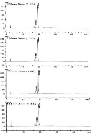
Fig 1.HPLC-DAD chromatograms of extract ofF. padanafruits at 500, 516, 530 and 550 nm
environmentally friendly and can also recover anthocyanins with good quality characteristics [30]. The use of acid stabilizes anthocyanins in the flavylium cation form, which is red at low pH [38]. However, solvent acidified with hydrochloric acid may hydrolyze acylated anthocyanins, which explains why it has been overlooked in the past that many anthocyanins are acylated with aliphatic acids. To avoid or at least minimize the breakdown of acylated anthocyanins, organic acids such as acetic, citric, or tartaric acids, which are easier to eliminate during anthocyanin concentration, have been preferred [39].
Test for Anthocyanins
Indo. J. Chem., 2014, 14 (4), 297 - 303
300
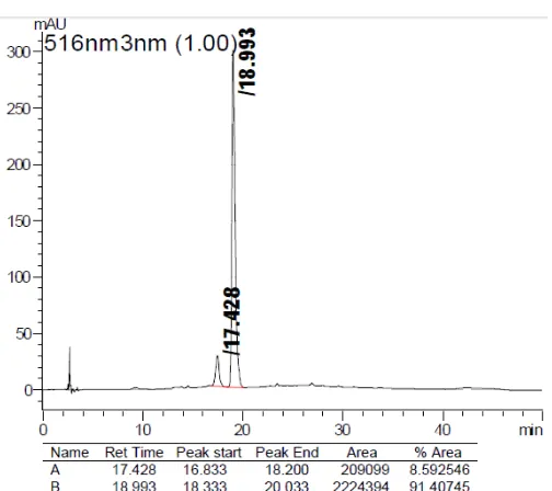
Fig 2.HPLC chromatograms of extract ofF. padanafruits (516 nm)
Fig 3.Structure and MS data of anthocyanin peak 1
extract and 3 mL HCl were mixed in a test-tube and then placed in boiling water bath for 5 min. The mixture was stable and did not lose color when boiled indicated the presence of anthocyanins in the extracts.
Identification of Anthocyanins
Fig. 1 showed the anthocyanin profile of the extract using the HPLC-DAD chromatograms at 500–550 nm. Value of the wavelength used is a specific wavelength to group anthocyanin. As can be seen, in four different DAD chromatograms (500, 516, 530, 550) nm at visible area, there are only two major peaks in the
chromatogram at the retention time range of 17–19 min, indicating the presence of two different anthocyanins in fruit of F. padana. From the chromatogram data at 516 nm (Fig. 2) can be seen that the second peak is the most dominant compound with the percentage reaches up to 91% of the total anthocyanin content inF. padanafruits extract.
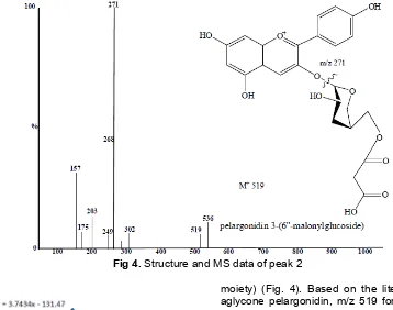
Fig 4.Structure and MS data of peak 2
Fig 5.Antioxidant activity of extract ofF. padanafruits
molecule and its fragmentation to prepared database (Table 1). Peak 1 and 2 showed same fragment ions at m/z 271 in MS analyses that could be tentatively identified as pelargonidin derivatives. Peaks 1 and 2 showed identical molecular ions at m/z 828 and 519, which concurs with that found in on line database [40] used for confirmation purposes. Peak 1 at m/z828 and MS2 fragments at 519 and 271 due to loss of coumarylglucoside (–309 amu) and Malonylglucoside (–248 amu), respectively, was identified as due to pelargonidin 3-(6”-p-coumarylglucoside)-5-(4”’-Malonyl-glucoside) (Fig. 3).
Peak 2 as the major anthocyanin in extract of F. padana fruits was attributed to pelargonidin 3-(6''-malonylglucoside), by MS data (m/z 519, MS2 fragments at 271, corresponding to a first loss of 248 amu (malonyl
moiety) (Fig. 4). Based on the literature data for the aglycone pelargonidin, m/z 519 for molecular ion was more suitable than the molecular ion m/z 536, therefore the m/z 519 molecular ion are used as the data.
Antioxidant Activity
In order to evaluate antioxidant activity of chosen theF. padanafruits, DPPH assay was applied and the results are presented in Fig. 5. IC50 determined by
DPPH was 55.70 mg/L. Specifically, a compound said to be a very potent antioxidant if IC50 values of less
than 50 ug/mL, a powerful antioxidant if IC50
50-100 ug/mL, medium strength if the IC50-value
151-200 mg/mL [41]. The antioxidant activity of the extract samples, giving IC50values of 55.7 mg/mL. This
indicates that the sample has a strong antioxidant activity.
CONCLUSION
Indo. J. Chem., 2014, 14 (3), 297 - 303
302
most experimental conditions is delphinidin, cyanidin, petunidin, pelargonidin, peonidin and malvidin. For different glycoside and/or acylated groups with the same anthocyanidin. By comparing their elution orders and mass spectrometric characteristics with published data in the literature one anthocyanidin were tentatively identified in F. padana samples. The anthocyanins showed pelargonidin aglycon and pelargonidin 3-(6’’-Malonylglucoside) was the major anthocyanin pigment that accounting for 91.4% of total anthocyanin. The results of the antioxidant activity of anthocyanin extracts showed that the extracts have high antioxidant activity with IC 50 values of 55.7 ppm.
REFERENCES
1. Gautier-Hion, A., Duplantier, J.-M., Quris, R., Feer, F.,Sourd, C., Decoux, J.-P., Dubost, G., Emmons, L., Erard, C., Hecketsweiler, P., Moungazi, A., Roussilhon, C., and Thiollay, J.-M., Oecologia, 65 (3), 324–337.
2. Gamble, J., Jaeger, S.R., and Harker, F.R., 2006, Postharvest Biol. Technol.,41 (1), 38–47.
3. Gould, K.S., McKelvie, J., and Markham, K., 2002, Plant Cell Environ., 25 (10),1261–1269.
4. Manetas, Y., 2006,Flora, 201 (3), 163–177.
5. Steyn, W.J., Wand, S.J.E., Holcroft, D.M., and Jacobs, G., 2002,New Phytol, 155 (3), 349–361. 6. Stintzing, F.C., and Carle, R., 2004, Trends Food
Sci. Technol.,15 (1), 19–38.
7. Huang, W., Zhang, S-L., Gai-hua Qin, G-H., Wenquan, L., and Wu, J., 2012,Afr. J. Agric. Res., 7 (26), 3772–3780.
8. Andersen, Ø.M., 1985, J. Food Sci., 50 (5), 1230– 1232.
9. Matysik, G., and Benesz, M., 1991, Chromatographia, 32 (1-2), 19–22.
10. Sherma, J., 2000,J Chromatogr. A, 880 (1-2), 129– 147.
11. Timberlake, C.F., and Bridle, P., 1976,Am. J. Enol. Vitic., 27, 97–105.
12. Bakker, J., Preston, N.W., and Timberlake, C.F., 1986,Am. J. Enol. Vitic., 37 (2), 121–126.
13. Hong, V., and Wrolstad, R.E., 1990, J. Agric. Food Chem., 38 (3), 708–715.
14. Desai, K.G.H., Olsen, K.F., Mallery, S.R., Stoner, G.D., and Schwendeman, S.P., 2010, Pharm. Res., 27 (4), 628–643.
15. Huang, Z., Wang, B., Williams, P., and Pace, R.D., 2009,LWT Food Sci. Technol., 42 (4), 819–824. 16. Li, P.C.H., Wong, M.C.K., Adomat, H., and Guns,
E.S.T., 2009, Can. J. Pure Appl. Sci., 3 (2), 765– 772.
17. Zheng, J., Ding, C., Wang, L., Li, G., Shi, J., Li, H., Wang, H., and Suo, Y., 2010,Food Chem.,126 (3), 859–865.
18. Wilson, I.D., Plumb, R., Granger, J., Major, H., Williams, R., and Lenz, E.M., 2005,J. Chromatogr. B, 817 (1), 67–76.
19. Lopes-da-Silva, F., de Pascual-Teresa, S., Rivas-Gonzalo, J., and Santos-Buelga, C., 2002, Eur. Food Res. Technol., 214 (3), 248–253.
20. DuPont, M.S., Bennett, R.N., Mellon, F.A., and Williamson, G., 2002,J. Nutr., 132 (2), 172–175. 21. Shoji, T., Masumoto, S., Moriichi, N., Kanda, T.,
and Ohtake, Y., 2006, J. Chromatogr. A, 1102 (1-2), 206–213.
22. Aaby, K., Ekeberg, D., and Skrede. G., 2007, J. Agric. Food Chem., 55 (11), 4395–4406.
23. Gil, M.I., Holcroft, D.M., and Kader, A.A., 1997,J. Agric. Food Chem., 45 (5), 1662–1667.
24. Wu, X., Beecher, G.R., Holden, J.M., Haytowitz, D.B., Gebhardt, S.E., and Prior, R.L., 2006. J. Agric. Food Chem., 54 (11), 4069–4075.
25. Gu, L., Kelm, M., Hammerstone, J.F., Beecher, G., Cunningham, D., Vannozzi, S., and Prior, R.L., 2002,J. Agric. Food Chem., 50 (17), 4852–4860. 26. Prior, R.L., Lazarus, S.A., Cao, G., Muccitelli, H.,
and Hammerstone, J.F., 2001, J. Agric. Food Chem., 49 (3), 1270–1276.
27. Garca-Beneytez, E., Cabello, F., and Revilla E., 2003,J. Agric. Food Chem., 51 (19), 5622–5629. 28. Oliveira, J., Azevedo, J., Silva, A.M., Teixeira, N.,
Cruz, L., Mateus, N., and de Freitas, V., 2010, J. Agric. Food Chem., 58 (8), 5154–5159.
29. Brand-Williams, W., Cuvelier, M.E., and Berset, C., 1995,LWT Food Sci. Technol., 28 (1), 25–30. 30. Kong, J., Chia, L.S., Goh, N.K., Chia, T.F., and
Brouillard, R., 2003, Phytochemistry, 64 (5), 923– 933.
31. Delgado-Vargas, F., and Paredes-Lopez, O., 2003, Natural Colorants for Food and Nutraceutical Uses, CRC Press, Boca Raton, FL, p. 326.
32. Oki, T., Masuda, M., Furuta, S., Nishiba, Y., Terahara, N., and Suda, I., 2002, J. Food Sci., 67 (5).1752–1756.
33. de Pascual-Teresa, S., Santos-Buelga, C., and Rivas-Gonzalo, J.C., 2002,J. Sci. Food Agric., 82 (9),1003–1006. Food Sci. Technol., 41 (1), 155–160.
38. Rivas-Gonzalo, J., 2003,“Analysis of polyphenols” in Methods in Polyphenols Analysis; Santos-Buelga, C., and Williamson, G., eds., Royal Society of Chemistry (Athenaeum Press, Ltd.), Cambridge, U.K., p. 95–98, 338–358.
39. Strack, D., and Wray, V., 1989, “Anthocyanins” in Methods in Plants Biochemistry, Dey, P.M., and Harbone, J.B., eds., Academic Press: San Diego, CA, Vol. 1: Plant Phenolics, p. 325–359.
40. http://www.lipidmaps.org/data/structure/LMSDSear ch.php?Mode=ProcessClassSearch&LMID=LMPK 12, accessed on November 10th2013.