Corresponding author: [email protected]
The effect of platelet rich fibrin (PRF) on
serum starved human dermal fibroblast
Sunardi Radiono*, Yohanes Widodo Wirohadidjojo, Arief Budiyanto,
Department of Dermatology and Venereology, Faculty of Medicine, Universitas Gadjah Mada, Yogyakarta, Indonesia
DOI: http://dx.doi.org/10.19106/JMedSci004802201605
ABSTRACT
Healing failure on chronic ulcers was suspected due to the decrease of Growth Factors
(GFs) supply caused by either GFs trapped in the ibrin, or degraded by protease, or
decreased level due to reduction of GFs gene expression. Administration of various GFs can stimulate healing of chronic ulcers. High level of GFs is available in biologic material called Platelet Rich Fibrin (PRF). This study was conducted to have in vitro evidence of
PRF effect on GFs-serum starved human dermal ibroblasts as representative cells of chronic ulcers. Human dermal ibroblasts (HDFs) were isolated from foreskin of six boys
aged 11- 14 years-old. After 24 hours of serum deprivation, HDFs were treated by 100, 50, and 25% PRF lysate diluted in cultured medium. Cellular migration was measured using scratch assay, while cellular viability was measured using MTT assay and collagen deposition was measured using Sirius Red assay. The HDFs of serum starvation group
showed signiicant impairment activities in terms of cellular migration (25%), cellular
proliferation (20%), and collagen deposition (10%) (p<0.05). Administration with various
levels of PRF lysate could signiicantly recover those activities (p < 0.05). Because cellular activities of serum starved HDFs is similar with ibroblasts isolated from the bottom of
chronic ulcers and administration of various levels of PRF lysate was capable to recover those activities, it can be concluded that PRF is a good biologic material to stimulate healing of chronic ulcers. However, in order to get better evidence based medicine, both pre clinical and clinical studies must be performed.
ABSTRAK
Kegagalan sembuh pada ulkus khronis disebabkan oleh rendahnya suplai growth factor
(GF) akibat GF terjebak dalam ibrin, atau dirusak oleh protease bakteri, atau akibat menurunnya ekspresi gena penyandi GF. Pemberian GF dapat memacu penyembuhan
ulkus khronis. Berbagai GF ternyata terdapat dalam material biologis seperti ibrin kaya
platelet (FKP). Penelitian ini dilakukan untuk memperoleh bukti in vitro bahwa FKP dapat
mengembalikan fungsi ibroblast tanpa serum sebagai model ibroblas dari ulkus khronis.
Fibroblas dermis manusia (FDM) diisolasi dari kulit kulup 6 anak laki-laki berumur 11-14 tahun. Setelah dibebaskan dari serum selama 24 jam, FDM kemudian diberi 100, 50, 25% lisat FKP dan medium pelarut sebagai kontrol. Selanjutnya, aktivitas selular berupa timbunan kolagen, proliferasi dan migrasi sel diukur berturut-turut dengan metode Sirius Merah, MTT, dan goresan.FDM tanpa serum ternyata secara mengalami penurunan kemampuan menimbun kolagen sebesar 10%, proliferasi sebesar 20% dan penurunan
ternyata dapat memulihkan aktivitas selular FDM tersebut. Dari eksperimen ini dapat disimpulkan bahwa FKP merupakan material biologis yang dapat dikembangkan untuk memacu penyembuhan ulkus khronis. Namun demikian, untuk memperoleh bukti-bukti klinis yang lebih baik, uji pra klinis dan uji klinis tetap diperlukan.
Keywords: chronic ulcers - human dermal ibroblasts - serum starvation - platelet rich
ibrin lysate – growth factors
INTRODUCTION
Chronic ulcers are dificult to heal spontaneously due to their characteristics of low cellular proliferation rates,1-3 low cellular
migration capacities,3-5 followed by cellular
senescence of ibroblasts on their wound
bed and edges.3,6 All of these may be caused
by lack of growth factor supplies7,8 as they
are indicated by the decrease of GF-gene expressions among diabetic patients who tend to have chronic ulcers,9 the entrapment of GFs
by ibrin that spill out when capillaries leak due to high venous pressure in patients with venous leg ulcers,7,10,11 and GFs degradation
by protease contained in bio-ilm on the
surface of chronic ulcer.2,12 The theory of GFs
supply deiciency will be strengthened when considering the indings of the clinicians who succeeded in stimulating chronic ulcers healing using either recombinant platelet derived growth factor (r-PDGF),13
or recombinant epidermal growth factor (r-EGF),14-16 or recombinant ibroblast growth
factor (r-FGF) that were applied topically.17-19
Platelets are the source of autologous GFs, which are released when platelets had been activated. Based on the technology of platelet-rich plasma (PRP), Eppley et al.,20
found that the level of PDGF, EGF, and FGF increased almost fourfold compared to the level of human plasma. Various levels of PRP supernatant have been able to restore activity of artiicial ibroblasts senescence not due to serum starvation but due to repeated Ultraviolet B exposure.21 Beside PRP technology, another
convenient technology to obtain GFs is PRF
technology22 and although it indicates slower
release of GFs,23,24 but its lysate contains high
levels of GFs.25
PRF often used by dentists to stimulate extraction socket healing,26 treatment of
maxillary sinus,27 and as grafting material for
intrabony periodontal defects.28,29 Eventhough
the application of PRF in dermatological ield has rarely been published, our dermatology experience indicates that it is effective to treat chronic ulcers which failed to heal under conventional PRF treatment. Unfortunately, in the absence of placebo in our cases, we could not make a conclusion that those healings occurred due to PRF administration. In order to obtain in vitro evidence about the effect of
PRF lysate in chronic ulcers, we performed HDFs cultivation in 24-hour-serum starvation and hope it will show characteristic similar to ibroblast isolated from the bottom of chronic ulcers. After that, we add various levels of PRF lysate and measured the changes in cellular activities compared to control.
MATERIALS AND METHOD
This study used in vitro experiment design
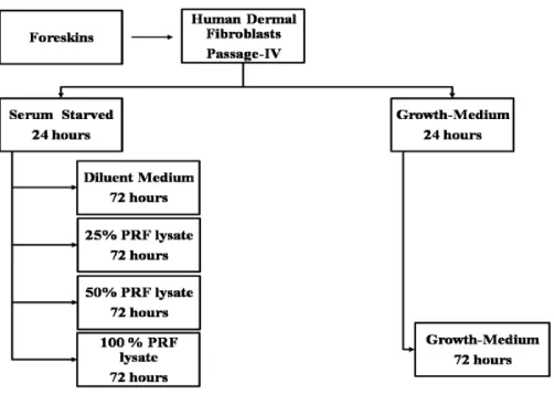
FIGURE 1. Experiment Scheme
Cell cultures.
Human ibroblasts from foreskin of
healthy six boys were cultured using the explants technique as described previously.30
The cells were cultured in Dulbecco Modiied Eagle Medium (DMEM) with 4.5 g/L D-glucose, 2mmol/L L-glutamine, 2.5mg/L amphotericin B, 100ug/L streptomycin, 100unit/L penicillin, and 10% fetal bovine serum. All materials were purchased from Gibco-USA. The forth passages or more were used for the experiments.
PRF lysate
PRFs were isolated based on a method by Dohan et al.22 Briely, 20 mL of blood
was taken from volunteers into sterile glass tube, centrifuged 400 g for 10 min, and after 5 min of room temperature incubation, PRF gel was separated from the red blood cells and transferred into new sterile tubes and kept at 40 C for 24 hr. Supernatants were
then collected and considered as 100 % PRF lysate as previously described by Yao Su et al.24 Additional dilution with DMEM was
performed to have inal concentration of 50
and 25%.
Experiment
Experiment was carried out with HDF re-seeding in wells of 200 uL cell suspension containing 103 cells/mL. After 72 hr of
incubation, various treatments carried out in accordance with a written protocol in FIGURE 1. DMEM enriched with 0.5% fetal bovine serum was used for serum starvation medium, diluent medium of PRF, and control.
Collagen deposition
Collagen deposition was calculated from optical density of insoluble collagen based on Sirius Red assay.31 Briely, after washing the
wells with phosphate buffer saline (PBS), cells were ixed with Bouin solution for an hour, and
The collagen deposition of normal HDFs was assumed to be 100% and collagen deposition of various treated groups was accounted as : (Unsoluble collagen of treated group /Unsoluble collagen of paired normal HDFs) x 100 %.
Proliferation index
Proliferation index was calculated from cellular viability that was determined in formasan-blue optical density of 570 nm iltered spectrometer. The proliferation index of normal HDFs was assumed to be 100% and the proliferation index of various treated groups was determined as :(Cellular viabilities of treated group/Cellular viabilities of paired normal HDFs) x 100 %.
For these purposes, all of medium was
aspirated and the wells were rinsed using PBS for 3 times, 10 min each. Two hundred μL of
fresh medium were illed into each well and 50 μL of 50 mg/mL MTT (MP Biomedicals-USA) were added. The plates were covered by aluminum foil and incubated for 6 hr in 37°C. Medium containing MTT were aspirated and 200 μL DMSO were then illed and followed
by 25 μL glycine buffer on the top. Optical
density was immediately read by 570 nm iltered spectrometer.
Fibroblast migration assay
Fibroblasts migration assay was perform-ed basperform-ed on method and computperform-ed basperform-ed on method of Liang, et al.,32 with slight
modiication. Briely, after serum starvation, all of wells were linearly scratched with the blunt tip of a 32G sterile needle through the centre of the well bottom. The cells were then cultivated in the experimental medium for 72 hr. The cells were then stained with Meyer’s haematoxylin and microscopic photo images were taken using a Moticam350
(China) camera in JPG format. By using Adobe Photoshop, both blue color’s pixels of ibroblasts along scratch line and white color’s pixels of empty space can be measured.
Fibroblast migration capacity was counted as: (pixel of ibroblasts along scratch line/total pixel along scratch line) x 100%.
Statistical analysis
All datas were presented as mean. Because of the data was not normally distributed, we used Wilcoxon rank sign test for testing the paired data and p<0.05 was considered as signiicant different.
RESULTS
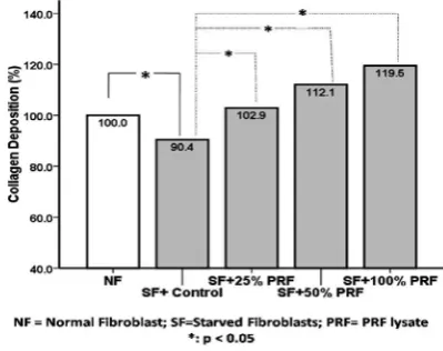
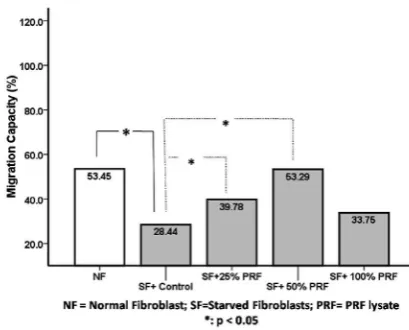
Serum starved HDFs revealed various decreases cellular activity in: collagen deposition (from 100% to 90.4%), proliferation index (from 100% to 80.9%), and in migration capacity (from 53.45% to 28.44%). Administration of various levels of PRF lysate affected on those variables as in following FIGURE 2-4.
FIGURE 2.Collagen deposition among various groups
its response was paralel with lysate’s level (FIGURE 2).
FIGURE 3. Proliferation index among various groups
It appears that serum starvation caused in suppression of proliferation index and administration of PRF lysate in various levels were able to ameliorate this activity signiicantly (p<0.05), even 25% PRF lysate could induce starved ibroblast better than normal ibroblasts in this variable (FIGURE 3).
FIGURE 4. Migration capacity among Various Groups
It shows that serum starvation caused depression of migration capacity of ibroblasts and various levels of PRF lysate were able to restore starved ibroblasts in migration capacity, mainly among starved ibroblasts
that had administration of 50% PRF lysate (FIGURE 4).
DISCUSSION
As shown on FIGURE 2 to 4, serum starvation for 24 hr was able to suppress HDFs in collagen synthesis (FIGURE 2), proliferation index (FIGURE 3), and migration capacity (FIGURE 4). Those are similar with the characteristic of ibroblasts isolated from the edge of venous ulcers that it is published
by Herrick et al.33 On the FIGURE 2, our
experiment revealed a restoration of collagen deposition of serum starved HDFs from 90.4% to the highest achievement of 113.8% due to administration of 100% PRF lysate. The lowest level of PRF lysate (25%) was still had improvement effect to 102.9%. These phenomena may be caused by level of GFs in this lysate. A study showed that PRF lysate contains a rich variety of growth factors which required for the synthesis of collagen such as PDGF.34 This effect may not be found among
fresh wound with normal GFs supplies as it is indicated by an experiment where collagen synthesis in tubes inserted from the edge of the new wound incision for ten days has not been shown better compared to albumin as control.35
FIGURE 4 of migration capacity. In wound healing processes, ibroblast migrations are crucial events and the researchers have found that it has under responsibility of PDGF,36
EGF and extracellular matrix component such as collagen type I and ibronectin.37,38
Naturally, PDGF and EGF are released by platelets immediately after the blood clotting,39 contained in the serum as important
supplement for ibroblast migration and proliferation on regular HDFs cultivation,40,41
and they have been found in high level of PRF lysate.25
These indings also indicate that platelet’s products are important for human dermal cells proliferation and migration, such as in newest creation of cultured human dermal substitutes without medium and animal products, but human serum or platelet’s product was still required.42-44 Compare to PRP technology,
isolating autologous GFs by PRF technology is considered simpler; it also contains various GF types which required for stimulation of healing processes as shown in our experiment. Similar result has been reported by Jørgensen
et al.,45 in their pilot study where PRF has also
effective for recalcitrant chronic wound. The weakness of our study is on angiogenesis variable as an important factor in healing processes. Another study has shown the effect of platelet rich ibrin matrix, another PRP based technology, capable to stimulate angiogenesis via endothelial cell proliferation.45 Differ with PRP technology
which has been shown capable to stimulate the proliferation of normal HDFs46 as well
as UV-induced HDFs senescence,21 research
on PRF application in dermatology ield is very limited. However, in order to achieve a better level of evidence based medicine of PRF application, various in-vitro and clinical researches are still needed.
CONCLUSION
PRF as a simple method to isolate autologous GF and it indicates a therapeutic effect in stimulate-healing of chronic ulcers. Research in large scale is needed to get a better level in evidence based medicine.
ACKNOWLEDGMENTS
This study is sponsored by Universitas Gadjah Mada research funding 2012.
REFERENCES
1. Loots MA, Lamme EN, Mekkes JR, Bos JD, Middelkoop E. Cultured ibroblasts from chronic diabetic wounds on the lower extremity (non-insulin-dependent diabetes mellitus) show disturbed proliferation. Arch Dermatol Res 1999; 291(2-3):93-9.
2. Schmidtchen A, Holst E, Tapper H, Björck L. Elastase-producing Pseudomonas aeruginosa degrade plasma proteins and extracellular products of human skin and ibroblasts, and inhibit ibroblast growth. Microb Pathog 2003; 34(1):47-55.
3. Harding KG, Moore K, Phillips TJ. Wound chronicity and ibroblast senescence-implications for treatment. Int Wound J 2005; 2(4):364-8.
4. Lerman OZ, Galiano RD, Armour M, Levine JP, Gurtner GC. cellular dysfunction in the diabetic ibroblast: impairment in migration, vascular endothelial growth factor production, and response to hypoxia. Am J Pathol 2003; 162(1):303-12.
6. Raffetto JD, Mendez MV, Phillips TJ, Park HY, Menzoian JO. The effect of passage number on ibroblast cellular senescence in patients with chronic venous insuficiency with and without ulcer. Am J Surg 1999; 178(2):107-12.
7. Drinkwater SL, Smith A, Sawyer BM, Burnand KG. Effect of venous ulcer exudates on angiogenesis in vitro. Br J Surg 2002; 89(6):709-13.
8. Labler L, Rancan M, Mica L, Harter L, Mihic-Probst D, Keel M. Vacuum-assisted closure therapy increases local interleukin-8 and vascular endothelial growth factor levels in traumatic wounds. J Trauma 2009: 66(3):749-57. http://dx.doi.org/ 10.1097/ TA.0b013e318171971a.
9. Cianfarani F, Zambruno G, Brogelli L, Sera F, Lacal PM, Pesce M, et al. Placenta growth factor in diabetic wound healing: altered expression and therapeutic potential. Am J Pathol 2006; 169(4):1167-82.
10. Falanga V, Eaglstein WH. The “trap” hypothesi s of venous ulceration. The Lancet 1993; 341(8851):1006-8. al. Chronic wounds and the medical bioilm paradigm. J Wound Care 2010; 19(2):45-6. 13. Miller MS. Use of topical recombinant
human platelet-derived growth factor-BB (becaplermin) in healing of chronic mixed arteriovenous lower extremity diabetic ulcers. J Foot Ankle Surg 1999; 38(3):227-31. 14. Zhu JX, Zhang YM. Application of
recombinant human epidermal growth factor on chronic ulcer wound. Zhongguo Xiu Fu Chong Jian Wai Ke Za Zhi 2002; 16(1):42-3. 15. Mohan VK. Recombinant human epidermal
growth factor (REGEN-D™ 150): effect on
healing of diabetic foot ulcers. Diabet Res Clin Pract. 2007; 78(3):405-11.
16. Choi JS, Leong KW, Yoo HS. In vivo wound healing of diabetic ulcers using electrospun nanoibers immobilized with human epidermal growth factor (EGF). Biomatrials 2008; 29(5):587-96.
17. Kawai K, Suzuki S, Tabata Y, Nishimura Y. Accelerated wound healing through the incorporation of basic ibroblast growth factor-impregnated gelatin microspheres into artiicial dermis using a pressure-induced decubitus ulcer model in genetically diabetic mice. Brith J Plas Surg 2005; 58(8):1115-23. 18. Kurokawa I, Hayami J, Kita Y. A
therapy-resistant chronic leg ulcer treated successfully with topical basic ibroblast growth factor. J Int Med Res 2003; 31(2):149-51.
19. Yang Y, Xia T, Zhi W, Wei L, Weng J, Zhang C, et al. Promotion of skin regeneration in diabetic rats by electrospun core-sheath ibers loaded with basic ibroblast growth factor. Biomat 2011; 32(18):4243-54. http://dx.doi. org/ 10.1016/j.biomaterials.2011.02.042. 20. Eppley BL, Woodell JE, Higgins J. Platelet
quantiication and growth factor analysis from platelet-rich plasma: implications for wound healing. Plast Reconstr Surg 2004; 114(6):1502-8.
21. Wirohadidjojo YW, Tresnowati N, Budiyanto A. Cellular viability and Collagen deposition among UVB irridiated human skin ibroblasts treated by platelets. J Clin Med Res 2012; 4(2): 29-33.
22. Dohan DM, Choukroun J, Diss A, Dohan SL, Dohan AJ, Mouhyi J, et al. Platelet-rich ibrin (PRF): a second-generation platelet concentrate. Part I: technological concepts and evolution. Oral Surg Oral Med Oral Pathol Oral Radiol Endod 2006;
101(3):E37-44.
of growth factors and thrombospondin-1 in Choukroun’s platelet-rich ibrin (PRF): a gold standard to achieve for all surgical platelet concentrates technologies. Growth Factors 2009; 27(1): 63-9. http://dx.doi.org/ 10.1080/08977190802636713.
24. Su CY, Kuo YP, Tseng YH, Su CH, Burnouf T. In vitro release of growth factors from platelet-rich ibrin (PRF): a proposal to optimize the clinical applications of PRF. Oral Surg Oral Med Oral Pathol Oral Radiol Endod 2009; 108(1):56-61. http://dx.doi. org/10.1016/j.tripleo.2009.02.004.
25. He L, Lin Y, Hu X, Zhang Y, Wu H. A comparative study of platelet-rich ibrin (PRF) and platelet-rich plasma (PRP) on the effect of proliferation and differentiation of rat osteoblasts in vitro. Oral Surg Oral Med Oral Pathol Oral Radiol Endod 2009; 108(5):707-13. http://dx.doi.org/10.1016/j. tripleo.2009.06.044.
26. Huei Zhao J, Hung Tsai C, Chao Chang Y. Clinical and histologic evaluations of healing in an extraction socket illed with platelet-rich ibrin. J Dent Sci 2011; 6(2):116-22.
27. Morandi G, Stocca T, Maglione M, Bertolotto M. Angiogenesis in a bilateral maxillary sinus elevation with or without platelet-rich ibrin. Dent Mats 2010; 26(S1):76.
28. Chao Chang Y, Chin Wu K, Huei Zhao J. Clinical application of platelet-rich ibrin as the sole grafting material in periodontal intrabony defects. J Dent Sci 2011; 6(3):181-8. http://dx.doi.org/ 10.1016/j.jds.2011.05.010. 29. Rosamma VJ, Arun Raghunath A, Sharma N.
Clinical effectiveness of autologous platelet rich ibrin in the management of infrabony periodontal defects. Singapure Dent J 2012; 33(1):5-12. http://dx.doi.org/10.1016/j. sdj.2012.10.003.
30. Viale MCL. Recontruction of human epidermis invitro. In: Shaw AJ, (ed); Epithelial cell culture. Oxford: Oxford University Press, 2000; 179-99.
31. Taskiran D, Taskiran E, Yercan H, Kutay FZ. Quantiication of total collagen in rabit tendon by the Sirius Red Methods. Tr J Med Science 1999;(29):7-9.
32. Liang CC, Park AY, Guan JL. In vitro scratch assay: a convenient and inexpensive method for analysis of cell migration in vitro. Nature Protocols 2007; 2(2):329-33.
33. Herrick SE, Ireland GW, Simon D, McCollum CN, Ferguson MW. Venous ulcer ibroblasts compared with normal ibroblasts show differences in collagen but not ibronectin production under both normal and hypoxic conditions. J Invest Dermatol 1996; 106(1):187-93.
34. Matsuda N, Lin WL, Kumar NM, Cho MI, Genco RJ. Mitogenic, chemotactic, and synthetic responses of rat periodontal ligament ibroblastic cells to polypeptide growth factors in vitro. J Periodontol 1992; 63(6):515-25.
35. Danielsen PL, Agren MS, Jorgensen LN. Platelet-rich ibrin versus albumin in surgical wound repair: a randomized trial with paired design. Ann Surg 2010; 251(5):825-31. http://dx.doi.org/ 10.1097/ SLA.0b013e3181d3548c.
36. Li W, Fan J, Chen M, Guan S, Sawcer D, Bokoch GM, et al. Mechanism of human dermal ibroblast migration driven by type I collagen and platelet-derived growth factor-BB. Mol Biol Cell 2004; 15(1):294-309. 37. Maheshwari G, Wells A, Grifith LG,
Lauffenburger DA. Biophysical integration of effects of epidermal growth factor and ibronectin on ibroblast migration. Biophys J 1999; 76(5):2814-23.
39. Antoniades HN, Scher CD, Stiles CD. Puriication of human platelet-derived growth factor. Proc Natl Acad Sci USA 1979; 76(4):1809-13.
40. Schreier T, Degen E, Baschong W. Fibroblast migration and proliferation during in vitro wound healing. A quantitative comparison between various growth factors and a low molecular weight blood dialysate used in the clinic to normalize impaired wound healing. Res Exp Med 1993; 193(4):195-205.
41. Mazlyzam AL, Aminuddin BS, Saim L, Ruszymah BH. Human serum is an advantageous supplement for human dermal ibroblast expansion: clinical implications for tissue engineering of skin. Arch Med Res 2008; 39(8):743-52. http://dx.doi.org/ 10.1016/j.arcmed.2008.09.001.
42. Morimoto N, Takemoto S, Kanda N, Ayvazyan A, Taira MT, Suzuki S. The utilization of animal product-free media and autologous serum in an autologous dermal substitute culture. J Surg Res 2011; 171(1):339-46. http://dx.doi.org/ 10.1016/j.jss.2009.11.724.
43. Grinnell F, Rocha LB, Iucu C, Rhee S, Jiang H. Nested collagen matrices: a new model to study migration of human ibroblast populations in three dimensions. Exp Cell Res 2006; 312(1);86-94.
44. Kim DH, Je YJ, Kim CD, Lee YH, Seo YJ, Lee JH, Lee Y. Can Platelet-rich plasma be used for skin rejuvenation? evaluation of effects of platelet-rich plasma on human dermal ibroblast. Ann Dermatol 2011; 23(4):424-31. http://dx.doi.org/ 10.5021/ad.2011.23.4.424. 45. Jørgensen B, Karlsmark T, Vogensen H,
Haase L, Lundquist R. A pilot study to evaluate the safety and clinical performance of Leucopatch, an autologous, additive-free, platelet-rich ibrin for the treatment of recalcitrant chronic wounds. Int J Low Extrem Wounds 2011; 10(4):218-23. http:// dx.doi.org/ 10.1177/1534734611426755. 46. Roy S, Driggs J, Elgharably H, Biswas S,