PATIENT INFORMATION—ANYTIME ANYWHERE
S
troke is the third leading cause of death in the Western World and the major cause of disability in adults. Carotid stenosis is the largest single etiologi-cal factor known to produce foetiologi-cal cerebral ischemia, but in those with carotid bifurcation disease only a minority gives warning symptoms, with the majority having their stroke from previously asymptomatic lesions. Since mor-bidity and mortality after acute stroke is unacceptably high, it is very important to recognize and to treat patients with carotid bifurcation disease before they develop symptoms [1].High-resolution ultrasound (duplex scanning and color flow imaging) can determine the degree of stenosis. Ultrasound image plaque characterization, ulceration, and brain CT scan-ning have been demonstrated to be individually associated with an increased risk of stroke. However, the combination of these findings with clinical data and risk factors has not been utilized to identify patients at high risk of stroke, making the management of these patients a difficult task.
High-resolution ultrasound has made possible the nonin-vasive visualization of the carotid bifurcation, and for that reason it has been extensively used in the study of arterial wall changes; these include measurement of the thickness of the intima media complex (IMT), estimation of the severity of stenosis due to atherosclerotic plaques, and plaque characterization [1], [2]. Ultrasound provides infor-mation not only on the degree of carotid artery stenosis but also on the characteristics of the arterial wall including the size and consistency of atherosclerotic plaques. Several studies have indicated that “complicated” carotid plaques are often associated with ipsilateral neurological symp-toms [1], [3]. These symptomatic plaques share common ultrasonic characteristics, being more echolucent (weak reflection of ultrasound and therefore containing echo-poor structures) and heterogeneous (having both echolu-cent and echogenic areas). In contrast, “uncomplicated” plaques that are often asymptomatic tend to be of uniform consistency (uniformly hypoechoic or uniformly hypere-choic) without evidence of ulceration [1], [3].
The objective of this article is to present an integrated system for the assessment of risk of stroke based on two modules: 1)
clinical risk factors and noninvasive investigations, and 2) carotid plaque texture analysis. The system supports the data collected from the Asymptomatic Carotid Stenosis and Risk of Stroke (ACSRS) natural history study that includes the follow-ing groups of data [1], [2], [4]: 1) clinical, biochemical, and electrocardiographic risk factors associated with cardiovascular deaths, and 2) whether any independent risk factors can be used to identify a high-risk group for cardiovascular death. Furthermore, the carotid plaque texture analysis module sup-ports the normalization, despeckling, segmentation, texture fea-ture extraction, and classification of ultrasound plaque images.
Clinical Risk Factors and Noninvasive Investigations Module
Material
The ACSRS is an ongoing international multicenter study under the auspices of the International Union of Angiology aiming to study patients with asymptomatic 50–99% stenosis in relation to the carotid bulb diameter as documented in the European Carotid Surgery Trial (ECST) [4] and to follow them up for at least five years to identify subgroups at high and low risk for future neurological events. Approval has been obtained from the Multiregional Research Ethics Committee (North Thames, London, United Kingdom) and local ethics committees. Patients were admitted to the study after informed consent. The methodology used in the ACSRS study, eligibili-ty of participating centers, and qualieligibili-ty control have been pub-lished in detail [2]. Patients with internal carotid artery (ICA) stenosis greater than 50% (ECST) on duplex scanning who had never had any ipsilateral hemispheric or retinal symptoms and did not have any neurological abnormality on examination were eligible for admission to the study [2], [4].
Risk Factors and Noninvasive Investigations
The following clinical risk factors and their duration and sever-ity were recorded for each patient [1], [2], [4]: age, gender, height and weight, hypertension, cardiac status, diabetes, smok-ing, fibrinogen blood level, fasting total cholesterol, HDL-cho-lesterol, LDL-choHDL-cho-lesterol, triglycerides, and creatinine blood levels and hematocrit; also, cardiac status including any history of myocardial infarction of cardiac failure, myocardial
An Integrated System
for Assessing Stroke Risk
Utilizing Clinical Risk Factors and Noninvasive
Investigations Plus Carotid Plaque Texture Analysis
BY EFTHYVOULOS C. KYRIACOU, CONSTANTINOS S. PATTICHIS, MINAS A. KARAOLIS, CHRISTOS P. LOIZOU,
CHRISTODOULOS I. CHRISTODOULOU, MARIOS S. PATTICHIS, STAVROS KAKKOS, AND ANDREW NICOLAIDES
© ARTVILLE, LLC
ischemia on ECG, and left ventricular hypertrophy (LVH) on ECG. Myocardial ischemia was considered to be present in one or more of the following as seen at any lead except aVR: flat T wave, negative T wave amplitude, and significant horizontal or downsloping S-T depression. The criteria of LVH included one or a combination of the following: the Sokolow-Lyon voltage criteria of sum of R wave amplitude in V5/V6 and S wave in VL greater than 35 mm, R wave amplitude in aVL greater than 13 mm, R wave amplitude in V5/V6 greater than 27 mm, or S wave amplitude in V1/V2 grater than 25 mm.
Duplex scanning was performed on admission to the study and subsequently every six months to grade ICA stenosis and also to study plaque morphology (see next section). Velocity criteria for grading the degree of stenosis are summarized in references [1], [2], [4]. The entire duplex examination was recorded on videotape. Ultrasound images of the plaques were classified into the following types [2]. Type 1: Uniformly echolucent (black), where bright areas occupy less than 15% of the plaque area. Type 2: Mainly echolucent, where bright echoes occupy 15–50% of the plaque area. Type 3: Mainly echolucent, where bright echoes occupy 50–85% of the plaque area. Type 4: Uniformly echogenic, where bright echoes occupy more than 85% of the plaque area. Type 5: Calcified cup with acoustic shadow so that the rest of the plaque cannot be visualized.
Endpoints
Primary endpoints were ipsilateral hemispheric stroke (including fatal stroke) defined as a hemispheric neurological deficit lasting for more than 24 hours, any stroke, ipsilateral hemispheric transient ischemic attacks (TIA), amaurosis fugax (AF), and death from cardiovascular causes other than stroke [1], [2], [4].
Online Analytical Mining System
An online analytical mining (OLAM) system was developed in Microsoft SQL with HTML and JSP functionality based on the following components. 1) Data collection: The data were collected under the ACSRS study using forms covering the aforementioned groups of data. 2) Data cleaning: The fields were identified, duplications were extracted, and missing val-ues were filled. 3) Data coding: All the aforementioned risk factors were selected and coded. 4) Data mining. a) Classification by decision tree induction: The C4.5 algorithm [5], which uses a divide-and-conquer approach to decision tree induction, was employed. The algorithm uses the infor-mation gain criterion and the gain ratio. It works top-down, seeking at each stage an attribute to split on, that which best separates the classes, and then recursively processing the sub-problems that result from the split. The algorithm uses heuris-tics for pruning, derived from the statistical significance of splits. b) Rule extraction using the association algorithm: The original set-oriented mining of association rules (SETM) [6] algorithm that generates candidate item sets as the database is scanned was used [7]. After reading a transaction, the item sets that were found to be frequent in the previous pass con-tained in this transaction are derived. New candidate item sets are generated by extending the frequent item sets with other items in the transaction. A frequent item set L is extended with only those items that are frequent and occur later in the lexicographic ordering of items than any of the items in L. The candidates generated from a transaction are added to the
set of candidate item sets maintained for the pass. Each rule is characterized by confidence and support. 5) Pattern evalua-tion and knowledge representaevalua-tion: Following the run of the C4.5 and the SETM algorithms, rules extracted were sorted for p=1, … , maximum number of risk factors investigated, based on the frequency of stroke events. Rules with consecu-tive numbers of risk factors were compared and the impor-tance of the p+1 risk factor based on the frequency of stroke events was derived. Based on the above procedure, rules to enable the following pattern evaluation and knowledge repre-sentation were carried out:
➤ hierarchical classification of risk factors
➤ minimal number of risk factors that should coexist for a stroke episode to occur
➤ a specific risk factor can be modified in order to reduce the risk of stroke.
Extracted frequency (or percentage) per rule was assumed to represent the percentage of the risk of stroke.
Carotid Plaque Texture Analysis Module
Material
Ultrasound images from a cross-sectional study of 274 images (137 asymptomatic and 137 symptomatic) were collected for texture analysis. A subset of 80 images from this dataset was used for the plaque segmentation, as well as an additional 100 images that were captured for the IMT segmentation. Images for plaque segmentation and texture analysis were captured at the Irvine Laboratory for Cardiovascular Investigation and Research, St Mary’s Hospital, United Kingdom, whereas the IMT segmentation images were captured at the Vascular Clinic of the Cyprus Institute of Neurology and Genetics. Patients with cardioembolic symptoms or distant symptoms (> 6 months) were excluded from the study. Asymptomatic plaques were truly asymptomatic if they had never been associated with symptoms in the past. Carotid plaques were labeled as symptomatic after one of the following symptoms was identified: stroke, TIA, or AF. The ultrasound images were collected using an ATL (model HDI 3000, Advanced Technology Laboratories, Seattle, Washington) duplex scan-ner with a 5–10 MHz multifrequency probe. Longitudinal scans were performed using duplex scanning and color flow imaging [2]. Ultrasound was at right angles to the adventitia and the image was magnified or the depth adjusted so that the plaque would fill a substantial area of the image, giving approximately a resolution of 20 pixels/mm.
Normalization and Despeckle Filtering
adventitia was visible adjacent to the plaque, and that for image normalization a standard sample consisting of half of the width of the brightest area of adventitia was obtained.
In addition, to address noise issues, we applied despeckle fil-tering to the normalized images. Speckle is a form of multi-plicative noise that corrupts medical ultrasound imaging, making visual observation difficult. It limits the contrast reso-lution, thereby limiting the detectability of small, low-contrast lesions and making the ultrasound images generally difficult to interpret [8]. It was therefore important to despeckle the images prior to further image analysis. A linear scaling filter (lsmv) [8], utilizing the mean and the variance of a 7×7 pixel
neighborhood, was used in this study. The filter was applied iteratively five times on each image, as documented in [8].
Segmentation
Following image normalization and despeckling, the Williams and Shah and the Lai and Chin snakes for segmenting the IMT [9] and atherosclerotic plaque [10] were used, respective-ly. For the IMT segmentation, initial contour estimation was carried out by selecting the area of interest (carried out manu-ally by the user [9]), whereas, for the plaque segmentation, the initial contour estimation was carried out without user interac-tion using the blood flow image [10].
Feature Extraction and Selection
Following the segmentation of the atherosclerotic carotid plaque, the following texture feature sets were computed [11]: 1) statisti-cal features (SFs): a) mean, b) variance, c) median value, d) skewness, e) kurtosis, f) energy, and g) entropy. 2) spatial gray level dependence matrices (SGLDMs): a) angular second moment, b) contrast, c) correlation, d) sum of squares: variance, e) inverse difference moment, f) sum average, g) sum variance, h) sum entropy, i) entropy, j) difference variance, k) difference entropy, and l) information measures on correlation 1 and 2. For a chosen distance d(in this work d= 1 was used) and for angles
θ= 0◦, 45◦, 90◦, and 135◦, we computed four values for each of
the above 13 texture measures. In this work, the mean of these four values was computed for each feature and it was used for classification. 3) Gray level difference statistics (GLDSs): a) homogeneity, b) contrast, c) energy, d) entropy, and e) mean. The above features were calculated for displacements δ= (0, 1), (1, 1), (1, 0), (1, –1), where δ≡(x, y), and their mean values were taken. 4) Neighborhood gray tone difference matrix (NGTDM): a) coarseness, b) contrast, c) busyness, d) complexi-ty, and e) strength. 5) Statistical feature matrix (SFM): a) coarse-ness, b) contrast, c) periodicity, and d) roughness. 6) Laws texture energy measures (LAWS): LL – texture energy from LL kernel, EE – texture energy from EE kernel, SS – texture energy from SS kernel, LE – average texture energy from LE and EL kernels, ES – average texture energy from ES and SE kernels, and LS – average texture energy from LS and SL kernels. We averaged the matched pairs of energy measures in order to obtain a degree of invariance with respect to rotations. 7) Fractal dimension texture analysis (FRACTALS): The Hurst coeffi-cients (H) for dimensions 4, 3, and 2 were computed.
Principal component analysis (PCA) was carried out in order reduce the dimensionality of the feature vector. This method can be used in cases when the input features vector is large but the components of this vector are highly correlated. After applying PCA, the data set is represented by a reduced number of uncorrelated features, while retaining most of its
information content. In this study feature sets were reduced to smaller dimension sets by using only the components that contributed to 98% of the variance in the data set.
Classification
Classification models were developed based on the texture fea-tures for differentiating between asymptomatic plaques or symp-tomatic plaques associated with retinal or hemispheric symptoms (stroke, TIA, or AF); i.e., unstable plaques. The prob-abilistic neural network (PNN) and support vector machines (SVM) classifiers were used and are briefly described below.
PNN Classifier:A PNN classifier was used for developing
classification models for the problem under study. The PNN falls within the category of nearest-neighbor classifiers [12]. For a given vector wto be classified, an activation aiis computed
for each of the two classes of plaques (i=1, 2 ). The activation
ai is defined to be the total distance of wfrom each of the Mi prototype feature vectors x(i)
j that belong to the i th class. SVM Classifier:The SVM classifier was also investigated.
The method is initially based on a nonlinear mapping of the initial data set followed by the identification of a hyperplane that can separate the data into two categories [12]. The SVM network was investigated using Gaussian radial basis function (RBF) kernels. This was decided since the rest of the kernel functions could not achieve reasonable results. The SVM with RBF kernels was investigated using 10-fold cross validation in order to identify the best classification parameters, such as the spread of the RBF kernels.
The leave-one-out estimate was then used for validating the classification models. This method calculates the error or the classifications score by iteratively using n – 1 samples in the training set and testing or evaluating the performance of the classifier on the remaining sample. In the experiments carried out in this study, this was repeated for 274 subsets of size 273 for developing the PNN and SVM classification models. It is known that for large n, this method is computationally expen-sive; however, it is approximately unbiased, at the expense of an increase in the variance of the estimator [12].
The performance of the classifier systems was measured using the following receiver operating characteristics (ROC) measures: 1) the number of true positives (TPs), where the system classifies a plaque as symptomatic in agreement with medical diagnosis; 2) the number of false positives (FPs) deci-sions, where the system classifies a plaque as symptomatic while the subject is normal (asymptomatic); 3) the number of false negatives (FNs), where the system classifies a plaque as negative (asymptomatic) and the subject is symptomatic; 4) the number of true negatives (TNs), where the system identi-fies a plaque as asymptomatic in agreement with the subject’s condition; 5) sensitivity (SE), which is the likelihood that an event will be detected given it is present; and 6) specificity (SP), which is the likelihood that the absence of an event will be detected given that it is absent. In addition, we define the number of correct classifications (CCs) to be the sum of TP and TN, divided by the total number of cases.
Results and Discussion
Clinical Risk Factors and Noninvasive Investigations
and 52 were strokes. There had been a total of 162 deaths, of which 104 (64%) were due to cardiovascular causes [1], [4].
The only criterion that is now well established and currently used in clinical practice for the assessment of the risk of stroke is the degree of ICA stenosis [4]. It has recently been demon-strated that the stenosis in relation to the carotid bulb diameter is linearly related to the risk of stroke. This is not the case with the stenosis in relation to the diameter of the distal ICA [4]. Other risk factors include gender, the presence of cardioem-bolic silent infracts on the CT-brain scan, hypertension, and blood levels of creatinine [1], [4].
Recent work has demonstrated that the type of plaque is an additional risk factor. Uniformly hyperechoic plaques (types 4+5) are associated with a low risk in contrast to
heteroge-neous plaques (types 2+3),
which are associated with high risk [1], [4]. Uniformly hyperechoic plaques occupy an interim position [13]. We also believe that texture feature analysis of the plaques can also be used to assess the risk.
Traditional risk factors such as age, cholesterol, and smok-ing are not good predictors, probably because they are also present in the majority of plaques with asymptomatic carotid stenosis.
The OLAM system allows the user to select attributes of the ACSRS database along with the frequency of a rule. It then scans the database and generates frequent item sets based on the user’s input. Both the C4.5 and SETM algorithms were run for a selected number of risk factors and all their combinations. The classification outcome for each rule was the frequency of stroke and no stroke events expressed in percentage for-mat. The preliminary results of the OLAM system are in agreement with the above findings. However, further systemic evaluation is still needed. The system is simple to use and user friendly, secure, and it runs in real time.
Carotid Plaque Texture Analysis
The results of this study showed that the proposed IMT and plaque segmentation methods performed on the normalized despeckled images were very satisfactory without requiring manual correction in more than 90% of the cases. More specifically, it was shown in [9] that there was no significant difference in the IMT measure-ments between the manual and the snakes segmentation measurements. The IMTmean±standard deviation results for
the first expert were 0.67 ±0.16 mm, 0.68 ±0.17 mm, and for
the second expert were 0.65 ±0.18 mm, 0.61 ±0.17 mm on
the original and normalized images, respectively. The
IMTmean±standard deviation snakes segmentation results on
the normalized despeckled images were 0.68 ± 0.12 mm.
Better snakes segmentation results with smaller inter-observer variability, smaller coefficient of variation, and smaller Hausdorff distance were obtained for the normalized despeck-led images, when compared with the manually segmented images [9]. The plaque segmentation results showed that the
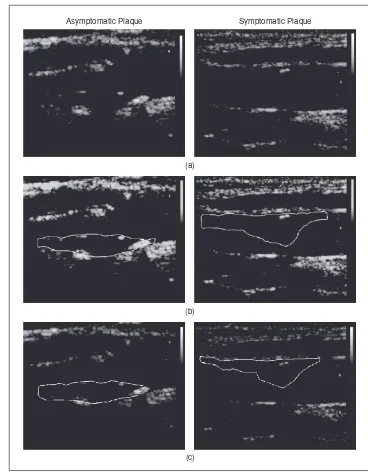
Fig. 1.Segmentation of atherosclerotic carotid plaque in asymptomatic (left column) and symp-tomatic (right column) cases using manual and automated segmentation. (a) Original image, (b) manual plaque delineation by the expert, and (c) automated snakes plaque segmentation.
(a)
Asymptomatic Plaque Symptomatic Plaque
(b)
Lai and Chin snakes segmentation method agrees with the expert by correctly detecting no plaque (TNF) in 80.89% of the cases, by correctly detecting a plaque (TPF) in 82.70% of the cases, disagrees with the expert by detecting no plaque (FNF) in 15.59% of the cases, and by detecting a plaque (FPF) in 5.86% of the cases [10]. Figure 1 illustrates two examples of the manual and automated segmentation methods of the ather-osclerotic carotid plaque.
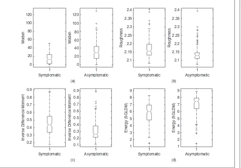
Statistical analysis and interpretation of the texture fea-tures extracted from the plaque ultrasound images (see also the box plots in Figure 2) showed that, in general, texture in symptomatic plaques tends to be darker [SF: median, see Figure 2(a)], with higher contrast, more rough [SFM: rough-ness, see Figure 2(b)], more heterogeneous (SGLDM: inverse difference moment, see Figure 2(c)], less periodical, less coarse (i.e., less local uniformity in intensity), and less complex [SGLDM: entropy, see Figure 2(d)], whereas in asymptomatic plaques texture tends to be brighter, with less contrast, more smooth, more homogeneous, more periodical, more coarse, and more complex with large areas with small gray tone variations [11]. These results are in agreement with the original visual observations that smooth surface, echogenicity, and homogenous texture are characteristics of
stable plaques, whereas irregular surface, echolucency, and a heterogeneous texture are characteristics of potentially unstable plaques.
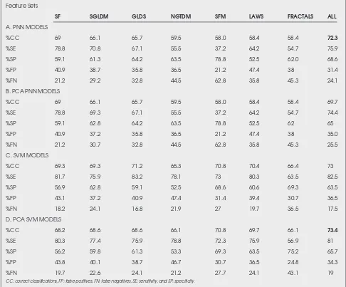
The SVM and PNN classifiers were investigated for developing models for differentiating between symptomatic and asymptomatic plaques based on texture features and PCA analysis. Results from all classifiers and texture sets are presented in Table 1. Results of SVM models were bet-ter than those achieved with PNN models. The highest per-centage of correct classifications score was 73.4% and was achieved using the SVM classifier on all texture features. For PNN, the highest percentage of correct classifications was 72.3% for all texture features. The system allows for both a personal workstation interface and a Web interface that is secure and runs in real time.
Concluding Remarks and Future Work
In this article, an integrated system for the assessment of the risk of stroke based on clinical risk factors and noninvasive investi-gations and carotid plaque texture analysis was presented.
the hands of a clinician dealing with subjects at risk of stroke. The results for each category could be used to predict and then (through clinical intervention) reduce the number of stroke episodes and hopefully the number of deaths as well. On the other hand, carotid endarterectomy could be avoided in cases where the risk of stroke is reduced after treating one or more of the treatable risk factors. However, more work is still need-ed in achieving this.
Both the IMT and plaque segmentation methods referred to in this study can reduce significantly the time required for image analysis, but they can also reduce the subjectivity that characterizes manual measurements. These methods will be further evaluated on ultrasound images of the carotid col-lected from a large-scale epidemiological study carried out by our group as well as for the segmentation and measure-ment of curved segmeasure-ments of the bifurcation or bulb of the carotid artery. Moreover, these methods will be validated by multiple experts.
Texture features provide useful information for differentiating between asymptomatic and symptomatic plaques. The investiga-tion of how morphological features [13] and clinical findings can
be utilized to enhance the diagnostic yield of the classification models proposed in this study is currently being performed.
We are also investigating video image analysis methods for accurate plaque motion estimation. We hope to find distin-guishing plaque motion characteristics that can be used to pre-dict rupture in the symptomatic cases. The new video motion estimation system will rely on an automated segmentation sys-tem to identify the plaque and the plaque surface. Furthermore, we will investigate the use of video image despeckling in providing more accurate motion estimation. Preliminary results from optical-flow methods for video motion estimation indicate that this approach can provide accurate motion estimates.
It is anticipated that the major outcome of this ongoing study will be the establishment of a standardized methodology facilitated via the integrated system for the evaluation of car-diovascular risk and carotid plaque texture analysis for the identification of subjects at high risk of stroke. However, more work is needed mainly for validating the rules generated by the OLAM system as well as in the integration of both mod-ules proposed in this study.
Table 1. Diagnostic performance of PNN and SVM models for classifying asymptomatic (N =137) and symptomatic (N =137) plaques using the leave-one-out method.
Feature Sets
SF SGLDM GLDS NGTDM SFM LAWS FRACTALS ALL
A. PNN MODELS
%CC 69 66.1 65.7 59.5 58.0 58.4 58.4 72.3
%SE 78.8 70.8 67.1 55.5 37.2 64.2 54.7 75.9
%SP 59.1 61.3 64.2 63.5 78.8 52.5 62.0 68.6
%FP 40.9 38.7 35.8 36.5 21.2 47.4 38 31.4
%FN 21.2 29.2 32.8 44.5 62.8 35.8 45.3 24.1
B. PCA PNN MODELS
%CC 69 66.1 65.7 59.5 58.0 58.4 58.4 69.7
%SE 78.8 69.3 67.1 55.5 37.2 64.2 54.7 74.4
%SP 59.1 62.8 64.2 63.5 78.8 52.5 62 65
%FP 40.9 37.2 35.8 36.5 21.2 47.4 38 35.0
%FN 21.2 30.7 32.8 44.5 62.8 35.8 45.3 25.5
C. SVM MODELS
%CC 69.3 69.3 71.2 65.3 70.8 70.4 66.4 73
%SE 81.7 75.9 83.2 78.1 73 80.3 63.5 82.5
%SP 56.9 62.8 59.1 52.5 68.6 60.6 69.3 63.5
%FP 43.1 37.2 40.9 47.4 31.4 39.4 30.7 36.5
%FN 18.2 24.1 16.8 21.9 27 19.7 36.5 17.5
D. PCA SVM MODELS
%CC 68.2 68.6 68.6 66.1 70.8 69.7 66.1 73.4
%SE 80.3 77.4 75.9 78.8 72.3 75.9 56.9 81
%SP 56.2 59.8 61.3 53.3 69.3 63.5 75.2 65.7
%FP 43.8 40.1 38.7 46.7 30.7 36.5 24.8 34.3
%FN 19.7 22.6 24.1 21.2 27.7 24.1 43.1 19
Acknowledgment
This study was partly funded through the Cardiovascular Disease Educational and Research (CDER) Trust (United Kingdom) with the objective to determine the value of nonin-vasive investigations in the identification of individuals with unstable carotid plaques. The online analytical mining system and the carotid plaque texture analysis systems were partly funded through the Integrated System for the Support of the Diagnosis for the Risk of Stroke (IASIS) and Integrated System for the Evaluation of Ultrasound Imaging of the Carotid Artery (TALOS) projects of the Program for Research and Technological Development 2003–2005 of the Research Promotion Foundation of Cyprus.
Efthyvoulos C. Kyriacou received his diploma in electrical and computer engineer-ing from the National Technical University of Athens (NTUA) in 1996 and his Ph.D. in biomedical engineering from the Department of Electrical and Computer Engineering, NTUA, in 2000. From 1996 to 2000 he was a research postgraduate student at the Institute of Communication and Computer Systems, NTUA, working in the areas of telemedicine, medical imaging, and medical informatics. From 2000 to 2001 he worked with the Athens Medical Center Group in Athens, Greece, in the fields of telemedicine and medical databases. He is a postdoctoral researcher at the Cyprus Institute of Neurology and Genetics in the fields of medical image processing and medical infor-matics. Since September 2002, he has been working as a visit-ing lecturer with the Department of Computer Science of the University of Cyprus. He has published several journal and conference papers in the fields of telemedicine, medical imag-ing, and medical informatics. Dr. Kyriacou is a member of IEEE Engineering in Medicine and Biology and Computer Societies, the Cyprus Association of Medical Physics and Biomedical Engineering, and the Hellenic Society of Biomedical Engineering.
Constantinos S. Pattichis received his diploma as technician engineer from the Higher Technical Institute in Cyprus in 1979, the B.Sc. in electrical engineering from the University of New Brunswick, Canada in 1983, the M.Sc. in biomedical Engineering from the University of Texas at Austin, U.S.A. in 1984, the M.Sc. in neurology from the University of Newcastle Upon Tyne, U.K., in 1991, and the Ph.D. in electronic engineering from the University of London, U.K. in 1992. He is an associate professor with the Department of Computer Science of the University of Cyprus. His current research interests include medical imaging, biosig-nal abiosig-nalysis, intelligent systems, and health telematics. He has published 47 refereed journal papers, 102 conference papers, and 18 chapters in books in these areas. He is coeditor of the book M-Health: Emerging Mobile Health Systems, published in 2006 by Springer Science. He was guest coeditor of the special issue on “Emerging Health Telematics Applications in Europe” of IEEE Transactions on Information Technology in Biomedicine, and general cochair of the Medical and Biological Engineering and Computing Conference (MEDICON’98) and the IEEE Region 8 Mediterranean
Conference on Information Technology and Electrotechnology (MELECON’2000). He was program cochair of the conference Information Technology in Biomedicine (ITAB06). He serves as an Associate Editor of
IEEE Transactions on Information Technology in Biomedicineand IEEE Transactions on Neural Networks. He served as chair of the Cyprus Association of Medical Physics and Biomedical Engineering (1996–1998), and the IEEE Cyprus Section (1998–2000). He is a Senior Member of the IEEE.
Minas A. Karaolisis a teaching assistant in the Computer Science Department of the University of Cyprus and is a Ph.D. candidate there. He received his diploma in computer science from the Technical University Carolo Wilhelmina Braunschweig, Braunschweig, Germany, and worked for four years as an assistant in the Computer Science Department there. He developed the automatic generation of loop plans for industrial buildings with the Preussag Company in Germany, using software engineering and databases. Worked for five years as a teacher of computers for the special education pro-grams of the Cyprus Ministry of Education. Current research interests include data mining applications and development of algorithms in medical diagnostic systems.
Christos P. Loizou received his B.Sc. in electrical engineering in 1986 and his M.Sc. in computer science and telecommunications in 1990 from the University of Kaiser-slautern, Germany, and his Ph.D. in 2005 from Kingston University in the United Kingdom. His research was on ultrasound image analysis of the carotid artery. He worked as a manager of a telecommunications company form 1990–1996 and was involved with research in medical image processing. From 1996 to 2000 he was a lecturer at the Computer Science Department at the Higher Technical Institute in Nicosia, Cyprus. He is now an assistant professor with Intercollege, and he has been lecturing at the Computer Science and Engineering Department there since 2000. He is also a researcher at the Institute of Neurology and Genetics in Nicosia, Cyprus. His research interests include medical and ultrasonic imaging, image analysis and medical signal analy-sis, pattern recognition, biosignal analyanaly-sis, and computer applications in medicine. He has received funding for five dif-ferent projects from the Institute of Promotion and Foundation in Cyprus. He has published one book chapter, three journal publications, and 11 conference papers.
research interests include intelligent information systems, arti-ficial neural networks, signal and image processing, pattern recognition, biosignal analysis, and computer applications in medicine and meteorology. His research work in the above areas has been published in eight refereed journal papers, four chapters in books and 27 conference papers.
Marios S. Pattichisreceived the B.Sc. (high honors and special honors) in computer sci-ences in 1991, the B.A. (high honors) in mathematics in 1991, the M.S. in engineering in 1993, and the Ph.D. in computer engineer-ing in 1998, all from the University of Texas at Austin. He is an assistant professor with the Department of Electrical and Computer Engineering at the University of New Mexico (UNM), Albuquerque, New Mexico. Since 2005, he has also been an adjunct assistant professor in radiology with the Department of Radiology at UNM. His research is in the areas of medical image and video processing, digital image and video models, radar image processing, SIMD, and reconfigurable computer architecture applications. He is an associate editor for Pattern Recognitionand the general chair of the 2008 IEEE Southwest Symposium on Image Analysis and Interpretation, to be held in Santa Fe, New Mexico. At UNM, he received the 2004 ECE distinguished teaching award and the 2006 School of Engineering Harrison Faculty Recognition Award.
Stavros Kakkos studied medicine at the University of Patras, Greece, between 1984 and 1990 and received his medical degree in 1990 with honors. In 1995 he was awarded a doctorate in medicine by the University of Patras, while in 1997 he finished his training in general surgery at the University Hospital of Patras, Greece, and received his licensed to practice general surgery. In 1998 he was awarded the Alexander S. Onassis Public Benefit Foundation scholarship to study for an M.S. in vascular technology and medicine, in London, United Kingdom. This degree was awarded by the University of London in 1999. Between 1999 and 2003 he performed research with the Department of Vascular Surgery, Imperial College, London, toward a Ph.D., which was granted by the University of London in 2004. Since 2004 he has been continuing his clinical training in vascular surgery, currently in the United States. Dr. Kakkos has received numerous awards and distinctions for his clinical and research activities. He has so far participated in four multicenter studies, authored over 50 papers in peer-reviewed journals, and also contributed in over 70 conference presentations and three book chapters.
Andrew Nicolaides is a graduate of the Pancyprian Gymnasium (Nicosia), a graduate of Guy’s Hospital Medical School (London University, 1962), and a fellow of the Royal College of Surgeons England and the Royal College of Surgeons Edinburgh (1967). His higher surgical training was at Oxford University, Kings College Hospital Medical School, and St. Mary’s Hospital Medical School, London. He was the professor of vascular surgery at the Imperial College School of Medicine (St Mary’s Hospital) and consultant
vascu-lar surgeon at St. Mary’s Hospital from 1983–2000 and medical director of the Cyprus Institute of Neurology and Genetics from 2001–2004. His research group is known internationally in sev-eral areas, including noninvasive vascular screening and diag-nostic investigation and early detection and prevention of cardiovascular and venous disease. His research is now directed toward the genetic risk factors for cardiovascular disease, identi-fication of individuals at risk, and the development of effective methods of prevention, especially stroke. He has received many awards and honorary memberships from many scientific soci-eties. He is editor-in-chief of International Angiologyand is on the editorial board of many vascular journals. He is coauthor of over 400 original papers and editor of 14 books.
Address for Correspondence:E. Kyriacou, Dept. of Computer Science, University of Cyprus, Cyprus. E-mail: [email protected].
References
[1] S.K. Kakkos, A. Nicolaides, M. Griffin, M. Sabetai, S. Dhanjil, D.J. Thomas, T. Sonecha, A.M. Salmasi, G. Geroulakos, N. Georgiou, S. Francis, E. Ioannidou, and C.J. Dore, “Factors associated with mortality in patients with asymptomatic carotid stenosis,” Int. Angiol., vol. 24, no. 3, pp. 221–230, 2005.
[2] A. Nicolaides, M. Sabetai, S.K. Kakkos, S. Dhanjil, T. Tegos, J.M. Stevens, D.J. Thomas, S. Francis, M. Griffin, G. Geroulakos, E. Ioannidou, and E. Kyriacou, “The Asymptomatic, Carotid, Stenosis and Risk of Stroke (ACSRS) study, ” Int. Angiol., vol. 22, no. 3, pp. 263–272, 2003.
[3] G. Belcaro, A.N. Nicolaides, G. Laurora, M.R. Cesarone, M. De Sanctis, L. Incandela, and A. Barsotti, “Ultrasound morphology classification of the arterial wall and cardiovascular events in a 6-year follow-up study,” Arterioscler. Thromb. Vasc. Biol., vol. 16, pp. 851–856, 1996.
[4] A.N. Nicolaides, S.K. Kakkos, M. Griffin, M. Sabetai, S. Dhanjil, T. Tegos, D.J. Thomas, A. Giannoukas, G. Geroulakos, N. Georgiou, S. Francis, E. Ioannidou, and C.J. Dore, “Severity of asymptomatic carotid stenosis and ipsilater-al hemispheric ischaemic events: Results from the ACSRS study,” Eur. J. Vasc. Endovasc. Surg., vol. 30, no. 3, pp. 275–284, 2005.
[5] J.R. Quinlan, C 4.5: Programs for Machine Learning. San Mateo, CA: Morgan Kaufmann, 1993.
[6] M. Houtsma and A. Swami, “Set-oriented mining of association rules,” Research Report RJ 9567, IBM Almaden Research Center, San Jose, CA, Oct. 1993.
[7] R. Rantzau, L.D. Shapiro, B. Mitschang, and Q. Wang, “Algorithms and appli-cations for universal quantification in relational databases,” Informat. Syst., vol. 28, no. 1–2, pp. 3–32, 2003.
[8] C.P. Loizou, C.S. Pattichis, C.I. Christodoulou, R.S.H. Istepanian, M. Pantziaris, and A. Nicolaides, “Comparative evaluation of despeckle filtering in ultrasound imaging of the carotid artery,” IEEE Trans. Ultrason. Ferroelect. Freq. Contr., vol. 52, no. 10, pp. 1653–1669, 2005.
[9] C.P. Loizou, C.S. Pattichis, M. Pantziaris, T. Tyllis, and A. Nicolaides, “Snakes based segmentation of the common carotid artery intima media,” Med. Biol. Eng. Comput., vol. 45, no. 1, pp. 35-49, Jan. 2007.
[10] C.P. Loizou, C.S. Pattichis, M. Pantziaris, and A. Nicolaides, “An integrated system for the segmentation of atherosclerotic carotid plaque,” IEEE Trans. Inform. Technol. Biomed.,to be published.
[12] E. Kyriacou, M. Pattichis, C. Christodoulou, C. Pattichis, S. Kakkos, M. Griffin, and A. Nicolaides, “Ultrasound imaging in the analysis of carotid plaque morphology for the assessment of stroke,” in Plaque Imaging: Pixel To Molecular Level, J.S. Suri, C. Yuan, D.L. Wilson, and S. Laxminarayan, Eds. (vol. 113 of Studies in Health Technology and Informatics). Amsterdam: IOS Press, 2005, pp. 241–275.
[13] G. Geroulakos, G. Ramaswami, A. Nicolaides, K. James, N. Labropoulos, G. Belcaro, and M. Holloway, “Characterization of symptomatic and asymptomatic carotid plaques using high-resolution real-time ultrasonography,” Br. J. Surg., vol. 80, no. 10, pp. 1274–1277, 1993.