Published online November 2007
Chukanovite, Fe
2(CO
3)(OH)
2, a new mineral from the weathered iron
meteorite Dronino
I
V. PEKOV1,*, N
PERCHIAZZI2, S
MERLINO2, V
N. KALACHEV1,M
MERLINI3and A
E. ZADOV41Faculty of Geology, Moscow State University, Vorobievy Gory, 119992 Moscow, Russia *Corresponding author, e-mail: [email protected]
2Dipartimento di Scienze della Terra, Università di Pisa, via S. Maria 53, 56126 Pisa, Italy
3Dipartimento di Scienze della Terra, Università degli Studi di Milano, via Botticelli, 23, 20133 Milano, Italy 4NPO Regenerator, 3rdPassage of Mar’ina Roshcha 40, 127018 Moscow, Russia
Abstract:The new mineral chukanovite, Fe2(CO3)(OH)2, occurs in cavities of weathered fragments of the Dronino ataxite iron meteorite found near the Dronino village, Kasimov district, Ryazan’ Oblast, Russia. It is a product of terrestrial alteration of meteorite iron. Associated minerals are goethite, akaganéite, hematite, hibbingite, reevesite, honessite, etc. Chukanovite forms acicular to fibrous individuals (up to 0.5 mm long and up to 2–3 µm thick) combined in spherulites up to 1 mm in diam-eter, botryoidal spherulitic clusters and parallel- or radial-columnar aggregates which form crusts up to 1 mm thick. Unal-tered chukanovite is transparent, pale-green or colourless. The surface of aggregates is brownish-green. Streak is white. Lus-tre is viLus-treous. Cleavage is perfect, probably on {0–21}, fracture is uneven. The mineral is brittle, the Mohs’ hardness is 3.5–4, the calculated density is 3.60 g/cm3. It is optically biaxial (–) with α1.673(3),β 1.770(5),γ 1.780(5), 2V
meas.10(5)◦.
Average chemical composition (wt. %; electron probe, H2O by modified Penfield method, CO2 by selective sorption) is: MgO 0.1, FeO 68.8, NiO 0.6, CO2 19.8, H2O 10.9, total 100.2. The empirical formula calculated on the basis of two metal atoms is (Fe2+
1.97Ni0.02Mg0.01)Σ2.00(CO3)0.93(OH)2.14·0.18H2O, ideally Fe2(CO3)(OH)2. Chukanovite is monoclinicP21/a, witha= 12.396(1) Å,b=9.407(1) Å,c=3.2152(3) Å,β= 97.78◦
. The strongest lines of the X-ray powder pattern [d(Å),I,(hkl)] are: 6.14, 40, (200); 5.15, 60, (231); 3.73, 80, (310); 2.645, 100, (230); 2.361, 40, (510); 2.171, 40, (520). The structure of chukanovite was refined on synchrotron data by the Rietveld method up toRp =3.43 %,wRp = 4.51 %,RBragg = 2.48 %. Chukanovite is
closely related to the minerals of the malachite-rosasite group. It was named in honour of Nikita V. Chukanov (b. 1953), Russian physicist and mineralogist. The holotype specimen is deposited in the Fersman Mineralogical Museum of the Russian Academy of Sciences, Moscow.
Key-words: chukanovite, new mineral, iron hydroxide-carbonate, malachite-rosasite group, Rietveld refinement, Dronino mete-orite.
Introduction
In the present paper, we describe a new mineral species, an iron hydroxide-carbonate closely related to pokrovskite, malachite and rosasite-group members. It was named
chukanovite(Cyrillic:QUK
a
NOVIT) in honour of NikitaVladimirovich Chukanov (b. 1953), Russian physicist and mineralogist, well-known specialist in the IR spectroscopy of minerals and synthetic compounds, a discoverer of many new mineral species, working at the Institute of Problems of Chemical Physics of the Russian Academy of Sciences, Chernogolovka, Russia. Both the new mineral and its name have been approved by the IMA Commission on New Min-erals and Mineral Names (IMA no. 2005-039). The holo-type specimen of chukanovite has been deposited in the Fersman Mineralogical Museum of the Russian Academy of Sciences, Moscow (catalogue no. 92013).
Occurrence and general appearance
Chukanovite occurs in cavities of weathered fragments of the Dronino meteorite which fell in prehistoric time and was found in 2000 near the Dronino village in Kasimov dis-trict, Ryazan’ Oblast, 350 km east-south of Moscow, Rus-sia (54◦44.8′N; 41◦25.3′E).
The Dronino meteorite is an ataxite iron meteorite mainly consisting of kamacite and containing minor amounts of taenite and chromite. Sporadically it is extremely enriched in troilite and poorly-studied Fe-Ni sulfides (Russellet al., 2004; Grokhovskyet al., 2005).
The Dronino meteorite shower fell approximatively 5000–8000 years ago, after the last glaciation on the ter-ritory of the present European Russia. Its numerous frag-ments have been found in glacial and post-glacial de-posits, mainly at a depth of 0.5–1 m under the surface. The
Chukanovite occurs as acicular to fibrous individuals, elongated along [001], up to 0.5 mm long and up to 2–3µm thick usually combined in spherulites up to 0.3 mm (rarely up to 1 mm) in diameter. Botryoidal spherulitic clusters and parallel- or radial-columnar aggregates forming crusts up to 1 mm thick (Fig. 1) are typical. Aggregates are usually porous, the core of some spherulites contains grains of ka-macite, taenite, sulfides or iron hydroxides. Tiny siderite ingrowths occur between chukanovite individuals. Aggre-gates of chukanovite are very similar in their morphology to well-known spherulitic aggregates of malachite, a struc-turally related mineral.
In weathered fragments of the Dronino meteorite, chukanovite was probably formed as a product of the reac-tion of iron (kamacite) with cold CO2-bearing sub-surface water. This reaction took place under local reducing condi-tions caused by the presence of iron in the mineral-forming medium inside a cavity. This mineral-forming system is isolated from common system of sub-surface water, which is oxidizing because of the saturation by atmospheric oxy-gen and leads to the alteration of iron meteorite fragments to Fe3+hydroxides and oxides from the outside. The reduc-ing medium seems necessary for the formation and preser-vation of this Fe2+ hydroxide-carbonate unstable under atmospheric conditions. Such instability seems the main cause for the rarity of chukanovite in nature (and probably the main cause of absence of other Fe2+hydrous carbonate and carbonate-hydroxide minerals), in spite of its simple composition, with widespread chemical constituents, and a common structure type. The compound Fe2+(CO
3)(OH)2 can be completely decomposed very fast, in several years or even several months, as a result of easy Fe2+ oxidation. We have observed the first stage of this process in room air. Formation and preservation of chukanovite in cavities of partially altered fragments of the Dronino meteorite was possible because of the conditions unusual for nature: an isolated system with abundant native iron that acts not only as reagent but also as reducing agent. Thus, chukanovite could be found also in the inner parts of the weathered zone of terrestrial native iron occurrences.
Physical and optical properties
Unaltered chukanovite (on fresh fracture) is transparent, pale-green or colourless with white streak and vitreous
parallel-columnar aggregate. Chukanovite is optically bi-axial (–) withα1.673(3),β1.770(5),γ1.780(5), 2V(meas.) 10(5)◦, 2
V(calc.) 34◦. Orientation:
X≈c. Under the micro-scope, the mineral is colourless and nonpleochroic.
Infrared spectroscopy
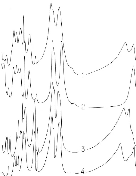
The infrared-absorption spectrum of chukanovite was ob-tained using a Specord 75 IR spectrophotometer (pow-der sample was prepared in KBr tablet, polystyrene and gaseous NH3 were used as frequency standards). The IR spectrum of the new mineral is close to the spectra of pokrovskite (the most similar), malachite and members of the rosasite group (Fig. 2). Absorption bands in the IR spectrum of chukanovite (in cm−1; frequencies of the most intensive bands are underlined, sh – shoulder) are: 3475, 3325, 1755, 1521, 1400sh, 1364, 1069, 1055sh, 955, 861, 837, 781, 710sh, 695, 655, 504, 452.
The IR spectrum of technogene “malachite-like basic iron carbonate” (Erdös & Altorfer, 1976) is practically identical to the spectrum of chukanovite as regards the wavenumbers of absorption maxima.
Chemical data
Contents of cations in chukanovite were determined from electron-microprobe data obtained with a Camebax microbeam instrument in wavelength-dispersion (WDS) mode using an operating voltage of 20 kV and an estimated beam-current of 20 nA. The electron beam was rastered over an area of 10 ×10µm2 to minimize damage to the sample. We used the following standards: diopside (Mg), FeO (Fe), Ni (Ni). Contents of Na, K, Ca, Mn, Cr, V, Ti, Co, Cu, Zn, Si, P, F, Cl are below detection limits. Some point analyses show the presence of S (up to 0.25 wt.%) proba-bly caused by micro-inclusions of iron sulfate-hydroxide. Several point analyses show 0.02–0.04 wt.% Al.
The H2O content was determined by Alimarin method adopted for micro-samples: heating to 1000 ◦C under
oxygen stream with H2O absorption in pipes filled with Mg(ClO4)2. The CO2 content was determined using a se-lective sorption method: heating to 1000◦C under oxygen
(a) (b)
(c) (d)
Fig. 1. Aggregates of chukanovite:a– spherulitic crust;b– separate spherulite as part of a crust;c– section of a spherulite;d– cluster of curved fibrous individuals. SEM images.
The average results (wt.%, ranges for sixteen analyses are given in parentheses) are: MgO 0.1 (0.05–0.2), FeO 68.8 (67.5–69.9), NiO 0.6 (0.5–0.8), CO219.8, H2O 10.9, total 100.2. The empirical formula calculated on the basis of two metal atoms, with (OH)/H2O ratio from charge balance, is: (Fe2+
1.97Ni0.02Mg0.01)Σ2.00(CO3)0.93(OH)2.14·0.18H2O, whe-reas the formula calculated on the ba-sis of five oxygen atoms is (Fe2+
1.93Ni0.02 Mg0.01)Σ1.96(CO3)0.91(OH)2.09·0.18H2O. The simpli-fied formula is: Fe2+
2 (CO3)(OH)2 which requires: FeO 69.85, CO2 21.39, H2O 8.76, total 100.00 wt.%. Samples used for H2O and CO2 determination were probably slightly contaminated with Fe hydroxides that could be the cause of higher H2O content and lower CO2 content in comparison with values calculated for the ideal formula.
The determination of the valence state of Fe was one of the problems when chukanovite was in study. The Möss-bauer study or wet-chemical analysis were not carried out because of scarcity of pure material. The presence of Fe2+ and absence of Fe3+ in unaltered chukanovite were confirmed using well-known colour reactions with
potas-sium hexaferricyanide, K3Fe3+(CN)6, and potassium hex-aferrocyanide, K4Fe2+(CN)6. For these tests, chukanovite was dissolved in dilute (3 vol.%) HCl bubbled with CO2 (10 min) for the protection of Fe2+ (when the mineral dissolves) from oxidation by atmospheric oxygen “dis-solved” in the solution. Solutions of K3Fe3+(CN)6 and K4Fe2+(CN)6 were prepared using the same dilute HCl bubbled with CO2. After the mixing of K3Fe3+(CN)6 so-lution with the “soso-lution of chukanovite”, strong blue colouring appears at once, which is a clear indica-tor of the presence of Fe2+: K
3Fe3+(CN)6 + Fe2+ → blue KFe2+Fe3+(CN)
6. Conversely, after the mixing of K4Fe2+(CN)6solution with the “solution of chukanovite”, no blue colouring appeared, which shows an absence of Fe3+. For the checking, the same tests were carried out with siderite, Fe2+CO
3, and goethite, Fe3+O(OH). Tests with siderite, a mineral with only Fe2+, show the same results as those with chukanovite. Tests with goethite, a mineral with only Fe3+, show a different result: no colouring with K3Fe3+(CN)6but strong blue colouring with K4Fe2+(CN)6 which is evidence for the presence of Fe3+: K
Fig. 2. IR spectra of chukanovite (1), pokrovskite (the type specimen from the Zlatogorskaya intrusion, Kazakhstan) (2), malachite (Nizh-niy Tagil, Central Urals, Russia) (3), and rosasite (Ojuela mine, Mapimi, Durango, Mexico) (4).
Fe3+→blue KFe2+Fe3+(CN)
6. Thus, these direct methods confirm that chukanovite contains only Fe2+, like struc-turally related minerals containing only bivalent cations: Cu2+, Mg, Co2+, Ni2+and Zn.
Chukanovite easily dissolves in cold dilute HCl with strong effervescence.
The Gladstone-Dale compatibility index (Mandarino, 1981) for chukanovite is 0.016 (superior).
X-ray crystallography and crystal
structure refinement
Experimental
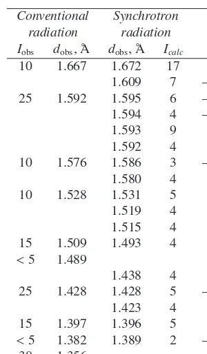
A preliminary X-ray powder diffraction pattern of chukanovite, reported in Table 1, was collected using a RKU Debye-Scherrer camera (114.6 mm in diameter) with FeKαradiation. Some diffraction lines corresponding
to minor additional phases, such as hibbingite (5.70 Å), goethite (4.18 Å), siderite (3.61, 2.80, 1.733 Å), taenite (3.38 Å), are present in the chukanovite powder pattern and are highlighted in italic.
Some fragments of fibrous pale-green microcrystalline chukanovite aggregates were carefully selected with the aid of both a polarizing and a binocular microscope, so as to minimize the presence of impurities, gently hand ground
10 4.18
80 3.73 3.754 17 310
3.734 49 220
35 3.61
<5 3.38
5 3.21 3.186 3 001
30 3.05 3.088 2 320
3.070 7 400
3.038 8 130
3.021 3 –111 2.998 6 –201 25 2.916 2.919 13 410
2.847 2 111
95 2.798 2.793 3 230
2.683 8 201
100 2.645 2.637 100 021
35 2.56 2.571 27 420
2.528 17 –221
2.521 3 121
2.459 5 330
40 2.361 2.377 12 510 2.377 7 –401 10 2.236 2.236 2 –131
2.235 5 031
2.196 5 240
40 2.171 2.177 9 520
2.167 15 –231 30 2.137 2.121 1 –421
2.075 5
2.047 6 600
20 2.04 2.039 7 231
2.037 4 2.000 4
15 1.966 1.934 4 530
<5 1.901 1.907 1 –521
1.887 6 331
10 1.875 1.877 2 620
5 1.85 1.850 5 –241
1.805 4 –611
20 1.797 1.798 4 250
1.796 7 511
10 1.766 1.768 12 241
50 1.733 1.737 5 –531
1.730 5 431
Table 1. continued.
Conventional Synchrotron radiation radiation
Iobs dobs, Å dobs, Å Icalc hkl 10 1.667 1.672 17 341 1.609 7 –711
and placed into a borosilicate Lindemann capillary 0.5 mm in diameter.
The synchrotron X-ray powder diffraction data were collected at the BM8-GILDA beamline (ESRF, Greno-ble, France). A monochromatic beam (λ = 0.79593 Å, calibrated against X-ray absorption of pure metal foils) was used and the diffractions were collected with a Fuji Imaging-Plate (IP) detector. The beam dimension on the sample was 0.2×0.2 mm. The sample to detector distance and the image plate tilt were calibrated with X-ray powder diffraction of standard LaB6(NIST-SRM 660a). Data were collected up to 48◦ 2θ, corresponding to ad-space
resolu-tion of 0.978 Å. Data were reduced with the Fit2D software (Hammersley, 1997).
Rietveld refinement of chukanovite
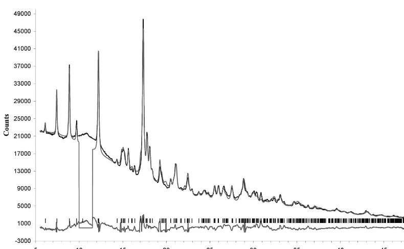
The close similarities, both in chemical formula and in X-ray powder pattern between pokrovskite, ideally Mg2(CO3)(OH)2, and chukanovite suggested a reliable starting model for this last phase, based on the cell pa-rameters and atomic coordinates obtained for pokrovskite (Perchiazzi & Merlino, 2006). The subsequent Rietveld re-finement was performed with the TOPAS-Academic pro-gram (Coelho, 2004). A preliminary Pawley refinement (Pawley, 1981) was performed to get starting values for background, modelled with a 6-term Chebyschev function, cell parameters and peak shapes. The refined region was from 5.5 to 48◦ 2θ, excluding from the refinement the
re-gion between 10 and 11.6◦ in 2θ, which presents a large
bump due to a poorly crystalline phase and only one small peak from chukanovite. It is worth noticing that this bump
is just centered on the 2θposition of the strongest peak of goethite.
In the early stages of the refinement, constraints on the Fe-O bonds were introduced and subsequently removed at the end of refinement; the carbonate group was refined as a rigid body, fixing the C-O distance to 1.284 Å (Zemann, 1981). Assuming as starting values for the atomic dis-placement parameters those coming from the single-crystal structure refinement of siderite, FeCO3, namely Beq = 0.44, 0.63, 0.44 Å2 for Fe, O and C respectively (Ef-fenbergeret al., 1981), isotropic displacement parameters were refined for all the atoms, constraining atoms of the same species to keep the same value. The isotropic dis-placement parameter of iron atoms obtained in this way was anomalously larger than the displacement parameter of oxygen atoms, therefore suggesting a possible small va-cancy in the iron sites. According to the indications of chemical data, a full occupancy was anyway assumed for the Fe1 and Fe2 sites, imposing for both Fe sites a common displacement parameter fixed to 70 % of the oxygens dis-placement parameter, namely to the ratio suggested by the siderite refinement.
The structure of chukanovite was refined up to Rp =
3.43 %,wRp =4.51 %,RBragg =2.48 %; a final Rietveld
plot of the refinement is reported in Fig. 3.
Structure description and discussion
Refined cell parameters for chukanovite are compared in Table 2 with the crystallographic data coming from structural refinements of the other phases of the rosasite-malachite group. For nullaginite, ideally Ni2(CO3)(OH)2, Nickel & Berry (1981) report the space group P21/m and cell parameters a = 9.236 Å, b = 12.001 Å, c =
3.091 Å, β = 90.48◦. Apparently, nullaginite is neither
straightforwardly related with a malachite-like cell nor with a rosasite-like cell; further investigations are needed to clearly define the true nature of this rare phase.
Final atomic coordinates and isotropic displacement pa-rameters are reported in Table 3, and the geometry of the two independent iron coordination polyhedra is given in Table 4.
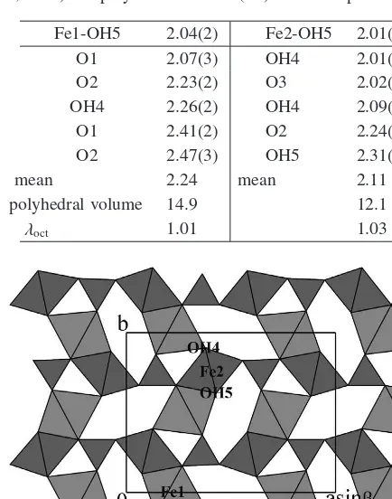
The crystal structure of chukanovite, projected along the c axis, is illustrated in Fig. 4. The Fe1 and Fe2 octahe-dra share edges to build octaheoctahe-dral “ribbons”, two-column wide and running along [001]. “Corrugated” layers parallel to (100) are formed through interconnection of those rib-bons by corner-sharing, with the carbonate groups inserted in the octahedral frame to strengthen the intralayer connec-tions, and also assuring an interlayer linking.
The octahedral environment of Fe1 is made up of four oxygen atoms, belonging to carbonate groups, and of two hydroxyls, whereas Fe2 is surrounded by four hydroxyls and two oxygen atoms. Bond lengths range from 2.04 to 2.47 Å for Fe1, and from 2.01 to 2.31 Å for Fe2 octahedra. The mean octahedral quadratic elongation parameter λoct
Fig. 3. Final Rietveld plots for the synchrotron diffraction pattern of chukanovite.
Table 2. Crystallography of the phases of the malachite-rosasite group (Å,◦) as resulting from the available crystal-structure refinements.
Phase Me2+ sp. gr. a b c β Ref.
Chukanovite Fe P1 21/a1 12.396(1) 9.407(1) 3.2152(3) 97.78(2) (1)
Glaukosphaerite (Cu,Ni) P1 21/a1 12.0613(4) 9.3653(4) 3.1361(1) 98.085(5) (2)
Kolwezite (Cu,Co) P1 21/a1 12.359(1) 9.451(1) 3.1814(3) 99.01(1) (3)
Mcguinnessite (Mg,Cu) P1 21/a1 12.1531(3) 9.3923(3) 3.1622(1) 97.784(4) (4)
Pokrovskite Mg P1 21/a1 12.2397(3) 9.3489(4) 3.1595(1) 96.422(6) (2)
Rosasite (Cu,Zn) P1 21/a1 12.2413(2) 9.3705(2) 3.1612(2) 98.730(3) (4)
Malachite Cu P1 21/a1 9.502 11.974 3.240 98.75 (5)
(1) This study; (2) Perchiazzi & Merlino (2006); (3) Perchiazzi & Merlini, in prep.; (4) Perchiazzi, (2006); (5) Ziganet al. (1977).
Table 3. Final fractional atomic coordinates and isotropic displacement parameters for chukanovite.
Fe1 Fe2 C O1 O2 O3 OH4 OH5
x 0.2114(5) 0.3983(5) 0.143 0.139 0.233 0.055 0.379(1) 0.427(1) y 0.0005(9) 0.7675(7) –0.265 –0.135 –0.332 –0.328 0.900(1) 0. 619(2) z 0.979(2) 0.554(2) 0.493 0.368 0.546 0.556 0.058(7) 0.132(9) Beq 1.12 1.12 3(1) 1.6(1) 1.6(1) 1.6(1) 1.6(1) 1.6(1)
As it may be seen from Table 4, the Fe1 octahedron is distinctly larger and more distorted than the Fe2 octahe-dron. Also in pokrovskite (Perchiazzi & Merlino, 2006) the corresponding Mg1 octahedron is larger than Mg2 octahe-dron, their polyhedral volumes being 13.1 and 12.2 Å3 re-spectively, but in pokrovskite the two polyhedra display the same mean octahedral quadratic elongationλoct=1.01.
Bond-valence balance, calculated according to Breese & O’Keeffe (1991), is reported in Table 5. In the calculations, full occupancy was assumed for both Fe1 and Fe2 sites. Hydrogen bonds were detected examining all O· · ·O dis-tances shorter than 3.1 Å and not belonging to the same coordination polyhedron. As it may be seen from Table 5,
their contributions to the valence balance, evaluated ac-cording to Ferraris & Ivaldi (1988), are critical for the structural stability of chukanovite. The balance can be con-sidered as satisfactory, with only small deviations from the expected values for the anions.
Table 4. Bond distances (Å) in Fe coordination polyhedra of chukanovite. Mean octahedral quadratic elongationλoct(Robinson
et al., 1971) and polyhedral volume (Å3) are also reported.
Fe1-OH5 2.04(2) Fe2-OH5 2.01(2)
Fig. 4. The chukanovite structure projected along [001]. Edge-sharing between octahedra form “ribbons”, two-column wide and running along c, interlinked through corner sharing to form corru-gated octahedral layers, parallel (100). Carbonate triangles are in-serted in the octahedral frame, assuring further intra- and inter-layer linking.
2006) and by chukanovite, as shown by the present work. A crystal structure refinement of kolwezite (Cu, Co)2(CO)3(OH)2, presently in progress, strongly supports a rosasite-like model (Perchiazzi & Merlini, in prep.).
The technogene Fe
2(CO
3)(OH)
2compound
Erdös & Altorfer (1976) reported the occurrence of a “malachite-like basic iron carbonate”, henceforth denoted with the acronym IHC (iron hydroxide-carbonate), from the corrosion product in the hot-water exchanger of an in-dustrial plant in Beringen, Switzerland. Actually, IHC was found in the steel valve of the exchanger, as the main con-stituent of a crust, associated with siderite and magnetite. Chemical data obtained by wet chemical analysis on a pu-rified fraction were FeO 61.2 %, Fe2O37.5 %, CO220.8 %, H2O % 9.3, MnO % 0.4, total 99.2 %, resulting in the chemical formula Fe2+
1.8Fe 3+
0.2(OH)2.2(CO3). About the oc-currence of Fe3+, the authors remark anyway that an ad-mixture with goethite cannot be excluded, and IHC was ideally considered as Fe2+
2 (OH)2(CO3).
The authors also stressed the close relationships between IHC, malachite and rosasite, on the basis of X-ray
powder-Table 5. Bond valence balance (v.u.) for chukanovite. O···O distances (Å) and hydrogen bond strengths (v.u.) are also reported.
Anion O1 O2 O3 OH4 OH5
Sum 2.14 1.98 1.94 0.94 0.87 Hydrogen bonds OH4· · ·O3 2.90(3) Å v.u.0.15
OH5· · ·O1 2.65(2) Å v.u.0.25
diffraction and infrared data; IHC powder-diffraction pat-tern was indexed on the basis of an orthorhombic cell (no space group assigned) with a = 9.39, b = 24.53,
c = 3.21 Å, V = 739.9 Å3, Z = 8; D(calc.) = 3.693, D(meas.)=3.59 g/cm3 (Erdös & Altorfer, 1976). We may now confidently maintain that IHC is the technogene ana-logue of chukanovite. The diffraction pattern presented by Erdös & Altorfer (1976) closely corresponds to that of chukanovite presented in Table 1; it is proper to remark the absence of the additional extraneous reflections reported in Col. 1 of Table 1. All the diffraction lines reported by these authors are easily indexed on the basis of a unit cell of chukanovite-type, and a least squares refinement converges to the following unit-cell parameters: a = 12.373(3) Å,
b = 9.390(2) Å, c = 3.220(1) Å, β = 97.65(3), fairly matching the corresponding parameters of chukanovite.
The study of the thermal behaviour in oxygen atmosphere (Erdös & Altorfer, 1976) shows that over 197◦C IHC
trans-forms (topotactical replacement) to Fe3+
2 O2(CO3), which decomposes (at∼600◦C) toα-Fe
2O3(hematite) and CO2. Slight dehydration and oxidation of the IHC was observed under room conditions in one year.
More recently, IHC was also observed as the transfor-mation product of biogenic magnetite (Kukkadapuet al., 2005).
Relation of chukanovite with pokrovskite
Chukanovite is a Fe2+-dominant member of the rosasite-malachite group of carbonate minerals, a group includ-ing several phases whose symmetry and cell constants are reported in Table 2. Very close relationships exist espe-cially with the Mg-dominant phase, pokrovskite, ideally Mg2(CO3) (OH)2 (Ivanovet al., 1984). Technogene ana-logue of pokrovskite was found as a corrosion product, in this case of an In-Mg alloy (Uszynski & Kubiak, 1995).
The chemical data of chukanovite suggest a possible ex-cess of water and a very slight deficiency of iron with re-spect to their ideal values in Fe2(CO3)(OH)2. A similar case is presented by pokrovskite. Both the chemical data (Fitzpatrick, 1986), and the structural result (Perchiazzi & Merlino, 2006), gathered on pokrovskite from Sonoma County, California, USA, pointed to a partial occupancy in the Mg sites and a corresponding substitution of hydroxyl anions by water molecules, resulting in the crystal chemi-cal formula (Mg1.770.23)(CO3)[(OH)1.54/(H2O)0.46].
chukanovite; to this aim we are presently trying to synthe-size the artificial analogue of chukanovite.
Acknowledgements: We are grateful to N.N. Kononkova and A.S. Astakhova for their assistance in obtaining the chemical data and to L.A. Pautov for his help with the SEM. We also thank J. Zemann and M. Wildner for their helpful reviews. This work was supported by MIUR (Min-istero dell’Istruzione, dell’Università e della Ricerca) and by University of Pisa through grants to the national project ‘Minerals to materials: crystal chemistry, microstructures, modularity, modulations’, and by a grant of the Russian Science Support Foundation (for IVP). Many thanks also to the GILDA beamline for the experimental data collec-tion.
References
Breese, N.E. & O’Keeffe, M. (1991): Bond-valence parameters for solids.Acta Cryst., B47, 192-197.
Coelho, A.A. (2004): TOPAS-ACADEMIC, http://pws.prserv.net/Alan.Coelho/
Effenberger, H., Mereiter, K., Zemann, J. (1981): Crystal struc-ture refinements of magnesite, calcite, rhodochrosite, siderite, smithsonite, and dolomite, with discussion of some aspects of the stereochemistry of calcite type carbonates.Zeit. Krist.,156, 233-243.
Erdös, E. & Altorfer, H. (1976): Ein dem Malachit ähnliches basisches Eisenkarbonat als Korrosionsprodukt von Stahl. Werkstoffe und Korrosion,27, 304-312.
Ferraris, G. & Ivaldi, G. (1988): Bond valencevs bond length in O· · ·O hydrogen bonds.Acta Cryst.,B44, 341-344.
netite.Am. Mineral.,90, 510-515.
Mandarino, J.A. (1981): The Gladstone-Dale relationship: Part IV. The compatibility concept and its application.Can. Mineral., 19, 441-450.
Nickel, E.H. & Berry, L.G. (1981): The new mineral nullagi-nite and additional data on the related minerals rosasite and glaukosphaerite.Can. Mineral.,19, 315-324.
Pawley, G.S. (1981): Unit-cell refinement from powder diffraction scans.J. Appl. Cryst.,14, 357-361.
Perchiazzi, N. (2006): Crystal structure determination and Rietveld refinement of rosasite and mcguinnessite.Zeit. Krist. suppl.23 505-510.
Perchiazzi, N. & Merlino, S. (2006): The malachite-rosasite group: crystal structures of glaukosphaerite and pokrovskite. Eur. J. Mineral.,18, 787-792.
Robinson, K., Gibbs, G. V., Ribbe, P. H. (1971) Quadratic elonga-tion: A quantitative measure of distortion in coordination poly-hedra.Science,172, 567-570.
Russell, S.S., Folco, L., Grady, M.M., Zolensky, M.E., Jones, R., Righter, K., Zipfel, J., Grossman, J.N. (2004): The Meteoritical Bulletin, No. 88, 2004 July.Meteoritics&Planetary Science, 39(supplement).
Uszynski, I. & Kubiak, R. (1995): On products of InMg corrosion in wet air and their conversion during annealing in vacuum.J. Alloys and Compounds,218, 259-262.
Zemann, J. (1981): Zur Stereochemie der Karbonate. Fortschr. Mineral.,59, 95-116.
Zigan, F., Joswig, W., Schuster, H.D., Mason, S.A. (1977): Verfeinerung der Struktur von Malachit, Cu2(CO3)(OH)2, durch
Neutronenbeugung.Zeit. Krist.,145, 412-426. Received 1 March 2007