Effect of duration of infusion of stress-like
concentrations of cortisol on follicular development
and the preovulatory surge of LH in sheep
q
M.S. Macfarlane, K.M. Breen, H. Sakurai,
B.M. Adams, T.E. Adams
∗Department of Animal Science, University of California Davis, CA 95616, USA
Received 15 September 1999; received in revised form 15 March 2000; accepted 26 April 2000
Abstract
Stress-like levels of cortisol suppress follicular growth and development and block or delay the preovulatory surge of LH when cortisol is continuously administered during the late luteal and early follicular phases of the ovine oestrous cycle. We postulated that cortisol infusion of shorter duration would have a similar effect. To test this hypothesis the oestrous cycles of mature ewes were syn-chronized using progestin-treated vaginal pessaries. Ewes were randomly assigned to one of four treatment groups. Animals received cortisol (0.1 mg/kg/h; n=8) or vehicle alone (n=8) beginning 5 days before, and continuing for 5 days after, pessary removal (PR). Additional groups received cortisol only during the 5 days period before (n=7), or the 5 days period after (n=8), PR. Continuous delivery of cortisol established stable serum concentrations of cortisol of 72.0±2.5 ng/ml within 6 h of initiation of infusion. Serum concentrations of oestradiol increased progressively during the period after PR in control animals receiving vehicle alone and the preovulatory surge of LH was evident in all control animals (eight of eight) 55.5±5.0 h after PR. In contrast, follicular develop-ment and the preovulatory surge of LH were evident during the period of cortisol infusion in only one of eight animals receiving stress-like levels of cortisol over the entire 10-day infusion period. Similarly, neither follicular development nor surge-like secretion of LH were evident during the infusion period in animals (zero of eight) receiving cortisol during the 5-day period after PR. This cortisol-dependent suppression of ovarian activity in sheep receiving stress-like levels of cortisol during the 5 days after PR was temporary and follicular development, the ovulatory surge of LH, and subsequent luteal function were evident in six of eight ewes after cessation of cortisol delivery. Similarly, follicular development and the preovulatory surge of LH were noted within 5 days af-ter PR in four of seven ewes receiving cortisol only during the 5-day period prior to PR. Collectively,
q
Supported by USDA Grant 93-37203-9111 and the California Agricultural Experiment Station.
∗Corresponding author. Tel.:+1-530-752-1266; fax:+1-530-752-0175.
E-mail address: [email protected] (T.E. Adams).
these data indicate that stress-like levels of cortisol reduce fertility of sheep by suppressing follicular development and the preovulatory surge of LH. Additionally, cortisol delivery during the follicular phase has a more profound suppressive effect on follicular development than cortisol administration during the luteal phase. © 2000 Elsevier Science B.V. All rights reserved.
Keywords: Cortisol; Sheep; LH surge; Follicular development; Oestradiol; Stress
1. Introduction
Stressful stimuli reduce fertility in primates (Norman et al., 1994; Xiao et al., 1998) and domestic species (Welsh and Johnson, 1981; Wilson et al., 1998). Indeed, climatic extremes (Doney et al., 1973), transportation (Ehnert and Moberg, 1991; Smart et al., 1994) or laparoscopy (Martin et al., 1981) suppress or delay expression of behavioural oestrus and ovulation in sheep. In addition to reducing fertility, these stressors also stimulate the activity of the hypothalamic-pituitary-adrenal axis, and a marked increase in serum concentration of cortisol is commonly associated with management-related stressors (Martin et al., 1981; Ehnert and Moberg, 1991; Komesaroff et al., 1998).
Although the causal link between stress and infertility has not been precisely defined, several studies indicate that glucocorticoids may contribute to the anti-gonadal effect of stress (Dobson and Smith, 1995; Chrousos et al., 1998). Indeed, intra-hypothalamic or systemic administration of glucocorticoid blocks follicular development and ovulation in rodents (Smith et al., 1971; Baldwin and Sawyer, 1974). Exogenous glucocorticoid has a similar effect in primates (Cunningham et al., 1978; Hayashi and Moberg, 1990) and some domestic species (Barb et al., 1982; Stoebel and Moberg, 1982). Moreover, recent studies in our laboratory indicate that follicular development and ovulation are blocked or delayed in sheep receiving stress-like concentrations of cortisol during the late luteal and early follicular phases of the oestrous cycle (Daley et al., 1999a). The study presented here examines oestradiol production and LH secretion in sheep receiving stress-like levels of cortisol during the luteal and/or follicular phases of the oestrous cycle in sheep. We hypothesized that stress-like concentrations of cortisol would block or delay follicular maturation and suppress the preovulatory surge of LH.
2. Materials and methods
2.1. Animals
Thirty-one mature Rambouillet ewes of proven fertility were used to assess the effects of chronic stress-like concentrations of cortisol on follicular development and ovulation. Ewes were housed in an open-sided barn under natural lighting and were allowed free access to water and alfalfa pellets. This study was conducted during November, a period
of high reproductive activity for sheep at this latitude (38◦N). All experimental procedures
2.2. Cannulation
Intravenous cannulae (Intramedic PE 190, Clay Adams, Parsippany, NJ) were placed in the right and left jugular veins and used for hormone infusion and blood collection, respectively. All cannulae were passed through a protective plastic tubing sheath along a halter and lead rope to the exterior of the animal holding area. Animals were freely mobile at the end of a 1 m lead. The cannulae were inserted 2 days prior to initiation of treatment to permit acclimation to the conditions of experimentation.
2.3. Hormone delivery
Cannulae for the delivery of cortisol or vehicle (50% ethanol-saline) were connected to syringes placed in Harvard infusion pumps (Model 2265, Harvard Bioscience, South Natick, MA). Cortisol (0.1 mg/kg/h; Sigma Chemical Co., St. Louis, MO) or a comparable volume of vehicle was delivered by continuous infusion at a rate of 1.0–1.2 ml/h.
2.4. Experimental design
Prior to the initiation of infusion ewes received two injections of prostaglandin F2α
(Lutalyse, Upjohn Co., Kalamazoo, MI) given 10 days apart to synchronize oestrous activity. Vaginal pessaries (Chrono-gest, Intervet International, Boxmeer, Holland) impregnated with a synthetic progestin (40 mg flugestone acetate) were inserted 10 days after the second prostaglandin injection. Luteolysis was simulated by pessary removal (PR) 10 days after insertion.
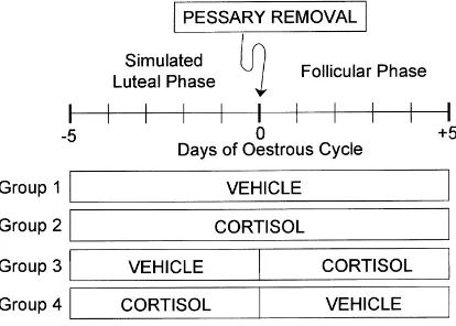
Animals were assigned at random to one of four treatment groups (Fig. 1). Animals in
Groups 1 (n=8) and 2 (n=8) received vehicle or cortisol, respectively, beginning 5 days
before, and continuing for 5 days after, PR. Animals in Group 3 (n=8) received vehicle prior to PR, with infusion of cortisol initiated at PR and continued for 5 days thereafter.
Conversely, animals in Group 4 (n=7) received cortisol during the 5-day period before PR
and vehicle thereafter. Blood samples were collected daily prior to PR and at 3 h intervals for 5 days thereafter. Additional blood samples were collected at 3 h intervals for 3 days after cessation of cortisol infusion from animals in Group 3. Luteal function (serum concen-tration of progesterone) was also monitored in blood samples collected at 2 days intervals during the post-infusion period. Serum was isolated by centrifugation within 12 h of
sam-ple collection. Serum samsam-ples were rapidly frozen and stored at−20◦C for later endocrine
analysis.
2.5. Hormone analysis
Serum concentrations of LH, cortisol, progesterone, and oestradiol were determined using previously validated radio-immunoassay procedures (Adams et al., 1975, 1988; Sakurai et al., 1992; Daley et al., 1999b). The LH (NIAMDD-oLH-26) reference standard was a gift from the National Hormone and Pituitary Program (Baltimore, MD). In all cases,
intra-and inter-assay coefficients of variation were<10%. The minimum sensitivity of the
LH, oestradiol, progesterone, and cortisol assays was 0.1 ng/ml, 0.5 pg/ml, 0.1 ng/ml, and 1 ng/ml, respectively.
2.6. Statistical analysis
Statistical significance of treatments was determined by ANOVA. Where significant treat-ment effects were noted, mean comparisons were made using Duncan’s Multiple Range
Test. Data are presented in the text as mean±SEM. χ2 analysis was used to determine
the significance of differences between treatments in number of ewes displaying surge-like secretion of LH (Gill, 1978). Preovulatory surge-like secretion of LH was defined as an abrupt, but short-lived increase in the serum concentration of LH (peak LH concentration >10 ng/ml) that was preceded by increased serum concentration of oestradiol (peak oestra-diol >4 pg/ml) and followed by sustained increase in progesterone lasting 10–14 days (peak progesterone >1 ng/ml).
3. Results
3.1. Cortisol
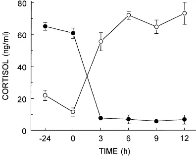
Continuous infusion of cortisol at a rate of 0.1 mg/kg/h elevated serum concentrations of
cortisol to 72.0±2.5 ng/ml within 6 h of the initiation of infusion (Fig. 2). Serum
concentra-tions of cortisol were maintained at this level for the remainder of the infusion period and
returned to the basal level (7.7±1.0 ng/ml) 3 h after the termination of cortisol infusion. In
contrast, serum concentrations of cortisol in control animals receiving vehicle alone were
Fig. 2. Serum concentrations of cortisol in sheep receiving exogenous cortisol (0.1 mg/kg/h) as a continuous infusion. Infusion of cortisol was initiated ((s); n=8 animals/group) or terminated ((d); n=7 animals/group) at time 0.
3.2. Follicular development and the preovulatory surge of LH
At PR serum concentrations of oestradiol were below the limit of detection (<0.5 pg/ml)
in all treatment groups. Serum concentrations of oestradiol increased progressively during the period after PR in animals (Group 1) receiving vehicle alone during the 10-day infusion period. Maximal serum levels of oestradiol (6–8 pg/ml) were noted 48–60 h after PR and a preovulatory surge of LH was evident in all control animals (eight of eight), with peak LH
noted 55.5±5.0 h after PR (Table 1).
In contrast, serum concentrations of oestradiol were maintained at basal (<0.5 pg/ml)
levels throughout the infusion period in sheep receiving stress-like levels of cortisol
Table 1
The effect of continuous infusion of stress-like levels of cortisol during the simulated luteal and/or early follicular phases of the oestrous cycle on the appearance and magnitude of the preovulatory surge of LH
Treatmenta n Animals displaying
ovulatory surge of LHb
Time of surge (h)c
Amplitude of surge (ng/ml)d
Vehicle 8 8 a 55.0±5.0 23.2±2.3
Cortisol–luteal and follicular phases 8 1 b 54 26.5
Cortisol–follicular phase 8 0 b – –
Cortisol–luteal phase 7 4 c 63.8±12.6 21.3±3.3
aAnimals received vehicle or stress-like levels of cortisol (0.1 mg/kg/h) beginning 5 days before and continuing for 5 days after pessary removal (PR). Additional animals received cortisol beginning at the time of PR and continuing for 5 days thereafter. A final group received cortisol during the 5-day period before PR and vehicle thereafter.
bSheep displaying a preovulatory surge of LH during the 5-day period after PR; values within the column that do not share a common letter are significantly different (P<0.05).
Fig. 3. Serum concentrations of progesterone in sheep receiving vehicle ((s); n=8) or stress-like levels of cortisol ((d); n=6) during the 5-day period after pessary removal (PR). Two animals from the cortisol-treated group (Group 3) did not show evidence of luteal function (serum progesterone maintained a concentrations below 0.2 ng/ml) during the post-treatment period. Data from these animals have not been included in the graph presented here.
beginning 5 days before and continuing for 5 days after PR (Group 2). Additionally, surge-like secretion of LH was evident in only one of eight animals receiving stress-like concentrations of cortisol throughout the 10-day infusion period.
Similarly, serum concentrations of oestradiol were maintained at basal levels throughout the infusion period in sheep receiving stress-like levels of cortisol beginning at PR and continuing for 5 days thereafter (Group 3). Although surge-like secretion of LH was not evident during the infusion period in sheep (zero of eight) receiving stress-like levels of cortisol during the 5-day period beginning at PR, surge-like secretion of LH was evident in six of eight sheep within 3 days (6–8 days after PR) of cessation of cortisol infusion. In addition, the development and subsequent regression of the corpus luteum in animals receiving stress-like levels of cortisol during the period after PR was delayed, relative to luteal activity in control animals (Fig. 3).
In contrast to the suppression noted in sheep receiving cortisol after PR, the preovulatory surge of LH was evident within 5 days after PR in four of seven ewes receiving cortisol only during the 5-day period prior to simulated luteolysis (Group 4).
4. Discussion
infusion indicates that stress-like levels of cortisol may arrest follicular development prior to that stage of maturation. Alternatively, cortisol may selectively impair oestrogen synthesis without compromising other aspects of follicular development.
Similarly, administration of stress-like levels of cortisol beginning at the onset of the follicular phase also suppressed follicular maturation and the ovulatory surge of LH. Con-versely, cortisol delivery only during the simulated luteal phase compromised oestradiol secretion and the preovulatory surge of LH during the subsequent follicular phase in some, but not all, ewes so treated. This anti-ovulatory effect of cortisol is consistent with the response to exogenous glucocorticoid noted in women (Cunningham et al., 1978), rodents (Baldwin and Sawyer, 1974), and other domestic species (Barb et al., 1982; Stoebel and Moberg, 1982). However, these results differ from a previous report that indicated that daily administration of a synthetic glucocorticoid (dexamethasone) did not compromise follicular development or ovulation in sheep (Phillips and Clarke, 1990). The physiologic basis for this discrepancy between our studies and this prior work is not clear, however, difference in the type of glucocorticoid, or dose and mode of administration, may contribute to these divergent observations.
In the work presented here we have attempted to approximate the increase in serum levels of cortisol noted in sheep during exposure to repetitive or persistent stress by infusion of the natural glucocorticoid. Continuous delivery of cortisol at a rate of 0.1 mg/kg/h established serum concentrations of cortisol comparable to those noted in sheep subject to moderate stressors, such as repeated laparoscopy, isolation and restraint, or bacterial infection (Martin et al., 1981; Minton, 1994; Battaglia et al., 1998). The use of the natural glucocorticoid at physiological concentrations strengthens our conclusion that stress-like concentrations of cortisol impair fertility in sheep. However, it should be noted that stress-induced increase in cortisol secretion is generally short-lived, suggesting that cortisol alone may not account in full for the infertility associated with stress.
Cortisol-dependent suppression of follicle maturation may reflect direct actions of the glucocorticoid at ovarian sites or, alternatively, action at hypothalamic or hypophyseal sites to limit secretion of the gonadotrophins. Recent studies indicate that cortisol-dependent arrest of follicular development in sheep is reversed by episodic administration of GnRH (Daley et al., 1999a). This suggests that stress-like levels of cortisol restrain pulsatile GnRH release during the follicular phase. Although, stress-like levels of cortisol do not affect the frequency or amplitude of episodic release of LH in ovariectomized sheep (Breen et al., 1999), LH pulse frequency is markedly reduced in sheep treated concurrently with cortisol and low concentrations of oestradiol (Breen et al., 1999; Daley et al., 1999b). This suggests that stress-like levels of cortisol may synergize with the low concentration of oestradiol characteristic of the early follicular phase to suppress GnRH pulse frequency and, thereby, limit gonadotrophin secretion.
concentrations of cortisol (∼75 versus∼65 ng/ml) and indicates that the suppressive effect of cortisol increases with increasing serum concentration over the physiological range.
In addition, we note here that cortisol-dependent suppression of follicle development and the LH surge is expressed even when cortisol delivery is initiated at PR. This suggests that stress-like levels of cortisol act relatively rapidly to suppress follicular maturation. However, the suppressive effect of cortisol appears to be temporary since four of seven sheep showed normal follicular development and ovulation within 5 days of PR when cortisol delivery was halted at PR. Similarly, luteal function was evident within 10 days of cessation of cortisol infusion in six of eight sheep receiving cortisol during the 5-day period after PR. These observations also indicate that the cortisol-dependent block to ovulation is temporary and follicular development, ovulation and subsequent luteal activity in most sheep are reinstated shortly after release from the inhibitory effects of stress-like levels of cortisol.
Taken together, these data are consistent with our working hypothesis that the infer-tility induced by stress or stress-like concentrations of cortisol is due, at least in part, to cortisol-dependent increase in the negative feedback potency of oestradiol. During the fol-licular phase of the oestrous cycle the augmented negative feedback potency of oestradiol would be expected to suppress the frequency and/or amplitude of episodic release of GnRH and decrease gonadotrophin secretion below the threshold required to maintain the normal progression of follicular development.
References
Adams, T.E., Kinder, J.E., Chakraborty, P.K., Estergreen, V.L., Reeves, J.J., 1975. Ewe luteal function influenced by pulsatile administration of synthetic LHRH/FSHRH. Endocrinology 97, 1460–1467.
Adams, T.E., Quirke, J.F., Hanrahan, J.P., Adams, B.M., Watson, J.G., 1988. Gonadotrophin secretion during the periovulatory period in Galway and Finnish Landrace ewes and Finnish Landrace ewes selected for high ovulation rate. J. Reprod. Fertil. 83, 575–584.
Baldwin, D.M., Sawyer, C.H., 1974. Effects of dexamethasone on LH release and ovulation in the cyclic rat. Endocrinology 94, 1397–1403.
Barb, C.R., Kraeling, R.R., Rampacek, G.B., Fonda, E.S., Kiser, T.E., 1982. Inhibition of ovulation and LH secretion in the gilt after treatment with ACTH or hydrocortisone. J. Reprod. Fertil. 64, 85–92.
Battaglia, D.F., Brown, M.E., Krasa, H.B., Thrun, L.A., Viguie, C., Karsch, F.J., 1998. Systemic challenge with endotoxin stimulates corticotropin releasing hormone and arginine vasopressin secretion into hypophyseal portal blood: coincidence with gonadotropin releasing hormone suppression. Endocrinology 139, 4175–4181. Bjersing, L., Hay, M.F., Kann, G., Moor, R.M., Naftolin, F., Scaramuzzi, R.J., Short, R.V., Younglai, E.V., 1972. Changes in gonadotrophins, ovarian steroids and follicular morphology in sheep at oestrus. J. Endocrinol. 52, 465–479.
Breen, K.M., Macfarlane, M.S., Sakurai, H., Adams, B.M., Adams, T.E., 1999. Stress-like levels of cortisol increase the negative feedback potency of estradiol in female sheep. Biol. Reprod. 60 (Suppl. 1) 276 (abstract 588).
Carson, R.S., Findlay, J.K., Clarke, I.J., Burger, H.G., 1981. Estradiol, testosterone, and androstenedione in ovine follicular fluid during growth and atresia of ovarian follicles. Biol. Reprod. 24, 105–113.
Chrousos, G.P., Torpy, D.J., Gold, P.W., 1998. Interactions between the hypothalamic-pituitary-adrenal axis and the female reproductive system: clinical implications. Ann. Int. Med. 129, 229–240.
Cunningham, G.R., Goldzieher, J.W., de Ia Pena, A., Oliver, M., 1978. The mechanism of ovulation inhibition by triamcinolone acetonide. J. Clin. Endocrinol. Metab. 46, 8–14.
Daley, C.A., Sakurai, H., Adams, B.M., Adams, T.E., 1999b. Effect of stress-like concentrations of cortisol on gonadotroph function in orchidectomized sheep. Biol. Reprod. 60, 158–163.
Dobson, H., Smith, R.F., 1995. Stress and reproduction in farm animals. J. Reprod. Fertil. 49 (Suppl.) 451–461. Doney, J.M., Gunn, R.G., Griffiths, J.G., 1973. The effect of premating stress on the onset of oestrus and on
ovulation rate in Scottish blackface ewes. J. Reprod. Fertil. 35, 381–384.
Ehnert, K., Moberg, G.P., 1991. Disruption of estrous behavior in ewes by dexamethasone or management-related stress. J. Anim. Sci. 69, 2988–2994.
Gill, J.L., 1978. Design and Analysis of Experiments in the Animal and Medical Sciences. Iowa State University Press, Ames, IA.
Hayashi, K.T., Moberg, G.P., 1990. Influence of the hypothalamic-pituitary-adrenal axis on the menstrual cycle and the pituitary responsiveness to estradiol in the female rhesus monkey (Macaca mulatta). Biol. Reprod. 42, 260–265.
Huet, C., Monget, P., Pisselet, C., Monniaux, D., 1997. Changes in extracellular matrix components and steroidogenic enzymes during growth and atresia of ovarian follicles in the sheep. Biol. Reprod. 56, 1025–1034. Komesaroffet, P.A., Esler, M., Clarke, I.J., Fullerton, M.J., Funder, J.W., 1998. Effects of estrogen and estrous
cycle on glucocorticoid and catecholamine responses to stress in sheep. Am. J. Physiol. 275, E671–E678. Martin, G.B., Oldham, C.M., Lindsay, D.R., 1981. Effect of stress due to laparoscopy on plasma cortisol levels,
the preovulatory surge of LH, and ovulation in the ewe. Theriogenology 16, 39–44.
Minton, J.E., 1994. Function of the hypothalamic-pituitary-adrenal axis and the sympathetic nervous system in models of acute stress in domestic farm animals. J. Anim. Sci. 72, 1891–1898.
Norman, R.L., McGlone, J., Smith, C.J., 1994. Restraint inhibits luteinizing hormone secretion in the follicular phase of the menstrual cycle in rhesus macaques. Biol. Reprod. 50, 16–26.
Phillips, D.J., Clarke, I.J., 1990. Effects of the synthetic glucocorticoid dexamethasone on reproductive function in the ewe. J. Endocrinol. 126, 289–295.
Sakurai, H., Adams, B.M., Adams, T.E., 1992. Pattern of gonadotropin-releasing hormone (GnRH)-like stimuli sufficient to induce follicular growth and ovulation in ewes passively immunized against GnRH. Biol. Reprod. 47, 177–184.
Smart, D., Forhead, A.J., Smith, R.F., Dobson, H., 1994. Transport stress delays the oestradiol-induced LH surge by a non-opioidergic mechanism in the early postpartum ewe. J. Endocrinol. 142, 447–451.
Smith, E.R., Johnson, J., Weick, R.F., Levine, S., Davidson, J.M., 1971. Inhibition of the reproductive system in immature rats by intracerebral implantation of cortisol. Neuroendocrinology 8, 94–106.
Stoebel, D.P., Moberg, G.P., 1982. Effect of adrenocorticotropin and cortisol on luteinizing hormone surge and estrous behavior of cows. J. Dairy Sci. 65, 1016–1024.
Welsh, T.J., Johnson, B.H., 1981. Stress-induced alterations in secretion of corticosteroids, progesterone, luteinizing hormone, and testosterone in bulls. Endocrinology 109, 185–190.
Wilson, S.J., Marion, R.S., Spain, J.N., Spiers, D.E., Keisler, D.H., Lucy, M.C., 1998. Effects of controlled heat stress on ovarian function of dairy cattle. 1. Lactating cows. J. Dairy Sci. 81, 2124–2131.