www.elsevier.com / locate / bres
Research report
Cellular determinants of reduced adaptability of the aging brain:
neurotransmitter utilization and cell signaling responses after MDMA
lesions
a ,
*
a bT.A. Slotkin
, F.J. Seidler , S.F. Ali
a
Department of Pharmacology and Cancer Biology, Duke University Medical Center, Durham, NC 27710, USA
b
Neurochemistry Laboratory, Division of Neurotoxicology, National Center for Toxicological Research, Jefferson, AR 72079, USA
Accepted 25 July 2000
Abstract
Senescence is accompanied by the loss of neurons and synapses, and the maintenance of function depends on adaptive change at the levels of synaptic activity and cellular responsiveness. In the current study, we administered the neurotoxin MDMA, to young and aged mice and assessed the effects on indices of neuronal activity and cell signaling mediated through adenylyl cyclase. Young mice given MDMA showed 80% depletion of dopamine in the caudate and 30% depletion in the cerebral cortex; measurements of dopamine turnover indicated a compensatory upregulation of the activity of the remaining neurons in the caudate but downregulation in the cerebral cortex. Serotonin levels were comparatively less affected but serotonin turnover was decreased significantly in both regions. At the level of cell signaling, the young mice showed heterologous upregulation of adenylyl cyclase activity and a consequent enhancement of responses mediated through neurotransmitter receptors. In aged mice, MDMA treatment produced the same degree of lesioning but substantially different changes in neuronal activity and cell signaling. In the cerebral cortex, dopamine turnover was increased, and serotonin turnover decreased, effects opposite in direction to those seen in young mice. In the aged group, MDMA elicited heterologous loss of adenylyl cyclase responses instead of displaying the supersensitivity that had been seen in the young group. The aging brain thus displays maladaptation to the loss of monoaminergic input, effects that may augment the functional impairment associated with neurodegenerative disorders or stroke. 2000 Elsevier Science B.V. All rights reserved.
Theme: Development and regeneration
Topic: Aging process
Keywords: Adenylyl cyclase; Aging brain; Dopamine; Methylenedioxymethamphetamine (MDMA); Serotonin (5-HT)
1. Introduction the levels of cell signaling and a great deal of attention has been paid to the role of neurotrophic factors in the
Aging is associated with increased susceptibility to prevention of cell death or restoration of function. It is
neuronal loss and disruption of cerebral function, either as increasingly evident that neurotransmitters themselves play
a component of senescence, or as a consequence of a key role in this process. Stimulation of nicotinic
neurodegenerative diseases or stroke. The adaptability of cholinergic receptors can prevent neuronal death caused by
the brain to neuronal or synaptic loss involves plasticity at simulated hypoxic / ischemic injury or by deprivation of
growth factors [13,40]. Similarly, stimulation of catechol-aminergic receptors can offset aging-related deterioration
Abbreviations: AC, adenylyl cyclase; ANOVA, analysis of variance;
DA, dopamine; DOPAC, 3,4-dihydroxyphenylacetic acid; 5-HIAA, 5- of behavioral performance [5] and recovery after surgical
hydroxyindoleacetic acid; 5-HT, 5-hydroxytryptamine (serotonin); HVA, lesions depends on the function of catecholamine systems homovanillic acid; MDMA, methylenedioxymethamphetamine; PXT, [10]. It is thus important to note that lesions of catechol-paroxetine
aminergic pathways in the aging brain can produce a
*Corresponding author. Tel.: 11-919-681-8015; fax: 1
1-919-684-different spectrum of behavioral effects from those seen
8197.
E-mail address: [email protected] (T.A. Slotkin). after lesioning of the young brain [30].
The issue of impaired adaptability of the aged brain to 2. Methods
the loss of monoaminergic inputs is critical to our
under-standing of deterioration of function in Parkinson’s dis- Studies were carried out in accordance with the
declara-ease, recovery from stroke and, as is now evident, geriatric tion of Helsinki and with the Guide for the Care and Use
depression. Elderly depressives are less responsive to of Laboratory Animals as adopted and promulgated by the
monoamine reuptake inhibitors, the mainstay of antide- National Institutes of Health. Male C57BL / 6N mice were
pressant therapy [6,23,25], and animal models of geriatric bred and housed four per cage with free access to food and
depression that incorporate monoaminergic lesions show water. Animals were selected at 3 and 24 months-of-age
corresponding differences from effects in young animals and given saline or MDMA (four injections of 20 mg / kg
[33]. Accordingly, the current study addresses the issue of i.p., spaced 2 h apart; MDMA obtained from Sigma
whether there is a cellular basis for the aging-related Chemical Co., St Louis, MO). One week later, animals
impairment in the response to neuronal loss. We have were subjected to cervical dislocation and the brains were
concentrated on the effects of MDMA, a widely-abused rapidly removed and dissected into different regions,
neurotoxic amphetamine analog. Although MDMA was placed on dry ice, and stored at2708C. This dose regimen
once thought to target 5-HT neurons selectively [14], has been used previously to elicit neurobehavioral damage
evidence now suggests that its primary actions are directed in rodents [17,24].
toward DA systems [22,28]; the spectrum of actions
depends on a variety of factors, including age [4]. We 2.1. Neurotransmitters and metabolites
compared the degree of DA and 5-HT lesioning achieved
in young and aged mice and then focused on two aspects Concentrations of DA, 5-HT and their metabolites
of adaptation: neurotransmitter turnover and cellular sig- DOPAC, HVA and 5-HIAA were quantitated by high
naling cascades. Transmitter turnover measures the utiliza- performance liquid chromatography with a C18 column
tion of the remaining stores of DA and 5-HT, which need and electrochemical detection [1]. Briefly, each region was
to compensate for the partial loss of neuronal projections. weighed and deproteinized with 0.2 N perchloric acid
Similarly, denervation typically evokes supersensitivity of containing 3,4-dihydroxybenzylamine (Sigma) as an
inter-cell signaling response elements, a major adaptation to the nal standard. The tissue was then disrupted by
ultrasonica-loss of input. We focused on the AC signaling cascade for tion, sedimented at 15,0003g and the supernatant solution
several reasons. First, deterioration of AC signaling is an was filtered through a 0.2mm Nylon-66 microfilter (MF-1
important correlate of aging [11,19,27]. In the peripheral centrifugal filter, Bioanalytical Systems, W. Lafayette, IN).
sympathetic nervous system, the responsiveness of car- Aliquots of 25 ml, representing 2.5 mg of original wet
diovascular adrenergic target tissues to neurotransmitter weight of brain tissue, were then injected directly onto the
stimulation declines markedly in the elderly, effects which chromatographic system. Results were calculated using a
reflect loss of specific receptor coupling mechanisms as standard curve constructed with varying concentrations of
well as of cellular intermediates in the signaling cascade, the authentic compounds (Sigma), with correction for
such as G-proteins and AC itself [3,11,19–21,27,29,38,39]. sample recovery using the internal standard.
In the central nervous system, impaired DA and 5-HT
3
responses have also been reported [15], although the 2.2. [ H]PXT binding
underlying causes have not been fully characterized. In
addition, we have demonstrated age-related disparities in The cell membrane fraction was prepared from brain
the AC response of the aged brain to alterations of regions by techniques described previously [18];
mem-hormonal status or to lesions of sensory afferent inputs brane protein was analyzed with Folin reagent. The
(olfactory bulbectomy) that replicate the endocrine and suspension was then used immediately for determinations
3
monoaminergic alterations thought to underlie human of AC activity (see below) and [ H]PXT binding [18],
3
geriatric depression [7,31–36]. Accordingly, our underly- using paroxetine[phenyl-69- H] (specific activity 25.4 Ci /
ing hypothesis is that damage to cell signaling capabilities mmol, New England Nuclear Corp., Boston, MA) with or
may be particularly relevant in aging, where failure of without addition of 100mM serotonin (Sigma) to displace
compensatory upregulation of responsiveness would ex- specific binding. Incubations lasted 120 min at 208C, and
acerbate, rather than offset, the effects of neuron loss. We were stopped by addition of 5 ml of ice-cold buffer,
have tested this hypothesis in mice in a relatively ‘early’ followed by vacuum filtration and washing onto Whatman
stage of senescence. Neurodegeneration, synaptic dysmor- GF / C filters, pre-soaked in 0.05% polyethyleneimine
phology and neuronal loss are likely to be present when (Sigma). Non-specific binding was approximately 20% of
very old animals are used, obscuring primary effects of the total in forebrain and brainstem, which highly express
lesioning on synaptic or cellular function [16]. According- the 5-HT transporter, and 40% in the cerebellum, which
ly, we have concentrated on 24-month-old mice, rather has much lower transporter expression. Because of the
than examining animals at the very extreme of the life large number of tissues involved in this study, 2 treatment
was not practicable to run Scatchard analyses on each. differences in AC reactivity to lesions and drug treatments
3
Accordingly, we examined binding at 85 pM [ H]PXT, a [7,32,33,35,36].
concentration above the K [18,33], but nevertheless belowd
full saturation of the binding site. The strategy of using a 2.4. Data analysis
single, subsaturating ligand concentration enables the
detection of drug- or age-induced changes regardless of Data are presented as means and standard errors, with
whether the changes are in affinity or capacity. intergroup comparisons by multivariate ANOVA (data
log-transformed whenever variance was heterogeneous). For an
2.3. AC activity initial global test, the results were divided into five
categorical groupings: DA and metabolites, 5-HT and
AC activity was evaluated in the same membrane metabolite, PXT binding, AC activity, and membrane
preparations according to established protocols [35,36]. protein concentration. Each grouping was then subjected to
Membrane aliquots were incubated for 10 min at 308C ANOVA using factors of age, MDMA treatment, brain
with final concentrations of 100 mM Tris–HCl (pH 7.4), region, and the multiple determinations within each
cate-10 mM theophylline, 1 mM adenosine 59-triphosphate, 10 gory (a repeated measure, since more than one type of
mM MgCl , 1 mg / ml bovine serum albumin, and a2 determination was made on each sample). In each case,
creatine phosphokinase-ATP regenerating system consist- whenever we found a significant effect of MDMA
treat-ing of 10 mM sodium phosphocreatine, 8 I.U. phosphoc- ment or interaction of MDMA with the other variables,
reatine kinase, and 10mM GTP (all reagents from Sigma). data were then subdivided into the individual regions,
The enzymatic reaction was stopped by placing the sam- which were then re-evaluated using ANOVA. Finally,
ples in a 90–1008C water bath for 5 min, followed by individual differences were established using Fisher’s
sedimentation at 30003g for 15 min, and the supernatant Protected Least Significant Difference. For all tests,
signifi-solution was assayed for cAMP using radioimmunoassay cance was assumed at the level of P,0.05 for main
kits (Amersham Corp., Chicago, IL). Preliminary experi- treatment effects and at P,0.1 for interactions [37]. Some
ments showed that the enzymatic reaction was linear well data are presented as the percentage change from
age-beyond the 30-min time period and was linear with matched control groups but the statistical analyses were
membrane protein concentration; concentrations of cofac- conducted on the unmanipulated data.
tors were optimal and, in particular, the addition of higher concentrations of GTP produced no further augmentation
of activity. 3. Results
Enzyme activity was evaluated under several different
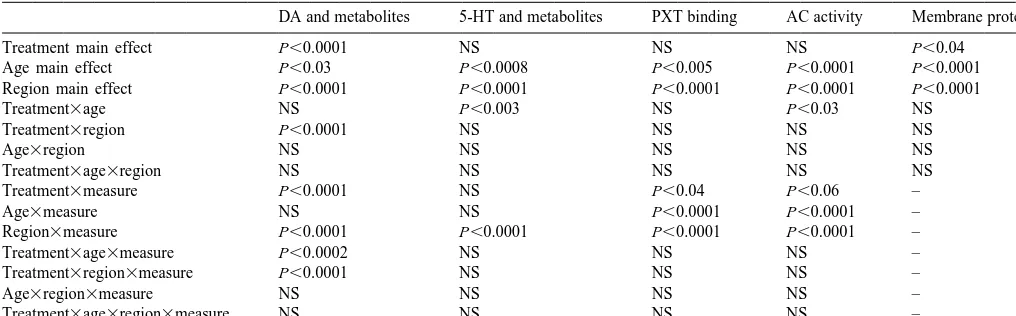
conditions. First, basal activity was evaluated. Second, the Global statistical analyses (Table 1) indicated significant
maximal G-protein-linked response was evaluated in sam- effects of age and MDMA treatment on all categories of
ples containing 10 mM NaF. Third, maximal total activity the measured variables, with significant
regionally-selec-of the AC catalytic unit, independent regionally-selec-of receptors or tive effects (main effects treatment, age and region,
G-proteins, was evaluated with 10 mM MnCl2 [12,42]. interactions among all three variables). Within each
cate-Finally, neurotransmitter receptor-mediated effects were gory, effects of aging and MDMA treatment were also
evaluated with either 100 mM 5-HT or DA [33,35]. The different for the various measures (interactions of
concentrations of all the agents used here have been found treatment3measure, age3measure, region3measure,
previously to be optimal for effects on AC and were treatment3age3measure, treatment3region3measure).
confirmed in preliminary experiments. For categories of neurotransmitters and their metabolites,
PXT binding and AC determinations were evaluated in this indicates selective effects on specific chemical entities,
terms of tissue concentration (binding per g tissue), and as whether the native biogenic amine or its catabolic
prod-specific activity relative to other membrane proteins (bind- ucts. For PXT binding, this denotes selective effects
ing per mg protein). directed toward absolute activity (activity per g tissue) or
Because of the larger amounts of tissue required for to the specific activity relative to other membrane proteins
3
determinations of [ H]PXT binding and AC activity, brain (activity per mg protein). For AC activity, the interaction
dissections for these determinations differed somewhat of main variables with the specific measures also denotes
from those used for the neurotransmitter and metabolite differential effects on absolute vs. specific activity, as well
determinations. Given the similarity of age- and drug- as differences among the five different types of AC
related effects on neurotransmitters and metabolites in the measurement (basal activity and effects of fluoride,
man-cerebral cortex and caudate (see below), we did not ganese, dopamine and serotonin).
separate these regions for PXT binding and AC determi- There were significant effects of both age and MDMA
nations, and instead used the intact forebrain. In addition, treatment on the membrane protein concentration, so that
we evaluated effects on the brainstem and cerebellum, two some of the differences in PXT binding and AC activity
Table 1
a
Global statistical analyses
DA and metabolites 5-HT and metabolites PXT binding AC activity Membrane protein
Treatment main effect P,0.0001 NS NS NS P,0.04
Age main effect P,0.03 P,0.0008 P,0.005 P,0.0001 P,0.0001
Region main effect P,0.0001 P,0.0001 P,0.0001 P,0.0001 P,0.0001
Treatment3age NS P,0.003 NS P,0.03 NS
Treatment3region P,0.0001 NS NS NS NS
Age3region NS NS NS NS NS
Treatment3age3region NS NS NS NS NS
Treatment3measure P,0.0001 NS P,0.04 P,0.06 –
Age3measure NS NS P,0.0001 P,0.0001 –
Region3measure P,0.0001 P,0.0001 P,0.0001 P,0.0001 –
Treatment3age3measure P,0.0002 NS NS NS –
Treatment3region3measure P,0.0001 NS NS NS –
Age3region3measure NS NS NS NS –
Treatment3age3region3measure NS NS NS NS –
a
For each category, the global analysis combined all related measures in a repeated-measures design. For DA and 5-HT, analyses assessed each amine and its related metabolites. For PXT binding, analyses assessed both the concentration and specific activity of binding sites. For AC, analyses assessed all five measures, both as concentration and specific activity. Interactions with ‘measure’ thus denote selectivities toward specific types of measurements within each category.
membranes. In some situations, evaluation of the mem- 3.1. Neurotransmitters and metabolites
brane protein concentration is necessary to correct for
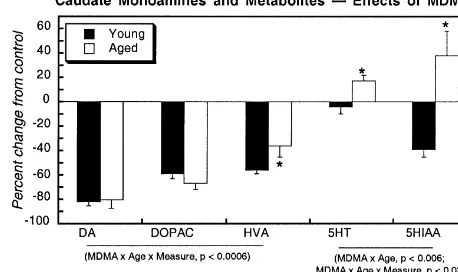
intersample variability in recovery of the cell membrane In control mice, there were only minor age-related
fraction. Here, samples from animals in the different differences in the levels of monoamines and their
metabo-treatment groups of a given age were all prepared simul- lites (Fig. 1, left panels). In the caudate nucleus, there were
taneously and processed in parallel, making it unlikely that significant overall differences in the two age groups
there are systematic differences in recovery of membranes; compared across all measurements, but for individual
in this case, systematic shifts in membrane protein con- measurements only DOPAC showed a slightly higher level
centration instead reflect changes in cell size and the in the aged group, at the margin of statistical significance.
attendant alteration of surface-to-volume ratio. Biologi- Cerebral cortical levels of DA and its metabolites were
cally-relevant effects on membrane protein concentrations indistinguishable in young versus aged mice, and levels of
are typical of aging and brain lesions, since changes in 5-HT and 5-HIAA were unchanged in all regions. In the
axon / synapse number, or replacement of damaged cells hippocampus, concentrations of DA and its metabolites
with glia, can all contribute to altered membrane protein were too low for reliable quantitation.
concentrations. Monoamine transporters, neurotransmitter Upon lesioning with MDMA, both young and aged mice
receptors, G-proteins and AC are all integral membrane showed equivalent, massive (80%) depletion of DA in the
proteins and are thus affected by alterations in cell size as caudate nucleus (Fig. 1, upper right panel). DA metabolites
well as by age- or drug-induced effects on specific were also reduced, although to a smaller extent than DA
expression of these proteins. Accordingly, we present PXT itself. Thus, the turnover of DA (ratio of total DA
binding and AC measurements both as activity per g tissue metabolites to DA) increased after lesioning, representing
and per mg protein to separate specific alterations in hyperactivity of the remaining nerve terminals (Table 2);
expression over-and-above effects on all membrane con- the lack of a significant interaction of treatment3age
stituents. From the standpoint of cell function (signal indicated that the increase in aged mice was not
dis-produced per mass of tissue), activity per g of tissue is the tinguishable from that seen in young mice. Despite the
relevant measure, whereas values adjusted for membrane equivalent loss of DA in the two age groups, aged mice
protein establish drug effects on the expression of in- showed a significantly smaller reduction in HVA (Fig. 1,
dividual proteins. upper right panel). The aged caudate also showed
sig-In light of the significant interactions of aging and nificantly different effects of MDMA on the 5-HT system:
MDMA treatment with region and measures within each the young animals showed little or no 5-HT depletion (Fig.
category, results are presented with subdivision into each 1, upper right panel) and a decrease in 5-HT turnover
region and measure. For clarity, comparisons are presented (Table 2); aged animals showed increases in both 5-HT
first between young and aged control mice, and then for and 5-HIAA and no change in turnover.
the effects of MDMA on the two populations, shown as the In the cerebral cortex (Fig. 1, middle right panel),
percentage of the corresponding age-matched control MDMA treatment elicited age-dependent differences in
Fig. 1. Effects of aging and MDMA treatment on levels of DA and its metabolites, DOPAC and HVA, and on 5-HT and its metabolite, 5-HIAA. Note the different units for caudate DA (top left panel). Data represent means and standard errors obtained from five to eight animals in each group. The left-hand panels depict the differences between young and aged control mice, whereas the right-hand panels depict the effects of MDMA as the percent change from the corresponding control values. ANOVA across all measures for each monoamine appears at the bottom of each panel and asterisks denote individual values where the aged group differs from the young group. Across all three regions, ANOVA indicates significant, regionally-selective and / or measure-selective (DA vs. DOPAC vs. HVA) differences in the DA system between young and aged controls: main effect of region (P,0.0001) and interactions of age3measure (P,0.06) and region3measure (P,0.0001). No age-dependent differences were seen for the 5-HT system in the control group: main effect of region (P,0.0001), interaction of region3measure (P,0.0001), but no interaction of age with the other variables. For the effects of MDMA, ANOVA across all regions indicates significant age-dependent effects for both DA and 5-HT systems. For DA and its metabolites, there were significant interactions of MDMA3region (P,0.0001), MDMA3age (P,0.008), MDMA3age3measure (P,0.0001), MDMA3region3measure (P,
0.0001), and MDMA3age3region3measure (P,0.07). For 5-HT and 5-HIAA, there were significant interactions of MDMA3age (P,0.0003) and MDMA3age3region (P,0.05).
but the effects of MDMA in general were of much smaller that effects in aged mice were distinguishable from those
magnitude. In the cortex, both DOPAC and HVA showed in young mice). In young mice, the ratio decreased
smaller reductions in the aged group than in young slightly, whereas in aged mice, the ratio increased after
animals. Most notably, DA turnover (Table 2) showed a treatment. Neither age group showed any significant loss
completely different reaction to MDMA in the two age of cerebrocortical 5-HT after MDMA treatment, but
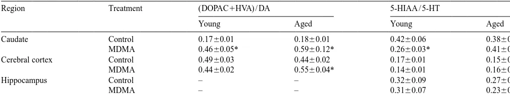
Table 2
a
Effects of MDMA on neurotransmitter turnover
Region Treatment (DOPAC1HVA) / DA 5-HIAA / 5-HT
Young Aged Young Aged
Caudate Control 0.1760.01 0.1860.01 0.4260.06 0.3860.06
MDMA 0.4660.05* 0.5960.12* 0.2660.03* 0.4160.07
Cerebral cortex Control 0.4960.03 0.4460.02 0.1760.01 0.1560.01
MDMA 0.4460.02 0.5560.04* 0.1460.01 0.1660.01
Hippocampus Control – – 0.3260.09 0.2760.06
MDMA – – 0.3160.07 0.2360.04
a
Data represent means and standard errors obtained from five to eight animals in each group.
Asterisks denote significant differences between MDMA-treated animals and their corresponding control group. In the caudate, two-factor ANOVA indicates no age-dependent difference for the increase of dopamine turnover after MDMA (no interaction of treatment3age) but the decrease in 5-HT turnover seen in young animals was not observed in aged animals (P,0.05 for interaction of treatment3age). In the cerebral cortex, two-factor ANOVA indicates age-dependent differences for the effects of MDMA on both DA turnover and 5-HT turnover (respectively, P,0.005 and P,0.05 for interaction of treatment3age). There were no significant age-dependent effects on 5-HT turnover in the hippocampus.
serotonergic systems (Fig. 1, middle right panel). 5-HIAA panels), binding was elevated (measured either as
con-tended to decrease in the young animals treated with centration or specific activity) after MDMA treatment of
MDMA, but to increase in the aged group, producing a young mice but no effect was seen in aged mice; although
statistically significant interaction of treatment3age (P, changes elicited by MDMA were proportionally larger in
0.03). When these data were used to calculate the metabo- the cerebellum, it should be kept in mind that the overall
lite ratio (5-HIAA to 5-HT) the differences between young concentration of these sites was very low.
and old animals became even more apparent (Table 2).
The ratio decreased in the young mice given MDMA but 3.3. AC activity
increased in the identically-treated aged animals
(treatment3age interaction, P,0.05). In control forebrain, there were small differences in AC
The hippocampus showed little or no MDMA-related between young and aged mice (Fig. 2). On a concentration
changes in 5-HT or 5-HIAA levels (Fig. 1, bottom right basis (activity per g tissue), activities for all measures
panel) or in the turnover ratio (Table 2). tended to be slightly higher in the aged group (upper left
panel); after adjustment for the concentration of other
3
3.2. [ H]PXT binding membrane proteins (upper right panel), values were higher
for young animals, in keeping with earlier studies with
In control mice, there were minor differences in PXT aged rats [35]. Similar, slight reductions in AC specific
binding between the young and aged cohorts (Fig. 2). The activity, were seen in brainstem (middle panels) and
concentration of sites (binding per g tissue) showed cerebellum (lower panels). AC stimulation by DA was
slightly higher values in the forebrain of aged mice (upper highly statistically significant (P,0.0001) and showed
left panel) and lower values in the brainstem (middle left preferential effects in the forebrain (P,0.0001 for
inter-panel). Because membrane protein concentrations were action of region3DA-induced stimulation), in keeping with
higher in the aged group, the specific activity of PXT the localization of 90% of DA projections in that region;
binding sites was actually lower in both regions when nevertheless, DA-induced stimulation was statistically
compared to other membrane proteins (upper and middle significant in all three regions (P,0.0001 for each). DA
right panels). As expected from the distribution of 5-HT caused a 35% increase in AC over basal values in the
projections, binding in the cerebellum (lower panels) was forebrain and a 7–15% increase in the other two regions.
much lower than in the forebrain or brainstem. DA stimulation over basal values did not differ with age
Lesioning with MDMA had only small (,10%) effects (3563% increase in young control forebrain, 3261% in
on PXT binding in the forebrain of either young or aged aged control forebrain). Although 5-HT also stimulated AC
mice. The concentration of sites (Fig. 3, upper left panel) significantly across all three regions (P,0.0001), the
was slightly elevated in the young group and reduced in response was limited to the cerebellum (P,0.0001),
the aged group; however, these differences were not whereas the forebrain and brainstem showed no significant
specific for the 5-HT transporter, since adjustment for stimulation. This is a likely consequence of the fact that
membrane protein (upper right panel) eliminated the 5-HT receptors both stimulate and inhibit AC, so that the
difference between the two age groups. In the brainstem net effect depends on the relative balance of positive and
(middle panels), neither young nor aged mice showed any negative inputs [33,35]. Alterations in AC signaling can
differences in PXT binding on either the basis of con- thus be elicited by changes in either stimulatory or
3
Fig. 2. Effects of aging on [ H]PXT binding and AC activity assessed either as absolute concentration (left panels) or as specific activity relative to membrane protein (right panels). Data represent means and standard errors obtained from six to seven animals in each group. ANOVA across all AC measures appears at the bottom of each panel and asterisks denote individual values where the aged group differs from the young group. Across all three regions, ANOVA indicates significant age-related differences for tissue weight (main effect, P,0.0002; age3region interaction, P,0.0001), membrane
3
protein (main effect of age, P,0.0001), [ H]PXT binding (main effect of age for binding per mg protein, P,0.006), and AC (main effect of age for activity per mg protein, P,0.0001; age3region interaction, P,0.06).
Despite the fact that young and aged mice showed activity and reactivity to DA or 5-HT (upper left panel). In
similar degrees of DA lesioning after MDMA treatment, this case, age-dependent differences were still evident after
there were profound differences in the effects of MDMA adjustment for membrane protein (upper right panel); after
on AC activity in the two age groups (Fig. 3). On a MDMA treatment, AC specific activity was relatively
concentration basis (activity per g tissue), young mice unchanged in young mice, but there was a global decrease
showed upregulation across all AC measures in the fore- in aged mice. Effects were much less notable in the
3
Fig. 3. Effects of MDMA on [ H]PXT binding and AC activity assessed either as absolute concentration (left panels) or as specific activity relative to membrane protein (right panels). Data represent means and standard errors obtained from five to six animals in each group. ANOVA across all AC measures appears at the bottom of each panel and asterisks denote individual values where the aged group differs from the young group. Across all three
3
regions, ANOVA indicates significant age-related differences for membrane protein (MDMA3age, P,0.04; MDMA3region, P,0.05), [ H]PXT binding (MDMA3age, P,0.05 for binding per g tissue, P,0.07 for binding mg protein), and AC (MDMA3age, P,0.01 for activity per g, P,0.05 for activity per mg protein; P,0.1 for MDMA3age3measure for activity per g).
as for the forebrain: a tendency toward increases in activity significantly smaller effect was obtained in aged mice,
per g tissue in young mice only. In the cerebellum (lower despite the equivalent degree of lesioning as evidenced by
panels), there were global increases in all measures of AC DA depletion.
after lesioning of young mice, and no change or a decrease in aged mice given the same treatment.
MDMA treatment caused an increase in the membrane 4. Discussion
protein concentration in the region (forebrain) possessing
the highest concentration of DA (Fig. 3, upper right panel). The current results indicate that lesioning of
in the aged brain as compared to young brain. These Because we found age-related differences in 5-HT
differences were apparent at two levels of organization: turnover after MDMA lesioning, we also explored the
first, patterns of neural activity as indicated by measure- possibility that changes in 5-HT transporter expression
ments of monoamine transmitter turnover, and second, cell might account for the apparent alterations in presynaptic
signaling responses mediated through AC. The age-related function. Measurement of the concentration of transporter
disparities were not apparent under basal conditions, as the sites, assessed with PXT binding per g tissue, indicated a
non-lesioned groups displayed similar transmitter utiliza- significantly greater decrease in the aged group, an effect
tion rates and signaling profiles. which would indeed increase 5-HT turnover by reducing
Administration of MDMA produced equivalent lesions the presynaptic recapture of 5-HT. However, it is unclear
in both age groups, evidenced by approximately 80% whether this represents a specific change in transporter
depletion of DA in the caudate. In young animals, the loss expression, since, with correction for MDMA-induced
of dopaminergic inputs showed two adaptive responses at effects in other membrane proteins (binding per mg
the level of neurotransmitter utilization. DA turnover protein), the age-related differences in PXT binding were
increased, indicating a rise in neuronal activity in the no longer evident. In fact, on this basis, MDMA produced
remaining dopaminergic projections, a compensatory small (|10%) reductions in 5-HT transporter binding in the
adaptation to the loss of DA inputs. Similarly, 5-HT forebrain without distinction between young and aged
turnover decreased in the young animals given MDMA; mice, an effect which would enhance 5-HT utilization in
since 5-HT receptors provide inhibitory control over AC both age groups. Furthermore, the largest proportional
[2,33,35], this effect also may compensate for the loss of changes in transporter binding sites were seen in the
stimulatory DA inputs. The response to lesioning on cerebellum, a region relatively sparse in 5-HT projections.
neuronal activity in the caudate can thus be viewed as a It is thus possible that alterations in transporter function
coordinated effort to augment stimulatory responses while contribute somewhat to the changes in net 5-HT utilization
suppressing inhibitory inputs. In contrast, the aged animals rates but is not clear whether measurements simply of
specifically were deficient in the adaptive suppression of transporter binding sites can distinguish the degree to
5-HT activity and instead, showed upregulation of 5-HT which they influence this outcome.
utilization. Although the aged group exhibited overall Differences between aged and young mice were not
increases in DA turnover equivalent to those in younger limited to effects on neurotransmitter utilization or
synap-animals, the result was accomplished through somewhat tic activity, but also were prominent at the level of cellular
different metabolic processes, involving a greater relative signaling as monitored by AC. Indeed, MDMA-induced
contribution of HVA. Since HVA is produced by deamina- lesions altered forebrain AC activity and reactivity to
tion of DA, this result is in keeping with earlier observa- stimulation in opposite directions at the two ages. In young
tions of greater monoamine oxidase activity in senescence animals, the predominant response (activity per g tissue)
[34]. Higher monoamine oxidase may also account for the was a global increase in all measures of AC activity, an
slightly greater DOPAC levels in aged controls. adjustment that would help compensate for the loss of DA
One major conclusion then, is that the adaptations to the input by increasing the efficiency of transmitter-generated
loss of DA inputs after MDMA lesioning involves adjust- responses. In contrast, the aged mice showed a decrease,
ments in neuronal activity extending to transmitter sys- which would worsen the functional effects of the lesion.
tems, like 5-HT, that are apparently spared (as evidenced Both the sensitization in young animals and the
desensiti-by a lack of transmitter depletion). If this is true, then zation in aged animals was heterologous, that is, the
age-related differences in the response to MDMA may adjustments were made at a post-receptor locus: the effects
21
encompass regions displaying much smaller outright loss are seen for basal, fluoride-stimulated and Mn
-stimu-of DA projections. In the cerebral cortex, we found a much lated AC activity, all of which do not require the
participa-smaller degree of DA depletion (about 30%) and again, tion of neurotransmitter receptors. This is in keeping with
there were no differences in the efficacy of lesioning in the recent studies in developing tissues and with congestive
two age groups. However, just as found in the caudate, the heart failure, that indicate that the major cellular
adjust-aged brain responded differently to the insult. Young mice ments to altered rates of transmitter input are at the level of
showed little or no change in cerebrocortical DA turnover AC expression [8,9,26,41,42]. In the present case, at least
but aged mice evinced hyperactivity. One implication is some of the increased AC in young, lesioned animals,
that the aged animals may suffer a more severe loss of DA represents effects on a wider variety of membrane proteins,
function at equivalent degrees of DA depletion, necessita- since the upregulation of AC was no greater than that of
ting a greater compensatory activation of the remaining overall membrane protein. The increase in membrane
nerve terminals. A potential explanation emerges when we protein in young forebrain after MDMA treatment may
consider effects on cell signaling (below), where the aged reflect either induction of multiple proteins, or a change in
animals, but not young animals, display a loss of signaling cell size consequent to the lesioning treatment; membrane
efficiency. Just as in the caudate, the aged animals showed protein increases as the surface-to-volume ratio increases,
activation of cerebrocortical 5-HT systems despite no indicating cell shrinkage, cell damage, reactive axonal
smaller in size. Since both aged and young mice had the References
same degree of lesion, assessed by DA depletion, this
[1] S.F. Ali, G.D. Newport, R.R. Holson, W. Slikker, J.F. Bowyer, Low
implies that the young brain may exhibit greater axonal
environmental temperatures or pharmacological agents which
sprouting or gliosis after lesioning. Future studies can
produce hypothermia decreases methamphetamine neurotoxicity in
address this issue through evaluation of specific axonal and mice, Brain Res. 658 (1994) 33–38.
glial markers. In either case, though, the fact that the aged [2] T.M. Aune, K.A. Kelley, G.E. Ranges, M.P. Bombara,
Serotonin-brain uniquely shows a deficit in AC signaling after activated signal transduction via serotonin receptors on Jurkat cells,
J. Immunol. 145 (1990) 1826–1831.
lesioning is a likely contributor to the effects on transmitter
[3] O.E. Brodde, H.R. Zerkowski, D. Schranz, A. Broedesitz, M.
utilization. Given a smaller cell signaling response, aged
Michel-Reher, E. Schafer-Beisenbusch, J.A. Piotrowski, H. Oelert,
mice would have to increase impulse activity even more Age-dependent changes in theb-adrenoceptor-G-protein(s)-adenylyl
because of the maladaptive changes in AC. As already cyclase system in human right atrium, J. Cardiovasc. Pharmacol. 26
(1995) 20–26.
discussed, cerebrocortical DA and 5-HT utilization rates
[4] H.W. Broening, J.F. Bowyer, W. Slikker, Age-dependent sensitivity
were higher after lesioning in aged animals than in young
of rats to the long-term effects of the serotonergic neurotoxicant
animals. 3,4-methylenedioxymethamphetamine (MDMA) correlates with the
If global changes at the level of cell signaling provide magnitude of the MDMA-induced thermal response, J. Pharmacol.
the driving force for age-related differences in the effects Exp. Ther. 275 (1995) 325–333.
[5] J.T. Coull, Pharmacological manipulations of thea -noradrenergic
of MDMA on neuronal activity, then what is the mecha- 2
system: effects on cognition, Drugs Aging 5 (1994) 116–126.
nism underlying such widespread change? It is highly
[6] Danish University Antidepressant Group, Paroxetine: a selective
unlikely that synaptic adjustments are simply a localized serotonin reuptake inhibitor showing better tolerance, but weaker
response to focal damage. Indeed, in the cerebellum, which antidepressant effect than clomipramine in a controlled multicenter
is quite sparse in DA projections, we obtained the same study, J. Affect. Disord 18 (1990) 289–299.
[7] F. Fumagalli, S.R. Jones, M.G. Caron, F.J. Seidler, T.A. Slotkin,
type of increases in AC function in young animals given
Expression of mRNA coding for the serotonin transporter in aged
MDMA, whereas the aged group again showed either no
vs. young rat brain: differential effects of glucocorticoids, Brain Res.
adaptive increase, or even a decrease in AC signaling. 719 (1996) 225–228.
Accordingly, it is likely that multiple circuits, activated or [8] M.H. Gao, P.P. Ping, S. Post, P.A. Insel, R.Y. Tang, H.K. Hammond,
repressed by the lesion, converge on the AC signaling Increased expression of adenylylcyclase type VI proportionately
increases b-adrenergic receptor-stimulated production of cAMP in
pathway to regulate its function in a heterologous manner.
neonatal rat cardiac myocytes, Proc. Natl. Acad. Sci. USA 95
In earlier work, we obtained similar results after olfactory
(1998) 1038–1043.
bulb lesions, which compromise the function of cortical [9] Y.S. Gao, J.F. Tolsa, M. Botello, J.U. Raj, Developmental change in
monoaminergic circuits: aged rats showed a loss of AC isoproterenol-mediated relaxation of pulmonary veins of fetal and
reactivity whereas young rats showed upregulation [33]. It newborn lambs, J. Appl. Physiol. 84 (1998) 1535–1539.
[10] L.B. Goldstein, S. Bullman, Effects of dorsal noradrenergic bundle
is entirely possible that such heterologous changes are
lesions on recovery after sensorimotor cortex injury, Pharmacol.
triggered by hormonal differences. Both DA and 5-HT
Biochem. Behav. 58 (1997) 1151–1157.
exert major control over the functioning of the HPA axis [11] J.A. Heinsimer, R.J. Lefkowitz, The impact of aging on adrenergic
and glucocorticoids evoke differential effects on AC-based receptor function: clinical and biochemical aspects, J. Am. Geriatr.
signaling in young vs. aged brain [35,36]. Again, future Soc. 33 (1985) 184–188.
[12] J.H. Hurley, Structure, mechanism, and regulation of mammalian
studies will need to address this issue.
adenylyl cyclase, J. Biol. Chem. 274 (1999) 7599–7602.
Regardless of the mechanism underlying the
maladap-[13] S. Kaneko, T. Maeda, T. Kume, H. Kochiyama, A. Akaike, S.
tive signaling responses in aged brain, the global loss of Shimohama, J. Kimura, Nicotine protects cultured cortical neurons
responsiveness is likely to contribute to the net effect on against glutamate-induced cytotoxicity via a7-neuronal receptors
synaptic and behavioral function, augmenting the disrup- and neuronal CNS receptors, Brain Res. 765 (1997) 135–140.
[14] J.W. Langston, Methylenedioxymethamphetamine selectively
dam-tive consequences of neuronal loss. Although we studied
age central serotonergic neurons in nonhuman primates, J. Am.
animals at 24 months-of-age so as to avoid the
neurode-Med. Assoc. 260 (1988) 51–55.
generation associated with extreme aging [16], it is quite [15] M.H. Makman, H.S. Ahn, L.J. Thal, N.S. Sharpless, B. Dvorkin,
likely that study of even older animals will reveal greater S.G. Horowitz, M. Rosenfeld, Evidence for selective loss of brain
differences in the response to lesioning. Age-related differ- dopamine- and histamine-stimulated adenylate cyclase activities in
rabbits with aging, Brain Res. 192 (1980) 177–183.
ences in cell signaling, and their repercussions for
neuro-[16] B. Meister, H. Johnson, B. Ulfhake, Increased expression of
transmitter disposition and synaptic activation, are likely to
serotonin transporter messenger RNA in raphe neurons of the aged
play a key role in the outcome of stroke and neurode- rat, Mol. Brain Res. 33 (1995) 87–96.
generative disorders that are prominent in senescence. [17] D.B. Miller, J.P. O’Callaghan, The role of temperature, stress, and
other factors in the neurotoxicity of the substituted amphetamines 3,4-methylenedioxymethamphetamine and fenfluramine, Mol. Neurobiol. 11 (1995) 177–192.
Acknowledgements 3
[18] C. Moret, M. Briley, Platelet H-paroxetine binding to the serotonin transporter is insensitive to changes in central serotonergic
innerva-The authors thank E.C. McCook for technical assistance. tion in the rat, Psychiat. Res. 38 (1991) 97–104.
Biochemical correlates with myocardial aging, Cardioscience 3 patients with reduced platelet serotonin transport and resistance to (1992) 67–78. imipramine inhibition of transport, Depression Anxiety 6 (1997) [20] N. Narayanan, J.A. Derby, Alterations in the properties of b- 19–25.
adrenergic receptors of myocardial membranes in aging: impair- [32] T.A. Slotkin, E.C. McCook, J.C. Ritchie, B.J. Carroll, F.J. Seidler, ments in agonist-receptor interactions and guanine nucleotide regu- Serotonin transporter expression in rat brain regions and blood lation accompany diminished catecholamine-responsiveness of platelets: aging and glucocorticoid effects, Biol. Psychiat. 41 (1997) adenylate cyclase, Mech. Ageing Dev. 19 (1982) 127–139. 172–183.
[21] N. Narayanan, L. Tucker, Autonomic interactions in the aging heart: [33] T.A. Slotkin, D.B. Miller, F. Fumagalli, E.C. McCook, J. Zhang, G. age-associated decrease in muscarinic cholinergic receptor mediated Bissette, F.J. Seidler, Modeling geriatric depression in animals: inhibition of b-adrenergic activation of adenylate cyclase, Mech. biochemical and behavioral effects of olfactory bulbectomy in Ageing Dev. 34 (1986) 249–259. young versus aged rats, J. Pharmacol. Exp. Ther. 289 (1999) [22] J.F. Nash, J. Brokin, Microdialysis studies on 3,4-methylenediox- 334–345.
ymethamphetamine-induced dopamine release: effects of dopamine [34] T.A. Slotkin, F.J. Seidler, J.C. Ritchie, Effects of aging and uptake inhibitions, J. Pharmacol. Exp. Ther. 259 (1991) 820–825. glucocorticoid treatment on monoamine oxidase subtypes in rat [23] J.C. Nelson, C.M. Mazure, P.I. Jatlow, Desipramine treatment of cerebral cortex: therapeutic implications, Brain Res. Bull. 47 (1998)
major depression in patients over 75 years of age, J. Clin. Psycho- 345–348.
pharmacol. 15 (1995) 99–105. [35] T.A. Slotkin, L. Thai, E.C. McCook, J.L. Saleh, J. Zhang, F.J. [24] J.P. O’Callaghan, D.B. Miller, Neurotoxicity profiles of substituted Seidler, Aging and glucocorticoids: effects on cell signaling me-amphetamines in the C57BL / 6J mouse, J. Pharmacol. Exp. Ther. diated through adenylyl cyclase, J. Pharmacol. Exp. Ther. 279
270 (1994) 741–751. (1996) 478–491.
[25] S.P. Roose, A.H. Glassman, E. Attia, S. Woodring, Comparative [36] T.A. Slotkin, J. Zhang, E.C. McCook, F.J. Seidler, Glucocorticoid-efficacy of selective serotonin reuptake inhibitors and tricyclics in targeting of the adenylyl cyclase signaling pathway in the cere-the treatment of melancholia, Am. J. Psychiat. 151 (1994) 1735– bellum of young vs. aged rats, Brain Res. 800 (1998) 236–244.
1739. [37] G.W. Snedecor, W.G. Cochran, Statistical Methods, Iowa State
[26] D.M. Roth, M.H. Gao, N.C. Lai, J. Drumm, N. Dalton, J.Y. Zhou, J. University Press, Ames, Iowa, 1967.
Zhu, D. Entrikin, K. Hammond, Cardiac-directed adenylyl cyclase [38] K. Urasawa, T. Murakami, H. Yasuda, Age-related alterations in expression improves heart function in murine cardiomyopathy, adenylyl cyclase system of rat hearts, Japan. Circ. J. 55 (1991)
Circulation 99 (1999) 3099–3102. 676–684.
[27] P.J. Scarpace, N. Turner, S.L. Mader, b-Adrenergic function in [39] M. White, R. Roden, W. Minobe, M.F. Khan, P. Larrabee, M. aging: basic mechanisms and clinical implications, Drugs Aging 1 Wollmering, J.D. Port, F. Anderson, D. Campbell, A.M. Feldman, (1991) 116–129. M.R. Bristow, Age-related changes in b-adrenergic neuroeffector [28] M. Shankaran, B.K. Yamamoto, G.A. Gudelsky, Mazindol attenuates systems in the human heart, Circulation 90 (1994) 1225–1238.
the 3,4-methylenedioxymethamphetamine-induced formation of hy- [40] H. Yamashita, S. Nakamura, Nicotine rescues PC12 cells from death droxy radicals and long term depletion of serotonin in the striatum, induced by nerve growth factor deprivation, Neurosci. Lett. 213 J. Neurochem. 72 (1999) 2516–2522. (1996) 145–147.
[29] Y. Shu, P.J. Scarpace, Forskolin binding sites and G-protein im- [41] J.L. Zeiders, F.J. Seidler, T.A. Slotkin, Ontogeny of regulatory munoreactivity in rat hearts during aging, J. Cardiovasc. Pharmacol. mechanisms for b-adrenoceptor control of rat cardiac adenylyl 23 (1994) 188–193. cyclase: targeting of G-proteins and the cyclase catalytic subunit, J. [30] J. Sirvio, P. Riekkinen, A. Valjakka, J. Jolkkonen, P.J. Riekkinen, Mol. Cell. Cardiol. 29 (1997) 603–615.
The effects of noradrenergic neurotoxin, DSP-4, on the performance [42] J.L. Zeiders, F.J. Seidler, T.A. Slotkin, Agonist-induced sensitiza-of young and aged rats in spatial navigation task, Brain Res. 563 tion of b-adrenoceptor signaling in neonatal rat heart: expression (1991) 297–302. and catalytic activity of adenylyl cyclase, J. Pharmacol. Exp. Ther. [31] T.A. Slotkin, J.C. Hays, C.B. Nemeroff, B.J. Carroll, Dexametha- 291 (1999) 503–510.
![Fig. 2. Effects of aging on [ H]PXT binding and AC activity assessed either as absolute concentration (left panels) or as specific activity relative to3activity per mg protein,membrane protein (right panels)](https://thumb-ap.123doks.com/thumbv2/123dok/3140623.1382975/7.612.55.546.41.564/effects-activity-assessed-concentration-specic-activity-relative-membrane.webp)
![Fig. 3. Effects of MDMA on [ H]PXT binding and AC activity assessed either as absolute concentration (left panels) or as specific activity relative to3membrane protein (right panels)](https://thumb-ap.123doks.com/thumbv2/123dok/3140623.1382975/8.612.55.553.59.520/effects-activity-assessed-absolute-concentration-specic-activity-membrane.webp)
