BIODEGRADATION OF CHLORPYRIFOS BY A MARINE
BACTERIUM Bacillus firmus Strain BY6 ASSOCIATED
WITH BRANCHING CORAL Acropora Sp.
Agus Sabdono1,2 1
Department of Marine Science, Diponegoro University, Semarang – 50275, Central Java, Indonesia
2
Center for Tropical Coastal and Marine Studies, Diponegoro University, Widya Puraya, Semarang – 50275, Central Java, Indonesia
ABSTRACT
A bacterium which utilizes chlorpyrifos as a sole source of carbon and energy was
isolated from coral surface. The organism utilized chlorpyrifos up to 25 mg l-1. The lag phase and time for degradation, however, were severely prolonged at chlorpyrifos
concentrations above 10 mg/liter. The growth kinetics of coral bacterium was studied in
batch culture. Estimation of maximum growth rates ( max), obtained from turbidity measurements, was 0.14 h-1 and half-saturation growth constant (Cs) was 9.34 mg l-1 chlorpyrifos. This strain demonstrated greatest similarity to members of the order
Bacillales and was closest to members of the Bacillus firmus group.
Keywords : coral bacterium , chlorpyrifos, growth kinetics
INTRODUCTION
Chlorpyrifos is an insecticide (kill or control insect) that has been one of the
pesticides most used worldwide since 1965. Currently, over 850 registered chlorpyrifos
products are on the market (Anonymous, 2002). Contamination of soil by the pesticide
may occasionally lead to the contamination of surface and groundwater (Anonymous,
1998). Reports from the Environmental Protection Agency (1997) suggest that a wide
range of water and terrestrial ecosystems may be contaminated with chlorpyrifos. A
single application of chlorpyrifos poses risks to small mammals, birds, fish and aquatic
invertebrate species (Anonymous, 2002). Sabdono et al. (2007) reported that
Chlorpyrifos [O,O-diethyl O-(3,5,6-trichloro-2-pyridyl)phosphorothioate]is used
worldwide as an agricultural insecticide and and household pest. Its environmental fate
has been studied extensively, and the reported half-lifein soil varies from 10 to 120 days,
with 3,5,6-trichloro-2-pyridinol (TCP) as the major degradation product (Singh et al.,,
2003; Racke et al., 1988). Chlorpyrifosis characterized by a P-O-C linkage as in other
organophosphate pesticides, such as diazinon, parathion, methylparathion, and
fenitrothion. Unlike in the case of other organophosphates,however, there have been no
reports of enhanced degradationof chlorpyrifos since its first use in 1965 (Singh et al.,
2003). It has been suggested that the accumulation of TCP, which has antimicrobial
properties, prevents the proliferation of chlorpyrifos-degrading microorganisms in soil
(Racke et al.,1990).
The important role of microorganisms in the degradation of organic pollutant is
clearly understand. Eventhough the microorganisms capable of degradation of organic
pollutants and their catabolic pathways have been studied intensively, information on the
microbial degradation of organophosphate in marine environments is still very limited.
To date, there have been no reports on accelerated biodegradation of chlorpyrifos by
marine bacteria. In most of the studies chlorpyrifos has been reported to be degraded by
soil bacteria, such as Escherichia coli (Wang et al.,2002), Arthrobacter sp. (Mallick, et
al., 1999) Agrobacterium sp. (Horne et al., 2002) and Enterobacter asburiae (Singh et
al., 2004).
The molecular study of degradation of certain organophosphates has been studied
widely. A various organophosphate-degrading genes (opd genes) have been isolated from
different species, some of which could degrade chlorpyrifos (Horne et al., 2002; Mulbry
et al., 1986; Richnis et al., 1997; Serdar et al., 1982). In most of the studies, opd genes
were found to be plasmid based and had similar DNA sequences. However, Horne et al.
(2002) isolated an opd gene from Agrobacterium radiobacter, which was located on the
chromosome but had a similar sequence to the opd gene from other bacteria.
The experiments reported here were carried out to isolate a
chlorpyrifos-mineralizing bacteriumfrom the coral tissues, characterize chlorpyrifos-degrading isolate,
MATERIALS AND METHODS
Sampling and isolation of coral-associated bacteria
The coral was collected from Teluk Awur (06o37’02,5’’ N; 1 10o38’21,4” E), North Java Sea, Indonesia (Fig.1) by scuba diving and identified as Acropora sp.
according to Veron (1986). Upon collection coral fragments were put into sterile plastic
bags (Whirl-Pak, Nasco, USA) and immediately brought to the Marine Station of the
Diponegoro University where it was rinsed with sterile seawater and scraped off with a
sterile knife. The resultant tissues were serially diluted, spread on ½ strength ZoBell
2216E marine agar medium and incubated at room temperature for 48 hours. On the basis
of morphological features, colonies were randomly picked and purified by making streak
plates (Madigan et al., 2000).
Screening of chlorpyrifos-degrading coral bacteria
Experiments were conducted by using 50-ml erlenmeyer flasks containing 15 ml
of 10 mg l-1 chlorpyrifos in Zobell 2216E medium. Flasks were inoculated with approximately 0.25 g (dry weight) of strain and incubated at room temperature on a
rotary shaker at 120 rpm for 48 h. Samples of culture (1 ml) were removed and
centrifuged in a microcentrifuge (Microfuge 11; Beckman Instruments, Inc., Fullerton,
Calif.) at 12,000 rpm for 2 min, and supernatant was decanted into eppendorf. Samples
were analyzed immediately or were refrigerated until analysis.
Growth analyses
Biomass concentrations were measured by spectrophotometry at 600 nm using a
dry weight calibration curve. A fluorometer (Turner Model III) with a 10-mm square
cuvette was used. Controls were run using medium made with double-distilled water with
no carbon source to insure that the growth observed was not due to organic contamination
present in the ordinary distilled water.
GC-MS analyses
After extractions, the samples were then analyzed by gas chromatograph Model
the carrier gas. A 1 meter glass column (3 mm ID) packed with 10% Silikon DC200 on
80-100 mesh Supelcon was used. Gasflow at 50 ml /min, column temperature at 200 oC, detector temperature at 230 oC and the injector temperature at 230 oC were maintained.
Bacterial strain
A coral bacterium strain BY6 was used in this study This strain was selected
based on the best growth and degradation among coral isolates. This strain was grown at
room temperature in Zobell 2216E medium suplemented with 10 mg l-1 chlorpyrifos.
Degradation of chlorpyrifos by isolate BY6
Experiments were conducted by using 250-ml erlenmeyer flasks containing 100
ml of 5, 10, 15, 20, and 25 mg l-1 chlorpyrifos in Zobell 2216E medium. Flasks were inoculated with approximately 0.25 g (dry weight) of strain and incubated at room
temperature on a rotary shaker at 120 rpm. Samples of culture (1 ml) were removed
periodically and centrifuged in a microcentrifuge (Microfuge 11; Beckman Instruments,
Inc., Fullerton, Calif.) at 12,000 rpm for 2 min, and supernatant was decanted into
eppendorf. Samples were analyzed immediately or were fixed with 10 l of 40% H2SO4
and refrigerated until analysis.
DNA extraction, PCR amplification and sequencing of 16S rRNA gene fragments
DNA extraction, PCR amplification of partial 16S rRNA gene of bacterial strain,
purification of PCR product and subsequent sequencing analysis was performed
according to the method of Radjasa et al. (2007). The determined DNA sequence of
strain was then compared for homology to the BLAST database. Phylogenetic tree was
constructed by using
Phylogenetic analysis.
A phylogenetic tree was constructed using maximum-likelihood analysis.
Alignment positions at which less than 50 % of sequences of the entire set of data had the
same residues were excluded from the calculations to prevent uncertain alignments
within highly variable positions of the 16S rDNA. CLUSTAL X was used for multiple
was performed with the PAUP*4.0 (Phylogenetic Analysis Using Parsimony) software
package (Swofford, 1998).
RESULTS AND DISCUSSION
Chlorpyrifos degradation by isolate BY6
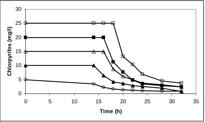
Chlorpyrifosdegradation of the five concentrations showed distinct patterns (Fig.
2 ). Strain BY5 degraded 25 mg l-1 chlorpyrifos rapidly. In this concentration approximately 50% of applied chlorpyrifos was degraded after 20 hours, following
which there was a period of rapid loss,with almost constant degradation after 32 hours.
Similarly, approximately 75-80% of the 5, 10, 15 and 20 mg l-1 chlorpyrifos concentration had been degraded in medium after24 hours. The bacterium isolated in
this present research was not so strong in its degradable ability. Singh et al. (2004)
reported that Enterobacter Strain B-14 hydrolyzed 35 mg l-1 concentration of chlorpyrifos within 24 h. So far, strain BY6 isthe first strain of coral bacterium reported
to be able to degrade chlorpyrifos. It was not surprisingly that this bacterium could not
degrade chlorpyrifos completely. It seems that cooperative metabolic activities in bacteria
of different cultures is needed to degrade chlorpyrifos. Mallick et al. (1999) and Wang et
al. (2002) reported that chlorpyrifos were to be degraded cometabolically in liquid media
by Flavobacterium sp. and also by an Escherichia coli clone with opd gene.
Cell growth and substrate utilization
The relationship of concentration of chlorpyrifos to growth rate strain BY6 was
determined by using batch culture methods. Values of specific growth rate ( ) obtained
from turbidity measurements ranged from 0.0509 h-1 to 0.0877 h-1 . Furthermore, the highest growth rate of 0.0877 h-1 occurred at 20 mg l-1 chlorpyrifos. Above this level, growth was strongly inhibited. As shown in Fig. 3, the estimate of half-saturation growth
constatnt (Cs) was 9.34 ppm chlorpyrifos and the maximum growth rates ( max) was 0.14 h-1. No report could be compared with this kinetic result.
Batch cultures of the coral bacterium strain BY6 on chlorpyrifos demonstrated
concentration of chlorpyrifos (Figure 4). Tyler and Finn (1974) stated that the duration
of lag phase depended on substrate concentration and on adaption of the inoculum. In
addition, the finite lag phase as predicted by extrapolation of the curves to zero
concentration could be due to osmotic shock. Dapaah and Hill (1992) stated that
frequently the lag phase is simply modelled by a pure time delay where nothing is
assumed to happen. However, microbial cell are fabricating new enzymes and ribosomes
to be used in their exponential growth and metabolic pathway (Freifelder, 1987).
Furthermore, Esener et al. (1982) demonstrated that the RNA content of Klebsiella
pneumoniae increases with the specific growth rate.
Phylogenetic analysis
A comparison of the 16S rRNA gene sequence of strain BY6 with sequences from
GenBank demonstrated that this strain is affiliated to the family Bacillus within the order
Bacillales. The phylogenetic tree showed that isolate BY6 is most closely related with
Bacillus firmus with a homology of 96 %. Different species have been reported to
degrade chlorpyrifos, such as, Escherichia coli (Wang et al.,2002), Arthrobacter sp.
(Mallick, et al., 1999) Agrobacterium sp. (Horne et al., 2002) and Enterobacter asburiae
(Singh et al., 2004). However, this is the first report of chlorpyrifos degradation by a
Bacillus species.
CONCLUSION
The results of this investigation indicate that the use of coral bacteria to deplete
chlorpyrifos in contaminated marine sites has potential. The degradation rate of
chlorpyrifos is strongly related to chlorpyrifos concentration Characterizing the pathway
for degradation and identifying the genes and enzymes involved in this process represent
areas for further investigation.
ACKNOWLEDGEMENTS
Sincere thank is addressed to Dr. Ocky Karna Radjasa and his team for their
technical assistance. This research was supported by and International Environment
REFERENCES
Anonymous.2002. Chlorpyrifos Facts. EPA 738-F-01-006.
Anonymous, 1998. Fact sheet: chlorpyrifos. Pestic. News 41: 18-19.
Environmental Protection Agency. 1997. Review of chlorpyrifos poisoning data. EPA, Washington, DC.
Dapaah, S.Y. and G.A. Hill 1992. Biodegradation of chlorophenol mixtures by
Pseudomonas putida. Biotechnol. Bioeng. 40: 1353-1358.
Esener, A.A., T. Veerman, J.A. Roels, and N.W.F. Kossen 1982. Modelling of bacterial growth : formulation and evaluation of a structured model. Biotechnol. Bioeng. 24: 1749-1764.
Freifelder, D.M. 1987. Microbial genetics. Jones and Bartlett Publisher, Inc.
Horne, I., T. D. Sutherland, R. L. Harcourt, R. J. Russell, and J. G. Oakeshott. 2002. Identification of an opd (organophosphate degradation) gene in an Agrobacterium isolate. Appl. Environ. Microbiol. 68:3371-3376 Identification of a plasmid-borne parathion hydrolase gene from Flavobacterium sp. by southern hybridization with opd from Pseudomonas diminuta. Appl. Environ. Microbiol. 51:926-930.
Racke, K. D., J. R. Coats, and K. R. Titus. 1988. Degradation of chlorpyrifos and its hydrolysis products, 3,5,6-trichloro-2-pyridinol, in soil. J. Environ. Sci. Health B 23:527-539.
Racke, K. D., D. A. Laskowski, and M. R. Schultz. 1990. Resistance of chlorpyrifos to enhanced biodegradation in soil. J. Agric. Food Chem. 38:1430-1436.
Radjasa, O.K., T. Martens, H.P. Grossart, T. Brinkoff, A. Sabdono and M. Simon 2007. Antagonistic activity of a marine bacterium Pseudoalteromonas luteoviolacea TAB4.2 associated with coral Acropora sp. J. Biol. Sci. 7:239-246.
Richnis, R., I. Kaneva, A. Mulchandani, and W. Chen. 1997. Biodegradation of organophosphorus pesticides using surface-expressed organophosphorus hydrolase. Nat. Biotechnol. 15:984-987.
Sabdono, A., S. Kang, H.G. Hur, H.P. Grossart, M. Simon and O.K. Radjasa. 2007. Organophosphate Pesticide Concentration in Coral Tissues of Indonesian Coastal Waters. Pakistan J. Biol. Sci. 10 (11): 1926-1929
Singh, B. K., A. Walker, J. A. W. Morgan, and D. J. Wright. 2003. Effect of soil pH on the biodegradation of chlorpyrifos and isolation of a chlorpyrifosdegrading bacterium. Appl. Environ. Microbiol. 69:5198–5206.
Singh, B. K., Walker, A., Morgan, J. A. W., Wright, D. J. (2004). Biodegradation of Chlorpyrifos by Enterobacter Strain B-14 and Its Use in Bioremediation of Contaminated Soils. Appl. Environ. Microbiol. 70: 4855-4863
Swofford, D. L. 1998. PAUP*. Phylogenetic Analysis Using Parsimony (*and Other Methods). Version 4. Sinauer Associates, Sunderland, Massachusetts. Copyright © Smithsonian Institution, 1998
Thompson, J.D., T.J. Gibson, F. Plewniak, F. Jeanmougi, and D.G. Higgins. 1997. The Clustal X-Window interface: Flexible strategies for multiple alignment through sequence alignment aided by quality analysis tools. Nucl. Acids Res. 25: 4876-4882.
Tyler, J.E. and R.K. Finn 1974. Growth rates of a Pseudomonad on 2,4Dichlorophenxy -acetic acid and 2,4-Dichlorophenol. Appl. Microbiol. 28 (2): 181-184.
Veron J.E.N, 1986. Corals of Australia and the Indo-Pacific. University of Hawaii, Honolulu
Wang, A. A., A. Mulchandani, and W. Chen. 2002. Specific adhesion to cellulose and hydrolysis of organophosphate nerve agents by a genetically engineered
Escherichia coli strain with a surface-expressed cellulose-binding domain and
organophosphorus hydrolase. Appl. Environ. Microbiol. 68:1684–1689.
0
Fig. 2. Chlorpyrifosdegradation of the five different concentrations
(Symbols: □: 5 mg/l, ■ : 10 ppm, ∆: 15 ppm, ▲: 20 ppm ,○ :75 ppm ppm chlorpyrifos)
y = 0.28x + 15
Fig. 4. Effect of adaption concentration on the lag phase at various concentrations of
chlorpyrifos
Fig. 5. Phylogenetic tree based on comparative 16S rRNA gene sequence analysis of
Bacillus species showing the phylogenetic affiliation of strain BY6. Verrucomcrobium spinosum was used as outgroup.The bar indicates 2% sequence