www.elsevier.comrlocateranireprosci
The early fetal life of the equine conceptus
D.C. Sharp
)Department of Animal Science, UniÕersity of Florida, GainesÕille, FL 32601, USA
Abstract
This paper will discuss development of the equine conceptus, especially from the perspective of the maternal environment in which it develops and to which it has considerable influence.
q2000 Published by Elsevier Science B.V. All rights reserved.
Keywords: Horse; Reproduction; Embryo; Placenta; Embryo–maternal interaction
1. Introduction
There is much interest and concern about the early fetal life of the equine conceptus for many reasons. For one thing, it is a time of highly unique specialization and reproductive strategy. For another thing, it is a time of high pregnancy failure, and
Ž .
therefore deserving of a better understanding. Cossar Ewart 1897 stated that he had been asked by Lord Arthur Cecil if he could account for ‘‘ . . . so many mares breaking
Ž .
service from the sixth to the ninth week of pregnancy .’’ He further stated that he felt it was ‘‘ . . . in many cases due to the detachment and escape of an embryo.’’ These quotes from one of the first comprehensive study of equine embryology emphasized the occurrence of pregnancy losses during the early weeks of pregnancy, but neither Ewart or the breeders of that time could appreciate the relatively high incidence of embryonic loss in the first three or four weeks of pregnancy. That would have to wait for the development of diagnostic ultrasound almost 100 years later. Ewart’s approach to the problem was entirely appropriate, however, in that he studied the formation of placental membranes on the theory that the losses could be explained by some disruption or aberration in the supply of nourishment to the developing embryo. Perhaps he was not too far off.
)Corresponding author.
0378-4320r00r$ - see front matterq2000 Published by Elsevier Science B.V. All rights reserved.
Ž .
2. Extraembryonic membranes and their formation
Discussion of the formation of extraembryonic membranes is also covered elsewhere in this symposium, and only a relatively basic description will be provided here.
2.1. Yolk-sac stage
One of the key features of the equine conceptus is the persistent yolk sac. Although most domestic animals develop a yolk sac, it quickly becomes nonfunctional and apparently does not contribute greatly to embryonic nutrition. In Equids, however, the yolk sac is a predominant structure for the first three to four weeks of pregnancy and is thought to play an important, if not critical, role in early embryonic nutritional supply. The yolk sac is formed by endodermal tissue which emanates from the inner cell mass and expands spherically, lying just under the outer trophectoderm layer. The resultant bilaminar omphalopleure, or two-layered structure can be first observed by day 10 or 11, and persists, in ever diminishing size throughout pregnancy. Beginning about day 14, mesoderm from the embryonic disc begins developing interposed between the endoder-mal and ectoderendoder-mal layers. The three layered structure so formed, or trilaminar omphalopleure, develops spherically from the region of the inner cell mass towards the abembryonic pole. As it does so, it is accompanied by vascular development as primitive blood islets form and a primitive circulatory system begins to take shape. The distal border of the developing trilaminar omphalopleure is demarcated by a prominent collecting vein and becomes known as the sinus terminalis. The sinus terminalis moves toward the abembryonic pole as mesoderm continues to develop between the endoderm and ectoderm, setting the stage for apparently spatially critical events later in pregnancy, including the site of umbilical attachment. More on this later. The yolk sac can still be recognized at term, but the question of when it ceases to contribute to embryonic or fetal well-being is unanswered. When placed into tissue culture and incubated in the presence of tritium-labelled leucine as amino acid precursor, it is clear that yolk sac tissue obtained from day 100 of pregnancy is still capable of synthesizing and secreting
Ž .
proteins of similar size class as yolk sac from day 16 McDowell et al., 1990 . The role these proteins play in early or late pregnancy is not known, but it seems clear that the yolk sac does not become totally inactive despite its greatly reduced volume. The receding yolk sac gives way to the advancing allantois, first recognized around day 21 or so, and it is likely that yolk sac function, whatever its role, begins to recede at that time as well. During the first three to four weeks of pregnancy, then, equine placentation can be described as choriovitelline. As the allantois forms and enlarges by the fourth or fifth week of pregnancy, the placenta becomes chorioallantoic because of the predominant allantoic sac which consists of endoderm, mesoderm and ectoderm, constituting a
Ž .
chorio-allantois see Fig. 1 .
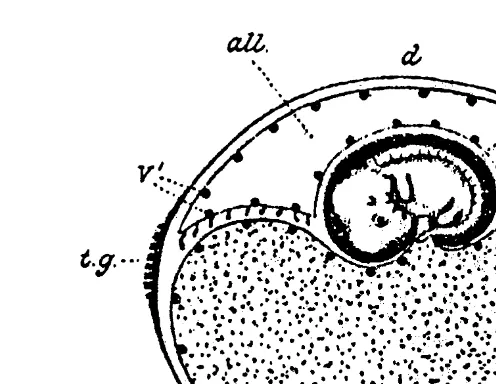
Fig. 1. Drawing of equine conceptus, 5 weeks, from Ewart, 1897. all, allantois; am amnion; cv, chorionic villi; tg chorionic girdle; v, vessels; st, sinus terminalis; ys, yolk sac; a, b, and c, absorbing area.
Ž .
conceptuses prior to day 16 or 17 Ginther, 1993; McDowell et al., 1998 . Perhaps it is the spherical nature of the equine conceptus that necessitates the development of a tough outer shell or acellular layer called the capsule. Again, the function is not well understood, but the capsule which surrounds the equine conceptus is a mucin-like network of glycoprotein which appears to originate largely from the underlying
trophec-Ž . Ž .
toderm Betteridge, 1989 . Readers are referred to Betteridge 2000 for more details on capsule formation and function. The argument could be made that the capsule provides a rigid structure for the equine conceptus, which enables it to shuttle back and forth in the uterine lumen in response to uterine contractions. Changes in the complex carbohydrate makeup of the capsule with stage of pregnancy could also reflect tissue remodeling
Ž
processes which permit fixation or other maternalrconceptus interactions Oriol et al., .
1993 . Likewise, presence of the capsule has been cited as a biological shield against Ž .
bacterial andror viral attack. Whatever the function s of this enigmatic structure, it is a familiar landmark to those who study this fascinating portion of equine pregnancy.
2.2. ConÕersion from chorioÕitelline to chorioallantoic placenta
to invade the maternal endometrium between days 36 and 38 where they phagocytize maternal epithelial cells. Then, after the phagocytic stage, the chorionic girdle cells continue to migrate, penetrating the basement membrane of the maternal epithelial cells to the stroma, where they locate between the uterine glands. Here they differentiate and hypertrophy to become mature endometrial cups, large raised structures that protrude above the uterine endometrial surface. They often form a concave or depressed surface that resulted in their description as ‘‘cups’’.
The mechanisms involved in formation of the chorionic girdle is a fascinating, as well as mysterious, aspect of equine early pregnancy. The proliferation of trophoblast cells over the exocoelom between the advancing allantois and the receding yolk sac
Ž .
suggests the action of a locally produced mitogenic stimulus. Stewart et al. 1995
Ž .
reported that hepatocyte growth factor–scatter factor HGF–SF was localized by in situ hybridization in the allantoic mesoderm, and its receptor, the proto-oncogene c-met was localized in invasive trophoblast of the chorionic girdle. These authors interpreted their findings to indicate that mesoderm-derived HGF–SF could play an important role in proliferation of the chorionic girdle cells. Because of the fact that the chorionic girdle is close to, but not fused with, the underlying allantoic mesoderm, the effect of a mitogenic stimulus would be to stimulate cell proliferation in an area that becomes somewhat
Ž
restricted, and the cells appear to ‘‘pile up’’ rather than expand linearly Stewart et al., .
1995 .
One of the primary functions of the endometrial cups is the production of equine
Ž .
chorionic gonadotropin eCG , although the primary purpose for this unique, highly glycosylated gonadotropin remains controversial. It is relatively clear that the secretion of eCG coincides temporally with the formation of multiple accessory corpora lutea, but is perhaps not so clear whether this is an essential event for maintenance of equine pregnancy. Likewise, it has been suggested that the secretion of eCG is timed so as to play a role in blunting or thwarting a maternal immune response to the interdigitation of chorionic villi with formation of the more advanced chorioallantoic placenta around day
Ž .
60 or so Samuel et al., 1974 . Both of these proposed functions have some supportive evidence, but neither has been proven beyond doubt.
2.3. Formation of microcotyledonary placenta
Ž
Between days 50 and 60, chorioallantoic macrovilli rudimentary irregularities or .
undulations begin to elongate and transform into the microcotyledons that constitute the
Ž .
major nutrient exchange mechanisms of the placenta Ginther, 1993 . Some maternal gaseous exchange apparently begins to take place. Vascularity of both maternal and fetal tissues increases, and the epithelium of maternal crypts becomes reduced. By day 100 or so, the structures begin to have a distinct cotyledon-like appearance, albeit in miniature.
2.4. Summary
Ž .
Ewart 1897 saw the changes in equine placentation as analogous to some of the
Ž .
was an atavistic remnant of the marsupial reproductive strategy in which migration out of the uterus was necessary to achieve the next level of nutrition for the embryo. As quaint as that sounds, it may provide focus on the dilemma of the developing equine embryo to maintain a nutritive supply. While we may not understand the evolutionary forces that created the peculiarities of the early equine placenta, it seems reasonable that the resulting structures, yolk sac placenta, capsule, relatively late forming chorioallantoic placenta, chorionic girdle, and the very late forming microcotyledonary placenta, reflect changes in the embryo’s efforts to obtain sufficient nutrition for growth and develop-ment.
3. Chorionic girdle as a luteotropin
The temporal association between eCG and formation of the accessory corpora lutea
Ž .
has been known for a considerable time Cole and Hart, 1930; Cole et al., 1931 .
Ž .
Furthermore, it is now widely accepted Ginther, 1993; Murphy and Martinuk, 1991 that eCG is indeed lueotropic; it has been demonstrated to stimulate progesterone
Ž .
production from luteal tissue in vitro Squires et al., 1974 , including tissue from the primary CL and the accessory CL that form later in pregnancy. Furthermore, Daels et al. Ž1998 have demonstrated that eCG significantly increased not only progestin secretion,.
Ž .
but estrogen secretion as well. Albrecht and Daels 1997 reported that this increase in estrogen secretion is associated with changes in expression of key steroidogenic
Ž .
enzymes as measured by immunolocalization. Albrecht and Daels 1997 stated that the
Ž .
intensity of immunolocalized staining for P450 17a-hydroxylase 17a-OH increased
after the onset of eCG, whereas the intensity of immunolocalization of P450 aromatase ŽAROM increased during pregnancy before the onset of eCG secretion and diminished. after the onset of eCG secretion. Thus the above certainly provide compelling evidence for a luteotropic function of eCG, but do not imply that such a luteotropic event is necessary for maintenance of pregnancy. On the other hand, the report of Hinrichs et al. Ž1985 that ovariectomized mares could serve as embryo transfer recipients if provided a. continuous source of progesterone argues against the need for a further boost in circulating progesterone concentrations at the time of accessory CL formation. Similarly,
Ž . Ž .
4. Chorionic girdle as immunomodulator
The dilemma faced by female mammals during early pregnancy is that an antigeni-cally foreign fetus will form intimate connections with the maternal endometrium necessitating long periods of modulation of the immune system so as not to reject the fetus and placenta, but maintain some degree of immune defense. In mares, the
Ž .
relatively prolonged time before such intimate attachments are formed about 60 days is unusual, and the temporal coincidence of placental attachment with chorionic girdle cell invasion and eCG secretion may not be accidental. The chorionic girdle cells, in addition to secreting eCG, appear to be the major source of fetal major histocompatibility
Ž . Ž .
complex MHC antigens Antczak and Allen, 1989 . This expression of MHC antigen by trophoblast tissue is unusual but is further complicated by the observation that mature endometrial cups do not appear to express paternal MHC antigen in appreciable quantity ŽAntczak and Allen, 1989 . Furthermore, the endometrial cups are ultimately lost due to. maternal leucocytic invasion.
5. Embryo—maternal interactions
The early life of the equine conceptus is taken up with maternal interactions for several reasons. However, the many unusual features of equine pregnancy make these interactions novel and interesting and certainly worthy of considerable study. The conceptus plays a role in controlling its own intrauterine environment, both by signaling its presence and assuring maternal recognition of pregnancy, and by contributing directly to the steroid environment of the intrauterine lumen. The equine conceptus migrates throughout the uterus prior to pregnancy recognition, likely serving to signal its presence uniformly, and to garner uterine secretions from throughout the entire uterus. The architecture of the equine conceptus likely also plays an important role in its orientation long before proper conceptus–maternal interdigitation occurs. Invasion of the en-dometrium by cells of the chorionic girdle certainly provides a unique conceptus–mater-nal interaction that is, as stated above, poorly understood.
6. Transport through the oviduct
The equine conceptus is unusual in that transport of ova through the oviduct occurs
Ž .
only if it is fertilized. This concept was reported by van Niekirk and Gerneke 1966 ,
Ž .
and further expanded by Betteridge and Mitchell 1972 . The latter authors flushed oviducts of mares and recovered unfertilized ova. They reported that the number of unfertilized ova recovered was well correlated with the number of ovulations
interven-Ž .
More recently, it has been shown that conceptuses at the morula or early blastocyst stage
Ž .
secrete prostaglandin E2 PGE , a uterotonic substance, which could contribute to tubal transport though induction of oviductal contractions locally, or through relaxation of the
Ž .
isthmic musculature Weber et al. 1995 . Furthermore, local administration of PGE to the oviduct appears to hasten tubal transport, resulting in early arrival of fertilized ova to
Ž .
the uterus Robinson et al., 1998 . Some authors refer to this discriminatory transport of fertilized ova through the oviduct as a form of maternal pregnancy recognition. Indeed,
Ž .
it is, but the term ‘‘maternal recognition of pregnancy,’’ as applied by Short 1967 has been classically used to describe the conceptus signal which prevents loss of the maternal corpus luteum at about 14 days after ovulation. Therefore, this author will retain the expression ‘‘maternal recognition of pregnancy’’ as a definition of luteal maintenance, not tubal transport.
7. Intrauterine environment
The early equine conceptus relies on its choriovitelline placenta to supply nourish-ment from uterine secretions for perhaps the first three to four weeks of pregnancy, if not beyond. That suggests that the uterine environment is extremely important to the developing equine conceptus, and merits discussion. It is clear that progesterone is essential to provide the appropriate intrauterine environment for conceptus development. It is also clear that the mare starts to prepare for a potential pregnancy with each and every luteal phase, regardless of whether or not she has been bred. Apparently it is a more acceptable trade-off, from the evolutionary standpoint, to prepare for a pregnancy that may not occur than to fail to prepare for one that does. The reproductive strategy of the mare is to make that decision at the time of maternal recognition of pregnancy.
Comparison of total intrauterine protein, as well as specific intrauterine proteins indicates the parallel production of such proteins up to the final day for maternal pregnancy recognition. Fig. 2 demonstrates the increase in a specific intrauterine protein, uteroferrin, in nonpregnant and pregnant mares. The increases in uterine luminal uteroferrin are not different in nonpregnant or pregnant mares until after day 14, the final deadline day for pregnancy recognition. By day 16, however, the differences
Ž .
between the two statuses are significant Zavy et al., 1982 . It seems reasonable to speculate that the differences in this one representative protein, uteroferrin, may reflect life or death differences as far as the conceptus is concerned.
The requirement for progesterone to drive uterine secretions has been shown in a variety of ways. For one, ovariectomized mares can be made suitable for pregnancy by
Ž .
simply administering progesterone, as demonstrated by Hinrichs et al. 1985 . For another, removal of progesterone from pregnant mares by surgical ablation of the corpus luteum, or by administration of prostaglandin, leads to pregnancy loss. However, reintroduction of progesterone can salvage the pregnancy. Interestingly, reintroduction of progesterone has a deadline as well. In experiments in which corpora lutea were
Ž regressed by administration of PGF on day 14, and progesterone administration 150
. Ž .
Ž .
Fig. 2. Changes in intrauterine luminal uteroferrin acid phosphatase in nonpregnant and pregnant mares.
Ž . Ž .
embryos survived Sharp, 1995 . A similar observation was reported by Ginther 1985 . One wonders if the few days of grace during which a lack of progesterone can be tolerated reflect a role for yolk sac nutrient stores.
8. Embryo mobility
It is now well accepted that the trans-uterine mobility of the equine conceptus is an
Ž .
important feature of the pregnancy recognition process. McDowell et al. 1998 demon-strated that restricting the trans-uterine mobility of equine conceptuses resulted in failure of the maternal recognition of pregnancy. In retrospect, the trans-uterine migration of equine conceptuses makes sense in light of the observations that uterine endometrium is
Ž .
capable of secreting PGF in vitro Vernon et al., 1981 , yet coincubation of conceptus membranes with endometrium from pregnant mares results in reduction in PGF
secre-Ž .
tion in vitro Berglund et al., 1982; Sharp and McDowell, 1985 This suggests that the PGF-inhibitory effect of the equine conceptus may be somewhat transient, and the trans-uterine mobility of the conceptus permits frequent, or intermittent interaction to reduce PGF secretion.
9. Uncoupling the oxytocinrrrrrPGF cascade
Ž .
Fig. 3. Endometrial prostaglandin synthesis inhibitory activity expressed as arbitrary inhibitory units in microsomal and cytosolic fractions of equine endometrium from day 16 pregnant and nonpregnant mares.
Fig. 4. Schematic representation of potential mechanisms of maternal recognition of pregnancy in mares.
Ž . Ž . Ž .
Oxytocin OT binds to its receptors OTR initiating the arachidonic acid AA cascade and production of
Ž .
prostaglandin F2a PGF . In pregnant mares, the oxytocin receptor numbers are reduced as is apparent affinity
Žrepresented by smaller font , leading to potentially reduced signal to arachidonic acid cascade. In addition, an.
Ž .
PGF. This has been demonstrated by administration of oxytocin to nonpregnant and Ž pregnant mares, with the resultant blockade of PGF release in pregnant mares Goff et
.
al., 1987 . Likewise, stimulation of the cervix of nonpregnant and pregnant mares results in oxytocin release in mares of both statuses, but PGF release, measured as PGF
Ž . Ž
metabolite PGFM occurred only in nonpregnant mares Sharp et al., 1997; Betteridge .
et al., 1985 . Furthermore, the latter workers demonstrated a reduction in oxytocin
Ž .
receptors in pregnant mares. Thatcher 1995 reported presence of an endometrial
Ž .
prostaglandin synthesis inhibitor EPSI isolated from bovine endometrium. There is
Ž .
also evidence that mares may express an EPSI as well. Watson and Sertich 1989 first demonstrated the presence of a conceptus-induced, endometrium-produced inhibitor of PGF. We have also observed such activity in the microsomal fraction of pregnant mares,
Ž .
although the chemical structure of the equine EPSI is yet to be determined see Fig. 3 . Interestingly, the discovery of an equine endometrial prostaglandin inhibitor begs the question of what it is about the presence of a conceptus that permits or stimulates its expression. It would seem that further work is required to understand the maternalrcon-ceptus interaction that permits continuation of pregnancy in mares. Fig. 4 indicates a current theory of maternal recognition of pregnancy in which occupancy of the endometrial oxytocin receptors activates the arachidonic acid cascade and prostaglandin
Ž . Ž
F2a PGF production. In pregnancy, reduced OT receptor number and affinity Sharp
.
et al., 1997 may reduce activation of the arachidonic acid cascade. Additionally, the
Ž .
pregnant mare endometrium contains a prostaglandin synthesis inhibitor EPSI which produces a potent blockade to the conversion of arachidonic acid to PGF. Likely, these two mechanisms work together to assure luteal maintenance.
References
Albrecht, B.A., Daels, P.F., 1997. Immunolocalization of 3 beta-hydroxysteroid dehydrogenase, cytochrome P450 17 alpha-hydroxylaser17,20 lyase and cytochrome P450 aromatase in the equine corpus luteum of dieoestrus and early pregnancy. J. Reprod. Fertil. 111, 127–133.
Antczak, D.F., Allen, W.R., 1989. Maternal immunological recognition of pregnancy in equids. J. Reprod.
Ž .
Fertil. Suppl. 37 , 69–78.
Berglund, L.A., Sharp, D.C., Vernon, M.W., Thatcher, W.W., 1982. Effect of pregnancy status and collection
Ž .
technique on prostaglandin F in the uterine lumen of pony mares. J. Reprod. Fertil. Suppl. 32 , 335–341. Betteridge, K.J., 1989. The structure and function of the equine capsule in relation to embryo manipulation and
Ž .
transfer. Equine Vet. J. Suppl. 8 , 92–100.
Betteridge, K.J., Mitchell, D., 1972. Retention of ova by the fallopian tube in mares. J. Reprod. Fertil. 31, 515. Betteridge, K.J., Eaglesome, M.D., Mitchell, D., 1979. Embryo transport through the Mre’s oviduct depends
Ž .
upon cleavage and is independent of the ipsilateral corpus luteum. J. Reprod. Fertil. Suppl. 27 , 387–394. Betteridge, K.J., Renard, A., Goff, A.K., 1985. Uterine prostaglandin release relative to embryo collection,
Ž .
transfer procedures and maintenance of the corpus luteum. Equine Vet. J. Suppl. 3 , 25–34.
Cole, H.H., Hart, G.H., 1930. The potency of blood serum of mares in progressive stages of pregnancy in effecting the sexual maturity of the immature rat. Am. J. Physiol. 93, 57.
Cole, H.H., Howell, C.E., Hart, G.H., 1931. Changes occurring in the ovary of the mare during pregnancy. Anat. Rec. 49, 199–210.
Daels, P.F., Albrecht, B.A., Mohammed, H.O., 1998. Equine chorionic gonadotropin regulates steroidogenesis in pregnant mares. Biol. Reprod. 59, 1062–1068.
Ginther, O.J., 1985. Embryonic loss in mares: nature of loss after experimental induction by ovariectomy or prostaglandin F2a. Theriogenology 24, 87–98.
Ginther, O.J., 1993. Reproductive Biology of the Mare. Equiservices, Cross Plains, WI.
Goff, A.K., Pontbriand, D., Sirois, J., 1987. Oxytocin stimulation of plasma 15 keto-13,13 dihydro
prosta-Ž .
glandin F2 alpha during the oestrous cycle and early pregnancy in the mare. J. Reprod. Fertil. Suppl. 35 , 253–260.
Hinrichs, K., Sertich, P.L., Cummings, M.R., Kenney, R.M., 1985. Pregnancy in ovariectomized mares
Ž .
achieved by conceptus transfer: a preliminary study. Equine Vet. J. Suppl. 3 , 74–76.
McDowell, K.J., Sharp, D.C., Fazleabas, A.T., Roberts, R.M., 1990. Synthesis and release of proteins by endometrium from pregnant and nonpregnant mares, and by conceptus membranes: characterization by two-dimensional gel electrophoresis. J. Reprod. Fertil. 89, 107–115.
McDowell, K.J., Sharp, D.C., Grubaugh, W.R., Thatcher, W.W., Wilcox, C.J., 1998. Restricted conceptus mobility results in failure of pregnancy maintenance. Biol. Reprod. 39, 340–348.
Murphy, B.D., Martinuk, S.D., 1991. Equine chorionic gonadotropin. Endocr. Rev. 12, 27–44.
Oriol, J.G., Sharom, F.J., Betteridge, K.J., 1993. Developmentally regulated changes in the glycoproteins of the equine embryonic capsule. J. Reprod. Fertil. 99, 653–664.
Robinson, S.J., Neal, H., Allen, W.R., 1998. Modulation of oviductal transport in the mare by local application of prostaglandin E2. In: Proc. 7th Int. Symp. Eq. Reprod., Pretoria, South Africa. .
Samuel, C.A., Allen, W.R., Steven, D.H., 1974. Studies on the equine placenta: I. Development of the microcotyledons. J. Reprod. Fertil. 41, 441–445.
Sharp, D.C., 1995. Unpublished observations.
Sharp, D.C., McDowell, K.J., 1985. Critical events surrounding the maternal recognition of pregnancy in
Ž .
mares. Equine Vet. J. Suppl. 3 , 19–22.
Sharp, D.C., Thatcher, M.-J., Salute, M.E., Fuchs, A.-R., 1997. Relationship between endometrial oxytocin receptors and oxytocin-induced prostaglandin F2a release during the oestrous cycle and early pregnancy
in pony mares. J. Reprod. Fertil. 109, 137–144.
Short, R.V., 1967. Reproduction. Annu. Rev. Physiol. 29, 373–400.
Squires, E.L., McKinnon, A.O., 1987. Hormone therapy for control of reproduction in mares and stallions. Vet. Clin. North Am.: Equine Pract. 3, 81–99.
Squires, E.L., Douglas, R.H., Steffenhagen, W.P., Ginther, O.J., 1974. Ovarian changes during the estrous cycle and pregnancy in mares. J. Anim. Sci. 38, 330–338.
Squires, E.L., Shideler, R.K., Voss, J.K., Webel, S.K., 1983a. Clinical applications of progestins in mares. Compend.: Equine 5, S16–S22.
Squires, E.L., Voss, J.K., Shideler, R.K., 1983b. Use of altrenogest for the broodmare. Proc. Ann. Conv. Am. Assoc. Equine Pract. Las Vegas, 431–434.
Stewart, F., Lennard, S.N., Allen, W.R., 1995. Mechanisms controlling formation of the equine chorionic girdle. Biol. Reprod. Mono. 1, 151–159.
Ž .
Thatcher, W.W., 1995. Maternal recognition of pregnancy. J. Reprod. Fertil. Suppl. 49 , 15–28.
van Niekirk, C.H., Gerneke, W.H., 1966. Persistence and parthenogenetic cleavage of tubal ova in the are. Onderstepoort J. Vet. Res. 33, 195–232.
Vernon, M.W., Zavy, M.T., Asquith, R.L., Sharp, D.C., 1981. Prostaglandin F2a in the equine endometrium:
steroid modulation and production capacities during the estrous cycle and early pregnancy. Biol. Reprod. 25, 581–589.
Watson, E.D., Sertich, P.L., 1989. Prostaglandin production by horse embryos and the effect of co-culture of embryos with endometrium from pregnant mares. J. Reprod. Fertil. 87, 331–336.
Weber, J.A., Woods, G.L., Lichtenwalner, A.B., 1995. Relaxatory effect of prostaglandin E2 on circular smooth muscle isolated from the equine oviductal isthmus. Biol. Repro. Mono. 1, 125–130.