In Vitro
Propagation of
Anthurium adreanum
cv. Nitta through Organogenesis
Rizka Tamania Saptari*), Masna Maya Sinta and Asmini Budiani
Indonesian Research Institute for Biotechnology and Bioindustry Jl. Taman Kencana no.1 Bogor West Java Indonesia
*) Corresponding author E-mail: [email protected]
Received: December 8, 2015 /Accepted: July 12, 2016 ABSTRACT
Anthurium adreanum Lindl. cv. Nitta is an ornamental
plant for cut lower industries. In vitro propagation enables a large scale production of high quality seedlings of A. adreanum quickly. The aim of this
experiment was to overcome some problems in
the in vitro propagation of A. adreanum, such as
contamination, lack of plantlets vigor and low rate of
survival in the acclimatization. In vitro propagation of
A. adreanum was conducted via organogenesis from
leaf explant. Explant sterilization using desogerm,
antioxidant, alcohol, rifampicin, and NaOCl was the best because they reduced browning and contamination
until 0 %. Organogenesis of A. adreanum was
successfully initiated using MS medium added with
2,4-D and BAP at 1 mg L-1, which produced 74 shoots
per explant. Shoots were best maturated in MS medium with a half strength of macro minerals added with 1 mg L-1 paclobutrazol. Survival rate increased
by 7 and 14 days hardening treatment using double
layer medium in the light culture room. Survival rate
reached 89.3 % at 8 weeks after acclimatization. Keywords: Anthurium adreanum; hardening; orga
nogenesis; paclobutrazol; sterilization
INTRODUCTION
Anthurium adreanum Lindl. is an ornamental
plant from the family of Araceae. This plant has been developed commercially in tropical and subtropical
countries. This plant can grow on the temperature of 16
oC – 30 oC (Dufour & Guérin, 2003). Global market of
Anthurium cultivars are the second among all tropical
cut lower after orchids. Flowers of A. adreanum are
used as cut lower because they are suficiently thick and long lasting (Agampodi & Jayawardena, 2007).
A. adreanum has lowers in various sizes and colors
with long lower stalks. A. adreanum cv. Nitta is one of A. adreanum cultivars from Indonesia which has
quite wide bright red lowers. It has a large market in
Indonesia because of its bright color.
Anthurium adreanum is an evergreen
plant that can be propagated by seeds. However,
propagation by seeds produced a high variable
offspring and very slow (Vargas, Mejías, Oropeza, & de García, 2004). Seed maturation takes about
three years from the pollination. Propagation by conventional cutting also requires considerable
time. Therefore, to sufice the market demand of these cut lowers, it can be done by tissue culture or
called in vitro propagation. By in vitro propagation, a high quality and pathogen-free of A. adreanum
seedling can be produced in a large scale and with the same quality eficiently.
Some in vitro propagation methods of A.
adreanum had been reported successfully by using
explants from leaves (Atak & Çelik, 2009; Bejoy,
Sumitha, & Anish, 2008; Yu, Liu, L., Liu, J. X., &
Wang, 2009), petiole (Viégas et al., 2007), shoot tips (Dhananjaya & Sulladmath, 2006), and nodes de Lima et al. (2006). In vitro propagation of A.
adreanum was done via indirect organogenesis,
which is initiating shoots from callus. Steps in this
method are sterilization, callus induction, shoot initiation, multiplication, maturation, rooting and acclimatization.
Sterilization is the process to eliminate the contaminant from the explant. In tropical countries, contamination is a serious problem that often occurs on the in vitro propagation (Nurhaimi-Haris, Sumaryono, & Carron, 2009). Sterilization using a single agent mostly did not succeed. So in this
research, we conducted experiment on sterilization
steps using more than one sterilant agents to get the best procedure to the sterilization of A. adreanum
leaves.
Callus of A. adreanum can be induced by the addition of auxin on the medium, or combination of auxin and cytokinin. Medium for callus induction
of A. adreanum from leaf explant was already
well established based on many reports. Bejoy,
Sumitha, & Anish (2008) succeeded in inducing
Cite this as: Saptari, R. T., Sinta, M. M., & Budiani, A. (2017). In vitro propagation of Anthurium adreanum cv. Nitta through organogenesis. AGRIVITA Journal of Agricultural Science, 39(2), 192–200. http://doi.org/10.17503/agrivita.
v39i2.752
callus by 53 % from leaf explants using MS medium supplemented with BAP (6-benzylaminopurine)
at 1 mg L-1 and 2,4-D (2,4-dichlorophenoxy acetic
acid) at 0.5 mg L-1. Zhao, Guo, Wang, & Wen (2004) succeeded in inducing callus from leaf explants by
using MS medium supplemented with BAP at 1
mg L-1 and 2,4-D at 0.1 mg L-1. Atak & Çelik (2009)
also succeeded in inducing callus by 81.25 % from
leaf explants of “Arizona” and “Sumi” cultivars by using BAP at 1 mg L-1 and 2,4-D at 0.6 mg L-1 on
the MS basal medium with a half strength of macro
minerals.
Initiation and multiplication of A. adreanum
shoots can be done by using cytokinin or its
combination with auxin. Medium for shoot initiation
of A. adreanum was also already well established
based on many researches. It used hormones such
as BAP (Vargas, Mejías, Oropeza, & de García,
2004), Thidiazuron (Yu, Liu, L., Liu, J. X., & Wang,
2009), and Kinetin (Dhananjaya & Sulladmath, 2006). The multiplication rate of A. adreanum
shootswas very high. About 33-44 shoots could be
produced per explant (Atak & Çelik, 2009; Vargas, Mejías, Oropeza, & de García, 2004).
The problem is that because of the high
multiplication, the shoot vigor was poor. Vigorous
plantlets are needed in the acclimatization in order to survive. Plantlet vigor can be maintained by using minerals and hormones in the right concentration
(Hardarani, Purwito, & Sukma, 2012), modifying
culture system, or using retardants such as paclobutrazol or ancimidol (Sumaryono & Sinta,
2011). There were also some reports regarding
the effect of carbon and light intensity on the
growth of A. adreanum plantlets (Gu et al., 2012;
Stancato & Tucci, 2010). However, the information
regarding the role of mineral concentration in
culture medium to the plant growth is still limited. A plant needs enough minerals to grow well. Macro minerals are needed in high concentration, while micro minerals are needed in low concentration but must exists. Plants with minerals deiciency will show many symptoms, such as lack of growth, chlorosis, and susceptible to disease. However,
over concentration of minerals can be toxic to the
plant growth (Hodges & Constable, 2010). In this
experiment, to increase the vigor of A. adreanum
plantlets we used paclobutrazol added in the MS basal media with various concentrations of macro
minerals and studied its effect on the vigor of A.
adreanum plantlets.
Beside maintained plantlets vigor, acclimatization is also the important and critical step on the propagation of A. adreanum by tissue
culture. Acclimatization is a step when plantlets are
transferred from in vitro culture in the laboratory
to ex vitro environment in the greenhouse. In vitro
environment is very humid. In the acclimatization, plantlets must adapt and survive in the ex vitro
environment with a low humidity. Hardening treatment
is needed for plantlets before acclimatization to increase the survival rate. Hardening can be done by planting plantlets on the gas-permeable vessels
(da Silva, Nagae, & Tanaka, 2005) or incubate
the culture in the greenhouse (Cardoso, Rossi, Rosalem, & da Silva, 2013) in order the plantlets to
adapt with the environment in a lower humidity. In this experiment, we studied the effect of hardening
treatment in laboratory and in the greenhouse and the optimum periods to increase survival rate of A.
adreanum plantlets.
MATERIALS AND METHODS
Preparation and Sterilization
Experiment was conducted in Laboratory of
Cell Culture and Plant Micropropagation, Indonesian Research Institute for Biotechnology and Bioindustry.
Newly opened leaves with light green color were used as explants. Mother plants were planted in the
greenhouse to reduce contamination before using
as explant source. Explants were cut and sterilized with three methods, (1) rinsed with desogerm,
soaked in antioxidant solution for 2 hours, soaked in
50 % Benomil solution for 15 minutes, and soaked in 20 % of NaOCl solution for 15 minutes; (2) rinsed with desogerm, soaked in antioxidant solution for
2 hours, alcohol for 3 seconds, rifampicin solution for 30 minutes, and then 20 % of NaOCl solution
for 15 minutes; (3) rinsed with desogerm, soaked
in alcohol for 3 seconds, rifampicin solution for 30
minutes, and then 20 % of NaOCl solution for 15
minutes.
Antioxidant solution was made from ascorbic
acid (1 mg L-1) and citric acid (1 mg L-1). Each sterilization step from one sterilant to other sterilant
was followed by rinsing with sterile aquadest. Afterwards, the sterile explants were cut (0.5 cm x 0.5 cm), then inoculated at medium for callus induction on the petridish. Medium were sterilized after pH adjustment on autoclave 1 kg cm-3 for
20 minutes. Each petridish contain 5 pieces of
Callus Initiation and Shoot Regeneration
In this experiment, callus initiation and shoot
regeneration were only used single composition of
media based on the established method reported. A
callus initiation followed the method by Atak & Çelik (2009). Sterile explants were inoculated on the MS basal medium with a half strength of macro minerals enriched with 2,4-D (0.6 mg L-1); BAP (1 mg L-1);
thiamine-HCl (0.5 mg L-1); folic acid (0.5 mg L-1);
biotin (0.05 mg L-1); and sucrose (20 mg L-1). The
culture was incubated in the dark room until callus was formed. The time of callus formation and color of callus were observed.
Formed callus was subcultured to the medium with the same composition as previous, but with the less concentration of 2,4-D. Culture was incubated on the light room with 36watt luorescent tube on the temperature of 25 ± 1 oC, and 12 hours photoperiod
until shoot was formed on the callus propagule. Number of shoots per propagule (2 x 1 cm) was calculated using handy counter with a destructive
observation. It used 20 propagules as a replication.
For shoot multiplication, propagules were cut in size of 0.5 x 0.5 cm and subculture to medium with the same compositions as those for shoot initiation on the bottle with 30 ml volume of medium. Culture bottle was incubated in the light room and continuously sub-cultured every 6 weeks.
Maintaining Plantlets Vigor
Plantlets with a minimum height of 2 cm were
separated from the propagule and sub-cultured
to maturation medium. Five plantlets were sub-cultured to MS basal medium with 0.5; 1.0; 2.0; 4.0
time(s) concentration of macro minerals, enriched
with sucrose (30 g L-1), and paclobutrazol (0; 1 mg L-1). Each treatment has ive of replications.
Culture was incubated in the light room for 4 weeks.
Parameters of plantlet vigor such as height, number of node, stem diameter, number, color, and size of
leaves were calculated from mature plantlets after 4 weeks of incubation on the maturation medium. Plantlets height was measured from the basal stem to the shoot tip. Stem diameter was measured about 0.5 cm from the basal stem using calipers. Size of leaf was the length multiplied with the width.
Hardening
Mature plantlets were hardened on the MS medium with sucrose (30 g L-1), IBA (indole-3- butyric acid) (1 mg L-1), and active charcoal (4 g L-1). It used double layer medium (10 ml of solid medium on the
bottom, and 2.5 ml of liquid medium on the top) on the test tube, covered with two layers of plastic wrap. Culture was incubated in the light culture room and in the green house for 0; 4; 7; 14 days. Each treatment
has 28 of replications.
Acclimatization
Plantlets from hardening medium were removed from the test tube and were cleared with tap water. Plantlets, which have been cleaned, were then soaked in fungicide solution for 5 minutes. Afterwards, plantlets were planted on multitray with
a hole size of 3 cm x 3 cm containing soil, dung
manure, cocopeat (1:1:1). The seedlings were then placed in the greenhouse and covered with UV plastic lid for 4 weeks. After 4 weeks, the lid was opened gradually and seedlings were then grown in the nursery. Survival rate was observed by the
number of plantlets survived for each treatment.
Statistical Analysis
Completely randomized design was used for this experiment. Data were then subjected to analysis of variance (ANOVA), followed by Duncan Multiple Range Test (DMRT), which differences at
p ≤ 0.05 considered to be signiicant. Analysis was
conducted using SPSS Statistics software package.
RESULTS AND DISCUSSION
Sterilization
Sterilization process on the in vitro culture
of Anthurium adreanum cv. Nitta was determined
by sterilant agent, steps of sterilization, and the
explants. It used newly opened young leaves
instead of leaf buds to ensure that all sterilant could reach the entire surface of explants. Young leaves
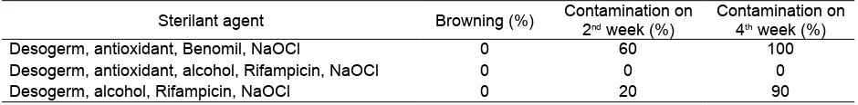
are susceptible to browning, so antioxidant solution was used on the sterilization to prevent browning. However, in this experiment browning did not occur whether antioxidant was used or not (Table 1).
Based on Table 1, the irst sterilization method
using desogerm, antioxidant, benlate fungicide and
NaOCl was not effective as 100 % contamination occurred after 4 weeks. However, the second method was the most effective for sterilization of A.
adreanum leaf explants as it reduced contamination
until 0 % after 2 weeks and 4 weeks. The second
method used desogerm, antioxidant, Rifampicin and 20 % NaOCl. The third method, although used Rifampicin, it could not reduce contamination. The
use of antioxidant with Rifampicin was effective for
Rifampicin is an antibiotic that is often used for sterilization on the in vitro culture (Eziashi et
al., 2014). Rifampicin was used for sterilization of
A. adreanum leaf explants to prevent contamination
from endogenous bacteria. Low concentration of antibiotics could prevent contamination without causing damage to explant cells. However, if it was used in high concentration more than 1 0.5
mg L-1 and long duration exposure, antibiotics
could inhibit the growth of explant cells. Therefore, in this experiment, explants were exposed on the rifampicin solution (0.5 mg L-1) only for 30 minutes.
Also, the use of antibiotics was better than a very
harmful sterilant agent such as HgCl2. da Silva,
Winarto, Dobránszki, & Zeng (2015) reported that
the sterilization of A. adreanum explants using 0.01
and 0.05 % HgCl2 could minimize contamination until
less than 10 %. However, because of its toxicity, it
needed to be extra careful using this sterilant agent. Therefore, the use of antibiotics instead of HgCl2
was safer in the explants sterilization.
Callus and Shoot Formation
Callus were formed after 8 weeks of incubation in the dark room. Callus were formed on the edge of
explants. Callus then transferred to the light culture
room soon proliferate into greenish propagule (Fig. 1a). Afterwards, shoots were formed from the propagule after 4 weeks (Fig. 1b). One hundred percent of propagule formed shoots. Shoot formation was induced
by a combination of auxin and cytokinin. Experiment
by Vargas, Mejías, Oropeza, & de García (2004) used
combination of NAA and BA could produce on average 44 shoots per explant of A. adreanum cv. Rubrun. While in this experiment, the combination of 2,4-D and BAP at 1 mg L-1 effectively stimulated shoot formation
up to on average 75 shoots per explant (2 x 1 cm) of
A. adreanum cv. Nitta (Fig. 1c). Experiment conducted
by Atak & Çelik (2009) reported that with the same
composition of medium for shoot formation, produced on average 34 shoots per explant of A. adreanum cv.
Arizona and 26 shoots per explant of A. adreanum
cv. Sumi. Thus, A. adreanum cv. Nitta produced twice
more shoots on the same medium composition. Table 1. Effect of sterilant agent on the sterilization of Anthurium adreanum cv. Nitta leaf explants
Sterilant agent Browning (%) Contamination on
2nd week (%)
Contamination on
4th week (%)
Desogerm, antioxidant, Benomil, NaOCl 0 60 100
Desogerm, antioxidant, alcohol, Rifampicin, NaOCl 0 0 0
Desogerm, alcohol, Rifampicin, NaOCl 0 20 90
Fig. 1. Organogenesis of A.adreanum cv. Nitta : Green callus (a), early shoot (b), shoot multiplication (c),
The shoots have poor vigor because of their
high rate of multiplication (Fig. 1d). The shoots have an average height of 1.69 cm, 0.07 cm of stem diameter, and 3 leaves with a size of 0.3 x 0.2 cm (0.06 cm2). That poor vigor affected survival rate in the acclimatization. Plantlet vigor related to
how well the plantlets grow in the culture medium and environment. Plantlets which were able to grow optimally in the in vitro culture, their organs also grew well such as having a larger stem diameter, more
nodes and relatively larger and greener leaves.
Good vigor was needed not only for acclimatization
purpose but also to keep in vitro culture stock remain sustainable. So, plantlet vigor of A. adreanum cv.
Nitta was maintained in this experiment by using
combination of macro minerals concentrations of the MS basal medium and paclobutrazol to increase plantlet vigor of A. adreanum cv. Nitta.
Plantlet Vigor
In this experiment, plantlet vigor was
maintained in the maturation medium using different concentrations of MS macro minerals and
paclobutrazol. MS basal medium with half (1/2x) concentration of macro minerals was used as control
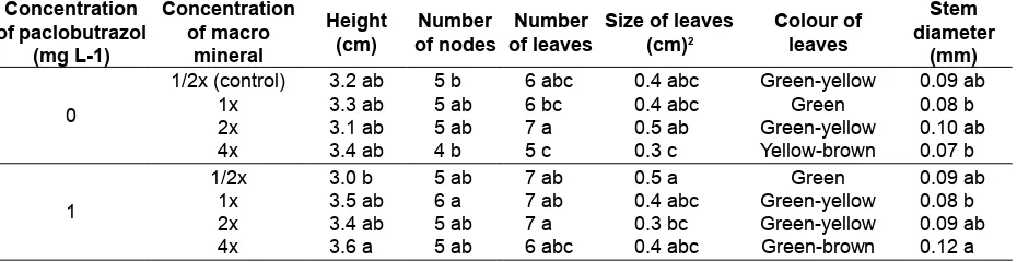
because it had been used for callus initiation until shoot formation. Concentration of macro minerals and paclobutrazol affected vigor of A.adreanum
plantlets. Plantlets which had a good vigor were
mainly indicated from the big green leaves and
wide stem. Paclobutrazol were used to inhibit stem
elongation to make plantlets more vigor. The use of paclobutrazol on the maturation medium only reduced plantlets height on the 1/2x concentration of MS macro minerals (Table 2).
The highest average number of nodes were grown on the full (1x) concentration of MS macro minerals with paclobutrazol. The highest average number of leaves were grown on the 1x concentration of MS macro minerals with or without paclobutrazol. The highest average size of leave was grown on the 1/2x concentration of MS macro minerals with
paclobutrazol, and 2x concentration of MS macro
minerals without paclobutrazol. However, on the
double (2x) concentration of MS macro minerals
without paclobutrazol, the leaves color was green to yellow, while the 1/2x concentration of MS macro minerals with paclobutrazol the leaves color was fresh green (Fig. 2). The yellow color of leaf could be the indication of leaf senescence, mineral deiciency,
or toxicity due to excess mineral on the plant.
The highest average diameters of stems
were grown on the 4x concentration of MS macro minerals with paclobutrazol. It has the highest
average height of plantlets and stem diameters, but
in the Fig. 2, showed that the leaves were mostly small and browning. Double concentration of MS macro minerals without paclobutrazol also increased
stem diameters, number and size of leaves, but
some leaves were yellow. Based on all parameters
measured, plantlets of A. adreanum cv. Nitta had
the best vigor when grown on the 1/2x concentration of MS macro minerals with 1 mg L-1 paclobutrazol.
Plantlets grown in this medium composition had the lowest plantlets height, but with relatively more
nodes, large stem diameters, highest number and size of leaves and greener.
Table 2. Effect of paclobutrazol and macromineral concentration on the Anthurium adreanum cv. Nitta
plantlets vigor after four weeks of culture
Concentration
1/2x (control) 3.2 ab 5 b 6 abc 0.4 abc Green-yellow 0.09 ab
1x 3.3 ab 5 ab 6 bc 0.4 abc Green 0.08 b
Half concentration of MS macro minerals
was an optimal concentration to maintain plantlet
vigor of A. adreanum cv. Nitta compared to
other concentrations. The result was similar to an experiment by Hardarani, Purwito, & Sukma
(2012) that plantlets of Hydrolea spinosa L.
planted on the MS medium with 1/2 concentration
of macro minerals have the highest biomass.
When paclobutrazol was supplemented to MS medium with 1/2 concentration of macro minerals,
it produced the best plantlet vigor of A. adreanum.
Paclobutrazol is a plant growth retardant which can cause dwarf in plants. Dwarf plants
usually have a better vigor than normal plants. Paclobutrazol inhibited biosynthesis of gibberellic acid (GA) that regulated stem elongation on plants
(Hedden & Graebe, 1985). The stem cannot
elongate by the inhibition of GA in its cells but it could be more vigor. Paclobutrazol has some
positive effects on plants. Nouriyani, Majidi, Seyyednejad, Siadat, & Naderi (2012) reported that application of paclobutrazol on the wheat increased
stem diameter, produced more green leaves by increasing the chlorophyll content. Hendrati (2008) also reported that in Eucalyptus, application of paclobbutrazol increased biomass and induced reproductive phase. The use of paclobutrazol on the in vitro culture to maintain plantlet vigor can increase survival rate on the acclimatization and keep sustainability of in vitro culture stock.
Survival Rate
Hardening on the double layer medium did
not have a signiicant effect on the survival rate of
A. adreanum cv. Nitta plantlets after four weeks
of acclimatization in the nursery (Fig. 3). Survival rate was about 75-100 %. After that, the survive seedlings from nursery were transferred to the greenhouse. On the 8 weeks after acclimatization, survival rate decreased for all treatments (Fig. 4). The lowest survival rate was from 4 days hardening
in nursery (32.2 %), in the culture room (42.9 %),
and 7 days hardening in nursery (46.5 %), while without hardening survival rate was 53.6 %. The highest survival rate was from 7 days hardening in
the culture room (89.3 %) and 14 days hardening in the culture room (82.1 %). Overall, hardening of
A. adreanum cv. Nitta plantlets in the culture room
produced higher survival rate than in the nursery. Hardening is a treatment to conditioning
in vitro planlets before acclimatization. In the in
vitro condition, sugar as an energy source for the
plant metabolism is already available in the culture medium. So that in vitro plants do not need to make sugar from CO2 and water to produce energy. Moreover, the environment of in vitro culture
has its temperature optimized for plant growth.
The microclimate on the vessels is very humid,
different from natural condition where the humidity is very luctuating. In the in vitro culture, plants do metabolism as heterotrophic organism. But, Fig. 2. Effect of paclobutrazol and macro mineral concentration on the A. adreanum plantlets vigor after 4
weeks of culture: without paclobutrazol (a) and with paclobutrazol (1 mg L-1) (b). Each picture, from
when in vitro plants or plantlets are acclimatizated to the ex vitro environment, they should start doing autotrophic metabolism and adapt to the
environment with high temperature and low
humidity that causes high evaporation. This different condition can make plantlets stressed and fail to survive on the nursery. Therefore, hardening can help plantlets to begin the adaptation to the ex vitro
environment. In this experiment, plastic wrap was
used as culture closures. Based on the experiment by Sinta, Riyadi, & Sumaryono (2011), the use of
plastic wrap as culture closures provided the best
aeration in the in vitro culture of oil palm. It made gas
exchange from the inside to the outside of culture vessels to be more intense, so that the moisture
could be reduced. However, to compensate with the high evaporation, double layer medium was used for hardening (Fig. 1e) so that plantlets were not
completely dried. Seven days of hardening in the
culture room was the best treatment to increase
survival rate of A. adreanum cv. Nitta plantlets. A longer time of hardening decreased survival rate
due to higher evaporation rate. Plantlets that were not completely adapted, experienced a water loss, some dropped their leaves, which caused a low
survival rate on the acclimatization.
Remarks: *) Same letters indicates no signiicant differences according to Duncan’s multiple range test (P = 0.05) Fig. 3. Survival rate of A.adreanum plantlets after four weeks of acclimatization
Fig. 4. Survival rate of A.adreanum plantlets after eight weeks of acclimatization
Remarks: *) Same letters indicates no signiicant differences according to Duncan’s multiple range test (P = 0.05) Without
hardening (control)
4 days 7 days 14 days
Hardening treatment
Culture room Nursery
Su
rv
iv
a
l
ra
te
(%
)
Without hardening
(control)
4 days 7 days 14 days
Culture room Nursery
Hardening treatment
Su
rv
iv
a
l
ra
te
(%
CONCLUSION
From the experiment it can be concluded that
organogenesis of A. adreanum cv. Nitta could be induced from leaf explant in the MS medium added
with 2,4-D and BAP (1 mg L-1); contamination of
explants could be reduced to 0 % with gradual
sterilization using desogerm, antioxidant, Benomil
and NaOCl; MS medium with half concentration of macro mineral added with 1 mg L-1 paclobutrazol produced the most vigorous A. adreanum plantlets; and survival rate of A. adreanum plantlets could be
increased by 7 and 14 days hardening treatment in
the culture room.
ACKNOWLEDGEMENT
This research was fund by PT. Riset
Perkebunan Nusantara in the Revitalization: Quick Yielding Research Program.
REFERENCES
Agampodi, V. A., & Jayawardena, B. M. (2007). Effect of coconut water in extending the vase life of Anthurium cut lower variety wild pink.
Tropical Agricultural Research, 19, 202-209.
Retrieved from http://dl.nsf.ac.lk/bitstream/
h a n d l e / 1 / 1 2 2 0 5 / P G I ATA R - 1 9 - 2 0 2 . pdf?sequence=2&isAllowed=y
Atak, Ç., & Çelik, Ö. (2009). Micropropagation of
Anthurium andraeanum from leaf explants.
Pakistan Journal of Botany, 41(3), 1155–
1161. Retrieved from http://www.pakbs.org/ pjbot/PDFs/41(3)/PJB41(3)1155.pdf
Bejoy, M., Sumitha, V. R., & Anish, N. P. (2008). Foliar
regeneration in Anthurium andraeanum
Hort. cv. Agnihothri. Biotechnology, 7(1), 134–138. http://doi.org/10.3923/biotech.20 08.134.138
Cardoso, J. C., Rossi, M. L., Rosalem, I. B., & da Silva, J. A. T. (2013). Pre-acclimatization in the greenhouse: An alternative to optimizing the micropropagation of gerbera. Scientia Horticulturae, 164, 616–624. http://doi.org/
10.1016/j.scienta.2013.10.022
da Silva, J. A. T., Nagae, S., & Tanaka, M.
(2005). Effect of physical factors on micro
propagation of Anthurium andreanum.
Plant Tissue Culture, 15(1), 1-6. Retrieved
from https://www.researchgate.net/publica
tion/280521670_Effect_of_Physical_ Factors_on_Micropropagation_of_
Anthurium_andreanum
da Silva, J. A. T., Winarto, B., Dobránszki, J., &
Zeng, S. (2015). Disinfection procedures
for in vitro propagation of Anthurium. Folia Horticulturae, 27(1), 3–14. http://doi.org/
10.1515/fhort-2015-0009
de Lima, F. C., Ulisses, C., Camara, T. R., Cavalcante, U. M. T., Albuquerque, C. C., & Willadino, L. (2006). Anthurium andraeanum Lindl. cv. Eidibel in vitro rooting and acclimatization
with arbuscular mycorrhizal fungi. Revista
Brasileira de Ciências Agrária, 1(1), 13-16.
Retrieved from http://www.redalyc.org/pdf/ 1190/119018241003.pdf
Dhananjaya, M. V., & Sulladmath, V. V. (2006). Rapid and eficient clonal propagation
of Anthurium andreanum cv. Singapore
hybrid. Indian Journal of Horticulture, 63(1), 59-61. Retrieved from http://www.
indianjournals.com/ijor.ijor:ijh &volume =63&issue=1&article=015
Dufour, L., & Guérin, V. (2003). Growth, developmental features and lower
production of Anthurium andreanum Lind. in tropical conditions. Scientia Horticulturae,
98(1), 25–35.
http://doi.org/10.1016/S0304-4238(02)00196-6
Eziashi, E. I., Asemota, O., Okwuagwu, C. O., Eke,
C. R., Chidi, N. I., & Oruade-Dimaro, E. A. (2014). Screening sterilizing agents and antibiotics for the elimination of bacterial contaminants from oil palm explants for plant tissue culture. European Journal
of Experimental Biology, 4(4), 111-115.
Retrieved from http://www.imedpub.com/ & Chen, J. (2012). Regeneration of
Anthurium andraeanum from leaf explants
and evaluation of microcutting rooting
and growth under different light qualities.
HortScience, 47(1), 88–92. Retrieved from
http://hortsci.ashspublications.org/content/
47/1/88.full
Hardarani, N., Purwito, A., & Sukma, D. (2012). Perbanyakan in vitro pada tanaman jeruju
(Hydrolea spinosa L.) dengan berbagai
micropropagation in jeruju (Hydrolea spinosa L.) with various of plant regulator
concentration]. Agroscientiae, 19(1), 6-10.
Retrieved from http://id.portalgaruda.f=
browse&mod=viewarticle&article=61083 Hedden, P., & Graebe, J. E. (1985). Inhibition of
gibberellin biosynthesis by paclobutrazol in cell-free homogenates of Cucurbita maxima
endosperm and Malus pumila embryos.
Journal of Plant Growth Regulation,
4(1), 111–122. http://doi.org/10.1007/BF
02266949
Hendrati, R. L. (2008). Pembungaan Eucalyptus
occidentalis pada perpanjangan masa
penyinaran dan paclobutrazol [Effect of extended light and paclobutrazol to
Eucalyptus occidentalis lowering]. Jurnal
Pemuliaan Tanaman Hutan, 2(3),
277-286. Retrieved from
http://ejournal.forda-mof.org/latihan/index.php/JPTH/article/
view/1785
Hodges, S. C., & Constable, G. (2010). Plant
responses to mineral deiciencies and toxicities. In J. McD. Stewart, D. M.
Oosterhuis, J. J. Heitholt, & J. R. Mauney (Eds.), Physiology of cotton (pp. 142-161).
Dordrecht, NL: Springer. http://doi.org/10.
1007/978-90-481-3195-2_14
Nouriyani, H., Majidi, E., Seyyednejad, S. M.,
Siadat, S. A., & Naderi, A. (2012). Effect of paclobutrazol under different levels of
nitrogen on some physiological traits of two wheat cultivars (Triticum aestivum L.). World
Applied Sciences Journal, 16(1), 01-06.
Retrieved from https://pdfs.semanticscholar.
org/8dae94be54aeb97d95b0e3528dab89.
Nurhaimi-Haris, Sumaryono, & Carron, M. P. (2009). Pengaruh bahan pra-sterilan, tutup tabung kultur, dan musim terhadap tingkat kontaminasi eksplan pada kultur microcutting karet [Effect of pre-sterilization agent, culture tube closure, and season on the contamination level of rubber microcutting culture]. Menara Perkebunan, 77(2), 89-99. Retrieved from http://mp. iribb
.org/index.php/mpjurnal/article/viewFile /96/75
Sinta, M. M., Riyadi, I., & Sumaryono. (2011).
Pengaruh jenis penutup botol kultur
terhadap pertumbuhan planlet kelapa
sawit (Elaeis guineensis Jacq.) [Effect of different culture vessel closures on the
growth of oil palm (Elaeis guineensis Jacq.) plantlets]. Menara Perkebunan, 79(1),
15-22. Retrieved from http://mp.iribb.org/index.
php/mpjurnal/article/view/68
Stancato, G. C., & Tucci, M. L. S. A. (2010). Monitoring the end of the in vitro phase
of Anthurium andreanum Lindl. plantlets.
Brazilian Journal of Plant Physiology, 22(1),
61-68. http://doi.org/10.1590/S1677-04202 010000100007
Sumaryono, & Sinta, M. M. (2011). Peningkatan
laju multiplikasi tunas dan keragaan planlet
Stevia rebaudiana pada kultur in vitro
[Increasing shoot multiplication rate and plantlet vigor of Stevia rebaudiana in vitro culture]. Menara Perkebunan, 79(2),
49-56. Retrieved from http://mp.iribb.org/index.
php/mpjurnal/article/view/59
Vargas, T. E., Mejías, A., Oropeza, M., & de García,
E. (2004). Plant regeneration of Anthurium
andreanum cv Rubrun. Electronic Journal
of Biotechnology, 7(3), 282–286. http://doi.
org/10.2225/vol7-issue3-fulltext-11
Viégas, J., da Rocha, M. T. R., Ferreira-Moura, I.,
da Rosa, D. L., de Souza, J. A., Corrêa, M.
G. S., & da Silva, J. A. T. (2007). Anthurium
andraeanum (Linden ex André) culture: In
vitro and ex vitro. Floriculture and Ornamental
Biotechnology, 1(1), 61-65. Retrieved from
http://www.globalsciencebooks.info/Online/ GSBOnline/images/0706/FOB_1(1)/FOB_ 1(1)61-65o.pdf
Yu, Y. X., Liu, L., Liu, J. X., & Wang, J. (2009). Plant regeneration by callus-mediated protocorm-like body induction of Anthurium
andraeanum Hort. Agricultural Sciences
in China, 8(5), 572–577. http://doi.
org/10.1016/S1671-2927(08)60248-5 Zhao, Y., Guo, W., Wang, G., & Wen, F. (2004).
Aseptic plantlet hardening of Anthurium andraeanum in vitro culture. Plant
Physiology Communications, 40(1),
48-50. Retrieved from http://europepmc.org/