Ultrastructure and morphology of
Chuaria circularis
(Walcott, 1899) Vidal and Ford (1985) from the
Neoproterozoic Visingso¨ Group, Sweden
Nina M. Talyzina *
Department of Earth Sciences,Historical Geology and Palaeontology,Uppsala Uni6ersity,Norby6a¨gen22, S-75236Uppsala,Sweden
Received 1 September 1999; accepted 11 January 2000
Abstract
The organic-walled microfossilChuaria circularis[Walcott, C.D., 1899. Precambrian fossiliferous formations. Geol. Soc. Am. Bull. 10, 199 – 244] Vidal and Ford [Vidal, G., Ford, T.D., 1985. Microbiotas from the Late Proterozoic Chuar Group (Northern Arizona) and Uinta Mountain Group (Utah) and their chronostratigraphic implications. Precambrian Res. 28, 349 – 389] from the Visingso¨ Group in Sweden have been re-examined using reflected and transmitted light, scanning electron and (SEM) transmission electron (TEM) microscopy. Specimens extracted from the rock matrix and those in situ, compressed on the bedding planes, were both studied. Besides previously described external wall surface, a psilate internal surface of Chuaria vesicle was observed. Transmission electron microscopy revealed a single-layered, electron-dense and homogeneous wall ultrastructure of the microfossil. Similar wall ultrastructure has been observed in other acid resistant microfossils from Lower Cambrian rocks. Various stages of the microfossils’ wall degradation were observed and are discussed in the paper. An overview of morphological interpretations and the history of research of C. circularis is presented. © 2000 Elsevier Science B.V. All rights reserved.
Keywords:Chuaria(acritarcha); Morphology; Ultrastructure; Neoproterozoic; Visingso¨ Group; Sweden
www.elsevier.com/locate/precamres
1. Introduction
The morphological and ultrastructural study of Chuaria circularis(Walcott, 1899) Vidal and Ford (1985) presented herein was carried out on abun-dant and well-preserved specimens from the
Vis-ingso¨ Group in Sweden. The species comprises
macroscopic organic-walled spheroid vesicles,
ranging between 70 and 3000 mm in diameter,
preserved as compressed carbonaceous discs on bedding planes. Though unnamed at the time, the microfossils from the Visingso¨ Group (Vidal, 1976, 1985), formerly referred to as ‘beds’, are one
of the first Chuaria specimens that have been
recorded and mentioned in geological literature * Fax: +46-18-471-2749.
E-mail address:[email protected] (N.M. Talyzina)
(Nathorst, 1879a,b; Linnarsson, 1880; Holm, 1885; Wiman, 1894). However, the microfossil was not described as a new species until the year
1899 when Walcott reported it as C. circularis,
being a ‘discinoid shell’ from the Late Precam-brian Chuar Group in the Grand Canyon, Ari-zona, USA (Walcott, 1899). In the perspective of the present knowledge of the Precambrian
macro-biotic records Walcott’s specimens of Chuaria
have been considered ‘the first true cellularly pre-served Precambrian organisms ever recorded’ (Shopf, 1999, p. 28). Similar independent discov-eries of unicellular microfossils from the Grand Canyon were made by White, cited by Powell (1876) (see also Ford and Breed, 1973; Sun, 1987), and almost simultaneously from the Visingso¨ shale of the same age by Swedish naturalist Nathorst (Nathorst, 1879a,b). Since then, the mi-crofossil has been frequently mentioned in the literature dealing with the Precambrian succes-sions of the southwest Sweden (Linnarsson, 1880; Nathorst, 1884, 1886, 1888, 1894; Holm, 1885; Wiman, 1894). Wiman (1894) was the first who illustrated the microfossil and recognized the acid-resistant nature of its vesicle referred to as ‘chitinous’.
The microfossils described a century ago as C.
circularis Walcott (1899) have been a subject of a continuous palaeontological debate concerning their systematic affiliation. Walcott considered them to be phosphatic shells of brachiopods (Wal-cott, 1899). Earlier, Holm (1885) wrote that the rounded objects from the Visingso¨ Beds look similar to either brachiopods from the genus Discina or plant remains of unknown affinity. In his opinion, circular folds indicated that the mi-croorganisms were globular objects flattened due to a compression. Wiman (1894) suggested that they might have been trilobite eggs considering their acid-resistant nature. Wenz (1938) attributed
rounded carbonaceous fossils to gastropods
whereas Brotzen (1941) consideredChuaria to be
a chitinous foraminiferan. Muir and Sarjeant
(1971) suggested that some Chuaria fossils may
have a close relationship with Tasmanites and
therefore probably belong to prasinophycean al-gae. Hofmann (1977, 1985) proposed that among other possible affinities this could be a medusoid
organism or photosynthesizing eukaryote of algal affinity. Sun (1987) classifiedChuariaas a colonial cyanobacterium.
At present,C.circularishas been placed among
acritarchs, an informal grouping of organic-walled microfossils mainly considered to be marine photoautotrophic plankters, but its closer biological affinity remains uncertain. During the last 20 years, two concepts concerning the
possi-ble affiliation of Chuaria have been discussed. In
the first concept it is assumed that the microfossil might be remains of eukaryotic organism, proba-bly of algal affinity (Tappan, 1980; Vidal and Knoll, 1983; Vidal, 1984). The other view is that this fossil could represent an external envelope of prokaryotic colonial cyanobacterium (Sun, 1987; Steiner, 1994, 1996). Jux (1977) reported the pres-ence of radial canals in the American specimens suggesting a prasinophycean algal affinity of Chuaria. Similar structures have been also ob-served by Amard (1992) in specimens from West Africa. However, the presence of pores had not been confirmed by Steiner (1994), who described a
fine-layered wall ultrastructure of Chuaria, and
compared it with the modern Nostoc
cyanobac-terium based on detailed light and electron mi-croscopy studies of its envelopes. The latter study
was mainly performed on the Chuaria specimens
from the Liulaobei Formation in China with a few additional scanning electron images of speci-mens from the Visingso¨ Group. However, as noted by Knoll (personal communication), the living nostocalean cyanobacteria do not show the tightly regular size frequency distribution as Chuaria and are absent in waters with full marine salinity.
The present paper presents analysis of the Chuaria microfossils from Sweden using reflected and transmitted light, scanning electron (SEM) and transmission electron (TEM) microscopy techniques in order to re-examine the monospe-cific genus from one of the classical localities.
2. Materials and methods
Fig. 1. The sketch-map of the Lake Va¨ttern area showing the locality studied. Modified from Vidal (1974).
occupied by the Lake Va¨ttern and they uncon-formably overlay the Proterozoic basement and are covered by the Quaternary deposits (Collini, 1951; Vidal, 1974, 1976, 1985). The contact with the Lower Palaeozoic sequence in the northwest-ern part of the Lake Va¨ttnorthwest-ern is not observed. The microfossils were collected from the locality Mull-skra¨den (Vidal, 1976) situated in the Omberg region (Fig. 1) on the eastern coast of the Lake Va¨ttern (Fig. 2) In this locality, the lithological succession is composed of grey to greenish silty shale in some places interbedded with thin arkosic, feldspathic sandy and silty layers. The samples have been collected from the silty grey – green shales. The detailed lithologic log of the Visingo¨ Group (previously called Beds) has been provided by Vidal (1976).
The Visingso¨ Group contains numerous or-ganic-walled microfossils described as acritarchs. Based on palynological analysis the pre-Varange-rian, late Riphean-early Vendian age for the Vis-ingso¨ sediments was assessed (Vidal, 1976, 1985)
The Chuaria specimens for transmitted light, SEM and TEM analyses were extracted from the rock matrix using HF and HCl acids and
filtra-tion in water using the 250 mm mesh filter. Then
the relatively big best-preserved Chuaria
speci-mens were picked up with a pipette. The isolated
specimens as well as those preserved in situ on the rock bedding planes were studied in reflected light under Wild Heerbrugg M 400 binocular
microscope. Permanent preparations for the
transmitted light microscopy were made using Held & Schyberg AB liquid glass. Specimens se-lected for SEM were mounted on a preparation stub and coated with a 22 nm layer of gold. Two specimens were sectioned for the TEM analysis. These specimens were dehydrated using ethanol and acetone and then embedded in a TAAB 812 Epoxy resin. The serial sections of 40 – 50 nm thick were cut perpendicularly to compressed surface of a specimen using a LKB Ultramicrotome system and placed on copper grids.
The studied preparations have a reference ab-breviation PMU-M-, followed by a number of a microscopic slide or a number of a stub for the scanning electron microscopy and are stored in the micropalaeontological collections of Palaeon-tologiska Museet, Uppsala. The cuper grids with fossil sections for TEM are kept at the
Depart-ment of Physiological Botany, Uppsala
University.
Observations in transmitted light were made under Leica DM IRBE microscope. SEM studies were carried out using Philips XL 30 microscope with PC computerized system attached and thin sections for TEM were examined using a trans-mission electron microscope Philips CM 10.
The colour of microfossils is red – brown, cor-responding to the thermal alteration index (TAI) 3 (Hayes et al., 1983) which means that the tem-perature affecting the sediment was approxi-mately 100 – 120°C.
The elemental analysis of C. circularis
(Wal-cott) Vidal and Ford was made using the EDAX energy dispersive system (EDS) attached to the SEM mentioned above.
3. Palaeontological descriptions
Group Acritarcha Evitt, 1963
Genus Chuaria Walcott, 1899
Chuaria circularis (Walcott, 1899) Vidal and Ford, 1985
Plates I, II and III
1894 Unnamed, Wiman, p. 109–113, pl. 5 (1–5).
1899 Chuaria circularis Walcott nov. g. and sp., p. 234–235, pl.27 (12–13).
1941 Chuaria wimani n. nom.; Brotzen, p. 258–259.
1966 Chuaria wimani Brotzen; Eisenack (1966), p. 52–55 (1–2).
1970 Kildinella magna Timofeev; Timofeev (1970), pl. 1 (A, B).
1973 Chuaria circularis Walcott; Ford and Breed, p. 539, pl. 61 (1–7), pl. 62 (1–6), pl. 63 (1–4).
1974 Chuaria circularis Walcott, 1899; Vidal, p. 6–8, pl. 1 (3–6).
1976 Chuaria circularis Walcott, 1899; Vidal, p. 18–19, pl. 8 (A–H).
1977 Chuaria circularis Walcott; Hofmann, p. 3–5, Fig. 2.
1977 Chuaria circularis Walcott; Ford and Breed (1977), p. 171–173, pl. 1 (1–6).
1979 Chuaria circularis Walcott, 1899; Vidal, p. 19–21, pl. 4 (a–b).
1981 Chuaria circularis Walcott, 1899; Vidal, p. 23–25, pl. 11 (J–K).
1985 Chuaria circularis Walcott emend. Vidal and Ford, p. 355–359, pl. 3 (A).
1987 Chuaria circularis Walcott; Sun, p. 115, pl. 1 (1–8), pl. 4 (1–2).
1990 Chuaria circularis (Walcott) Vidal (1990); Vidal, p. 488, Fig. 1.
1993 Chuaria circularis (Walcott) Vidal and Ford (1985); Vidal, Moczydlowska and Rudavskaya, p. 390–393, pl. 3 (A–D), pl. 4 (D).
1994 Leiosphaeridia wimanii (Brotzen, 1941), Butterfield, n. comb.; Butterfield, Knoll and Swett, p. 42–43, fig. 13 (D–F).
1994 Chuaria circularis Walcott 1899;
Butterfield, Knoll and Swett, p. 32–34, pl. 8 (G–H), pl. 13 (G–I).
1994 Chuaria circularis Walcott; Yin and Sun (1994), p. 99–100, pl. 4 (b).
Plate I. The reflected and transmitted light micrographs ofChuaria circularis (Walcott, 1899) Vidal and Ford (1985) from the Visingso¨ Group. (1 – 2) Reflected light micrographs. Sample PMU-V-Ch. (1) The abundant accumulation of specimens on the bedding plane. (2) Median rupture ofChuaria. Note large concentric folds in all threeChuariaspecimens. (3 – 6) Transmitted light micrographs. (3) Specimen PMU-M-2-E/44. (4) Specimen PMU-M-2-M/38/3. 5. Enlarged wall fragment of the specimen in 3 (see above). (6) Enlarged wall fragment of the specimen in 4 (see above).
1995 Chuaria circularis(Walcott, 1899) Vidal and Ford (1985); Hofmann and Rainbird (1995), p. 724–725, pl. 1 (1–6).
1997 Chuaria circularisWalcott, 1899, emend. Vidal and Ford (1985); Samuelsson, p. 173–174, pl. 7 (A, D).
Remarks. The genus Chuaria Walcott, 1899
includes a single species C. circularis (Walcott,
1899) Vidal and Ford (1985).
3.1. Emended diagnosis by Vidal and Ford (1985)
3.2. Description
The vesicle wall ofC.circularis (Walcott, 1899)
Vidal and Ford, 1985 is carbonaceus (Fig. 3). The external and internal surfaces of the vesicle wall are psilate or chagrinate (Plate I, 3 – 6) as observed in the transmitted light microscope. The wall is strongly folded due to compression (Plate I, 2 – 5; Plate II, 1 – 4 and Plate III, 3 – 4). Small irregular
folds are randomly distributed on the wall surface (Plate II, 3) and large concentric folds extend around the vesicle periphery (Plate I, 2; II, 2). Both kinds of folds are clearly recognized in transmitted light and SEM images. The holes within the fossil wall, which are observed in some specimens on parts of the vesicle (Plate II, 6 and Plate III, 1, 2) are caused by degradation. Median rupture of the vesicle is observed in some specimens (Plate I, 2).
Plate III. Transmission electron micrographs ofChuaria circularisfrom the Visingso¨ Group. (1 – 2) Wall fragments of the degraded, thin-walled specimen (1 is detail of Plate II, 6). Note the degradation holes inside the microfossil wall (a) and surface degradation (b). M-2-96. (3 – 6) Wall fragments of the well-preserved specimen. The specimen displayes some degradation of the wall surface (5, b; 7, b). Sample M-2-96.
The vesicle wall is single-layered. Under trans-mission electron microscope it appears to be elec-tron-dense and homogeneous (Plate III, 2 – 7). The cavities which are observed inside the wall of some specimens (Plate II, 6; Plate III, 1, 2) are caused by degradation (probably biodegradation) processes. The wall thickness varies from 0.5 to
2.5 mm in the degraded specimen (Plate II, 5, 6;
Plate III, 1, 2) and from 2.3 to 5.4 mm in the
could be compared to those of Tasmanites are observed. The diameter of the studied specimens
ranges 250 – 2000 mm.
3.3. Occurrence
The Chuar Group, Grand Canyon (Walcott, 1899; Ford and Breed, 1973; Vidal and Ford, 1985) and the Red Pine Formation, Uinta Moun-tains, USA (Hofmann, 1977); the Little Dal Group, Mackenzie Mountains, Canada (Hof-mann and Aitken, 1979; Hof(Hof-mann, 1985) and the Shaler Supergroup, Arctic Canada (Hofmann and Rainbird, 1995); the Eleonore Bay Group, East Greenland (Vidal, 1979) and the Thule Group, northern Greenland (Vidal and Dawes, 1980); the Rysso¨ and Hunneberg Formations, Svalbard (Knoll and Calder, 1983; Knoll, 1984); the Svan-bergfjellet Formation, Spitsbergen (Butterfield et al., 1994); the Visingso¨ Group, Sweden (Wiman, 1894; Vidal, 1976); the Vadsø, Tanafjord and Vestertana Groups, East Finnmark, (Vidal, 1981) and the Barents Sea Group, Varanger Peninsula,
Norway (Vidal and Siedlecka, 1983); the
Debengdin, Khajpakh, Maastakh and Khatyspyt Formations of Yakutia, eastern Siberia (Vidal et al., 1993); the Kildinskaya Group of the Kildin Island and the Sredni and Rybachi Peninsulas, and the Chapoma Formation, Kola Penisula, Russia (Samuelsson, 1977); the Liulaobei, Ji-uliqiao and Nanfen Formations, China (Duan, 1982; Sun, 1987; Steiner, 1994).
The relative age of all mentioned occurrences is
consistent with Early Neoproterozoic, late
Riphean, predating the Varangerian glacial event.
4. Discussion
The Chuaria specimens studied from the Vis-ingso¨ Group conform morphologically to the
di-agnosis ofC.circularisWalcott, 1899 emended by
Vidal and Ford (1985). The psilate external sur-face, and thick, solid and single-layered vesicle have been observed using different kinds of mi-croscopy. In addition to previously reported mor-phology of the external wall surface, the psilate or chagrinate internal surface is also observed. Some Chuaria vesicles from the Visingso¨ Group possess a median rupture (Plate I, 2). However, it is not clear whether this feature reflects an excystment structure or a rupture due to compaction.
The concentric wrinkles of the wall are abun-dant in the Visingso¨ specimens. This is probably
the most prominent feature of C. circularis that
has been noted in the first diagnosis by Walcott
(1899). Although very common in Chuaria, this
feature is certainly not unique for the genus. It is characteristic of thick-walled specimens of other taxa and probably indicates a thick vesicle wall. Similar kinds of folds have been observed in some
Precambrian Leiosphaeridia(Knoll, 1994, p. 6746,
Fig. 4, B), but this does not necessarily mean that these two genera are related.
The concentric folds and a large size of the vesicle have been treated as the main diagnostic
features of C. circularis by some researchers
(Duan, 1982; Butterfield et al., 1994). The reason is that although the folds are a secondary
tapho-nomic feature, they are very common in Chuaria
specimens (Butterfield et al., 1994) and, as men-Fig. 3. The elemental composition ofChuaria circularis
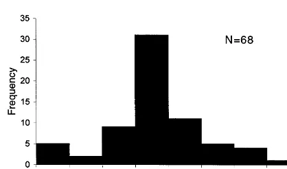
Fig. 4. Size distribution of Chuaria specimens, Visingso¨ Group, Sweden.
cies of the genus. The test on the wall resistance
is significant in the definition of C. circularis
and in contrary to suggestion of Duan (1982), it is more appropriate to exclude non-acid
resis-tant specimens from the genus of Chuaria.
A variation of transparency and the wall colour from light brown to dark brown was ob-served in the specimens studied. Apart from the thermal alteration index mentioned above, the intensity of colour depends on the wall thick-ness. Comparison of thin sections from one light brown and one dark brown specimen under TEM showed that the wall of the first specimen is thinner, and is penetrated by abundant sec-ondary degradational cavities. Furthermore, it has significantly distorted margins (Plate II, 5, 6; Plate III, 1, 2) resulting from a substantial degradation of the specimen. The dark brown specimen (Plate III, 3 – 7) is devoid of the degra-dational internal cavities and has better pre-served margins. Simple polygonal cavities, favus or compound faviform cavities and compound cribate cavities have been previously reported in Chuaria specimens from the Visingso¨ Group (Vi-dal, 1974) and explained as a result of a pyrite crystal growth caused by bacterial activity (re-ducing bacterial metabolism and precipitation of sulfides). Such a kind of degradation possibly caused the difference observed in the wall
thick-ness between C. circularis from the Grand
Canyon, USA and specimens from the Visingso¨
Group referred to as Chuaria wimani Brotzen
and revised as Leiosphaeridia wimanii (Brotzen,
1941) Butterfield, n. comb. (Butterfield et al., 1994).
No lamellar structure comparable to that re-ported by Amard (1992) in specimens from
West Africa has been observed in Chuaria from
the Visingso¨ Group. This structure could be taphonomic and caused by a degradation. Illus-trations of in situ specimens (Amard, 1992 on Plate I, 3, 4) show highly corroded surface of the fossil, with cracks following distortions in the rock matrix. The ‘radial canals’ first noted by Jux (1977) and illustrated by Amard (1992) are probably comparable to the cavities in the
wall of the Visingso¨ Chuaria, interpreted here as
biodegradational. tioned above, probably correspond to the wall
thickness and its flexibility. Arbitrarily chosen dimension limit alone has neither taxonomic nor systematic value. As noted by Vidal et al. (1993) ‘…From this lack of clearly diagnostic
charac-ters emanates the suspicion that C. circularis
may in fact be a taxonomic waste-basket con-taining true biogenic, as well as various non-bio-genic objects such as films of organic sapropel and carbonaceous intraclasts’. However, the
ar-tificial size limitation of Chuaria might be
con-venient, taken along with other features,
allowing distinction between specimens of the megascopic acritarch, which is abundant at a
certain level of Proterozoic, and numerous
leiosphaerids, which are widely distributed in successions throughout the geological time.
The distribution diagram of diameters of a
large in situ population of Chuaria (Fig. 4; Plate
I, 1) from the Visingso¨ Group basically corre-sponds to a standard size distribution for a taxon (see range from 0.5 to 2.0 mm). However, the presence of a mode at the range of 0.3 – 0.5 mm allows to suggest that the fossil probably represent a vegetative state which grows in the course of the life cycle of the organism. A
simi-lar polymodal size distribution of Chuaria
speci-mens from Uinta Mountain Group, USA is reported by Hofmann (1977, Fig. 3).
The Visingso¨ Group specimens have the vesi-cle wall resistant to HCl and HF. This property
of the acid-resistance of Chuaria wall was
spe-The SEM and TEM studies of specimens from the Liulaobei Formation in China (Steiner, 1994) did not reveal any canal structures. The reported ‘fine-layered’ ultrastructure in these specimens (Steiner, 1994, Plate 8, Fig. 2) was interpreted as a feature of an external envelope
of a colonial cyanobacterium Nostoc. At a
mag-nification of ×34 500 similar ultrastructure was
observed in the Visingso¨ Chuaria specimens.
However, this feature seems to be common for other acritarchs, which have homogeneous and electron-dense wall. A similar ultrastructure of
the wall has been observed in specimens of Tas
-manites tenellus Volkova from the Lower Cam-brian in Estonia (Talyzina and Moczydlowska, submitted, plate V, 5), the genus which is as-sumed to be a prasinophyte (Wall, 1962; Guy-Ohlson and Boalch, 1992). This makes the
association of Chuaria with the Nostoc-like
prokaryotes rather doubtful.
5. Conclusions
(1) C. circularis (Walcott, 1899) Vidal and Ford (1985) from the Visingso¨ Group possesses a single-layered and homogeneous vesicle wall. The external and internal wall surfaces are psi-late in well preserved specimens and may appear shagrinate due to degradational processes.
Nei-ther radial canals comparable to pores of Tas
-manites nor lamellar structure have been
observed. The latter observations are considered taphonomic.
(2) Morphological and ultrastructural features of C. circularis (Walcott, 1899) Vidal and Ford (1985) are insufficient for a firm biological clas-sification. Combination of morphological study with other methods may improve our
under-standing of the biological nature of this
presently enigmatic microfossil.
Acknowledgements
This study is a part of the Ph.D. project started under the late Professor G. Vidal, and later on supervised by Dr M.
Moczydlowska-Vi-dal, who are greatly acknowledged. The author thanks J. Johansson for a help in collecting the studied samples. A. Axen and G. Wife provided
technical expertise during the electron
mi-croscopy preparations and analysis. In addition to the doctoral scholarship from Uppsala Uni-versity, the work was supported by the research grant from the Swedish Natural Science Re-search Council (NFR) to M.
Moczydlowska-Vi-dal (G-AA/GU 09939-319). Reviews by Dr R.
Scherer, Professor A Knoll and Professor S. Golubic improved the report.
References
Amard, B., 1992. Ultrastructure of Chuaria (Walcott) Vidal and Ford (Acritarcha) from the Late Proterozoic Penjari Formation, Benin and Burkina-Faso, West Africa. Pre-cambrian Res. 57, 121 – 123.
Brotzen, F., 1941. Na˚gra bidrag till Visingso¨ formationens stratigrafi och tektonik. G.F.F. 63, 245 – 261.
Butterfield, N.J., Knoll, A.H., Swett, K., 1994. Paleobiology of Neoproterozoic Svanbergfjellet Formation, Spitsber-gen. Fossils and Strata, 34. 84 pp.
Collini, B., 1951. Visingso¨formationen. In: Geijer, P., Collini, B., Munthe, H., Sandgren, R. (Eds.), Beskrivning till kartbladet Gra¨nna. S.G.U., A, 193, pp. 27 – 37. Duan, C.-H., 1982. Late Precambrian algal megafossils:
Chuaria and Tawuia in some areas of eastern China. Alcheringa 6 (1 – 2), 57 – 68.
Eisenack, A., 1966. U8ber Chuaria wimani Brotzen. N. Jahrb. Geol. Pala¨ont. Monatsh. Stuttgart 1, 52 – 56.
Ford, T.D., Breed, W.J., 1973. The problematical Precam-brian fossilChuaria. Palaeontology 16, 535 – 550. Ford, T.D., Breed, W.J., 1977. Chuaria circularis Walcott
and other Precambrian fossils from the Grand Canyon. J. Palaeont. Soc. India 20, 170 – 177.
Guy-Ohlson, D., Boalch, G.T., 1992. Comparative morphol-ogy of the Genus Tasmanits (Pterospermales, Chloro-phyta). Phycologia 31, 523 – 528.
Hayes, J.M., Kaplan, I.R., Wedekine, K.M., 1983. Precam-brian organic geochemistry, preservation of the record. In: Schopf, J.W. (Ed.), Earth’s Earliest Biosphere, its Origin and Evolution. Prinston University Press, Lawrenceville, pp. 93 – 134.
Hofmann, H.J., 1977. The problematic fossil Chuaria from the Late Precambrian Uinta Mountain Group, Utah. Precambrian Res. 4, 1 – 11.
Hofmann, H.J., Aitken, J.D., 1979. Precambrian biota from the Little Dal Group, Mackenzie Mountains, northwest Canada. Can. J. Earth Sci. 16, 150 – 166.
Hofmann, H.J., Rainbird, R.H., 1995. Carbonaceous Megafossils from the Neoproterozoic Shaler Supergroup of Arctic Canada. Palaeontology 37 (4), 721 – 731. Holm, G., 1885. Om Vettern och Visingso¨formationen:
Bi-hang till Kungliga Svenska Vetenskapsakademiens Han-dlingar Bd. II (7), 1 – 49.
Jux, U.J., 1977. U8ber die wandstrukturen sphaeromorpher acritarchen: Tasmanites Newton, Tapajonites Sommar & Van Boekel, Chuaria Walcott. Palaeontographica Abt. B 160, 1 – 16.
Knoll, A.H., 1984. Microbiotas of the Late Precambrian Hunneberg Formation, Nordaustlandet, Svalbard. J. Pa-leontol. 58, 131 – 162.
Knoll, A.H., 1994. Proterozoic and early Cambrian protists. Evidence for accelerating evolutionary tempo. Proc. Natl. Sci. USA 91, 6743 – 6750.
Knoll, A.H., Calder, S., 1983. Microbiotas of the Late Pre-cambrian Rysso¨ Formation, Nordaustlandet, Svalbard. Palaeontology 26, 467 – 496.
Linnarsson, G., 1880. De a¨ldsta paleozoiska lagren i trakten kring Motala. G.F.F. 5, 23 – 30.
Muir, M.D., Sarjeant, W.A.S., 1971. An annotated bibliog-raphy of the Tasmanaceae and of related living forms (Algae: Pracinophyceae): in C.I.M.P. Microfossiles or-ganiques du Paleozoique, 3; les Acritarches (Se´cretaire de Re´daction: S. Jardine´): Editions du Centre National de la Recherche Scientifique, pp. 1 – 117.
Nathorst, A.C., 1879a. En egendomlig strukturvarietet af ler-haltig kalksten fra˚n Grennatrakten. G.F.F. 4, 216. Nathorst, A.C., 1879b. Om de aldre sandstens — och
skif-ferbildningarne vid Vettern. G.F.F. 4, 421 – 436.
Nathorst, A.C., 1884. Upplysningar till Geologisk O8fversigtskarta o¨fver Sverige. S.G.U. Ser Ba 4, 14. Nathorst, A.C., 1886. Na˚gra ord om Visingso¨serien. G.F.F.
8, h. 1.
Nathorst, A.C., 1888. In dem Jaresberichte der Universita¨t in Lund 1887 – 88, p. 27.
Nathorst, A.C., 1894. Jordens historia: 595. In: Wiman, C., 1894. Pala¨ontologiskche Notizen 1: ein Pra¨kambrisches Fossil. Bulletin of the Geological Institutions of the Uni-versity of Upsala, 2, 109 – 113.
Powell, J.W., 1876. Report on the geology of the eastern portion of the Uinta Mountains. U.S. Geol. Survey, p. 218.
Samuelsson, J., 1977. Biostratigraphy and Palaeobiology of Early Neoproterozoic Strata of Kola Peninsula, North-west Russia. Norsk Geologisk Tidsskrift 77, 1 – 28. Shopf, J.W., 1999. Cradle of Life. The Discovery of Earth’s
Earliest Fossils. Princeton University Press, Princeton, NJ, p. 367.
Steiner, M., 1994. Die Neoproterozoischen Megaalgen Siid-chinas. Berliner Geowissenschaftliche Abhandlungen (E9) 15, p. 146.
Steiner, M., 1996. Chuaria circularis Walcott 1899 — ‘Megasphaeromorph Acritarch’ of Prokaryotic Colony? In: Fatka, O., Servais, T. (Eds.), C.I.M.P. Acritarch in Praha. Acta Univ. Carolinae Geol. 40, 645 – 665. Sun, W., 1987. Palaeontology and biostratigraphy of Late
Precambrian macroscopic colonial algae:ChuariaWalcott and Tawuia Hofmann. Palaeontographica Abt. B 203, 109 – 134.
Talyzina, N.M., Moczydlowska, M., submitted. Morphologi-cal and Ultrastructural studies of some acritarchs from the Lower Cambrian Lu¨kati Formation, Estonia. Rev. Palaeobotany Palynol.
Tappan, H., 1980. The Paleobiology of Plant Protists. WH Freeman, San Francisco, p. 818.
Timofeev, B.V., 1970. Sphaeromorphida ge´ants dans le Pre´-cambrien avance´. Rev. Palaeobotany Palynol. 10, 157 – 160.
Vidal, G., 1974. Late Precambrian microfossils from the basal sandstone unit of the Visingso¨ Beds, South Swe-den. Geol. Palaeontol. 8, 1 – 14.
Vidal, G., 1976. Late Precambrian microfossils from the Vis-ingso¨ Beds in Southern Sweden. Fossils Strata 9, 57. Vidal, G., 1979. Acritarchs from the Upper Proterozoic and
Lower Cambrian of East Greenland. Grønlands Geolo-giske Undersøgelse Bull. 134, 55.
Vidal, G., 1981. Micropalaeontology and Biostratigraphy of the Upper Proterozoic and Lower Cambrian Sequence in East Finnmark, Northern Norway. Norges Geologiske Undersøkelse 362, 1 – 53.
Vidal, G., 1984. The Oldest Eukaryotic Cells. Sci. Am. 250 (2), 48 – 57.
Vidal, G., 1985. Prepaleozoisk sedimentberggrund. In: Persson, L., Bruun, A,., Vidal, G. (Eds.), Beskrivning till Berggrundskartan HJO SO.S.G.U., Ser. Af, 134, 77 – 91. Vidal, G., 1990. The Late Proterozoic acritarchChuaria cir
-cularis(Walcott). J. Palaeontol. 64, 488.
Vidal, G., Dawes, P.R., 1980. Acritarchs from the Protero-zoic Thule Group, North-West Greenland. Grønlands Geologiske Undersøgelse Rapport 1000, 24 – 29.
Vidal, G., Ford, T.D., 1985. Microbiotas from the Late Proterozoic Chuar Group (Northern Arizona) and Uinta Mountain Group (Utah) and their chronostratigraphic implications. Precambrian Res. 28, 349 – 389.
Vidal, G., Knoll, A.H., 1983. Proterozoic Plankton. Geol. Soc. Am. Memoir 161, 265 – 277.
Vidal, G., Siedlecka, A, 1983. Planktonic, Acid-resistant Mi-crofossils from the Upper Proterozoic Strata of the Barents Sea Region of Varanger Peninsula, East Finnmark, Northern Norway. Norges Geologiske Un-dersøkelse 382, 45 – 79.
Vidal, G., Moczydlowska, M., Rudavskaya, V.A., 1993. Biostratigraphic implications of a Chuaria–Tawuia as-semblage and associated acritarchs from the Neoprotero-zoic of Yakutia. Palaeontology 36 (2), 387 – 402. Walcott, C.D., 1899. Precambrian fossiliferous formations.
Geol. Soc. Am. Bull. 10, 199 – 244.
Wall, D., 1962. Evidence from recent plankton regarding the biological affinities of Tasmanites Newton 1875 and Leiosphaeridia Eisenack 1958. Geol. Mag. 99, 353 – 362. Wenz, W., 1938. Gastropoda. In: Schindewolf, D.H. (Ed.),
Handbuch der Pala¨ozoologie, vol. 6 (1). Borntraeger, Berlin, p. 240.
Wiman, C., 1894. Pala¨ontologiskche Notizen 1: ein Pra¨kam-brisches Fossil. Bull. Geol. Inst. Univ. Upsala 2, 109 – 113.
Yin, L., Sun, W., 1994. Microbiota from the Neoproterozoic Liulaobei Formation in the Huainan region, northern Anhui, China. Precambrian Res. 65, 95 – 114.