Radioprotective of Roselle Tea in Recurrent Radiodiagnostic
Ionizing Radiation on Peripheral Hemogram Characteristics
Mokhamad Fakhrul Ulum1*, Deni Noviana1, Sri Estuningsih2, Endah Mulia Ningsih3, Abas Kurniawan3, Andi Rahayu3, Ayip Fadil3, Erli Chandra3, Fitria Apriliani3, Hastin
Utami Damayantie3, Kurniawan Prasetya3
1
Division of Veterinary Surgery and Radiology, 2
Division of Veterinary Pathology, Department of Veterinary Clinic, Reproduction and Pathology, Faculty of Veterinary Medicine, Bogor Agricultural University (IPB)
3
Faculty of Veterinary Medicine, Bogor Agricultural University (IPB)
Address: Jalan Agatis, Faculty of Veterinary Medicine, Bogor Agricultural University (IPB), Dramaga, Bogor, INDONESIA 16680, www.bedahradiologi.fkh.ipb.ac.id
*Corresponding author: [email protected]
Abstract
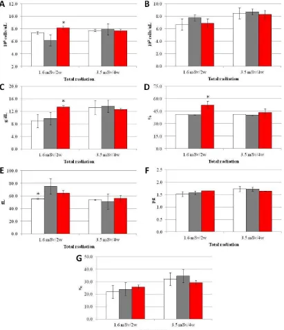
The aim of this study was to describe peripheral hemogram characteristics of mice treated with Roselle tea prior to exposure of recurrent radiodiagnostic ionizing radiation. Eighteen adult ddy male mice of Mus musculus were divided into three groups, i.e. control group (K, n=6), ionizing radiation group (P, n=6), and tea of roselle radiation group (RT, n=6). Blood samples were collected from blood venous at sinus retroorbitalis of anesthetized mice. These blood samples were examined for the numbers of leucocyte (WBC), erythrocyte (RBC), hemoglobin (Hb), hematocrit (PCV), RBC volume (MCV), Hb weight (MCH), and Hb concentration (MCHC). Blood samples were collected from mice which had been exposed of ionizing radiation for 2 weeks and 4 weeks periods. The result showed that there were no significant differences (P>0.05) on WBC, MCH, and MCHC. However, we found differences (P<0.05) on hematology characteristics for RBC, Hb, PCV, and MCV of mice after 2 weeks exposure of ionizing radiation.
Keywords: hematology, ionizing radiation, roselle tea.
INTRODUCTION
X-rays were first discovered on November 8, 1895 by the German physic-ist Wilhelm Conrad Roentgen. Soon afterward, x rays was used as a diagnostic tool. During its first 100 years of discovery, x-rays were widely used for the benefit of the medical picture of the disease (McCurnin & Bassert 2006). Nowadays, X-rays is commonly used to diagnose or as a component of therapy for a wide range of disease conditions. Radio-diagnostic were performed by imaging to diagnose degree of the disease such as the primary tumor, and subsequent metastasis can be visualized non-invasively by whole
body imaging through the patient (Bouvet
et al. 2002; Jeong et al. 2003). However, this procedures can develop to residual (or long-term) normal organ or tissue injury (Mauch et al. 1995). Ionizing radiation of X-ray is a hazard ionizing radiation. It causes damages on the exposured tissues (Thrall 2002). Low dose of ionizing radia-tion exposure from X-ray causes differences or destroys tissue (McCurnin & Bassert 2006).
110
of normal tissues to irradiation (Citrin et al. 2010). Many naturally occurring com-pounds found in medicinal plants, herbs, and spices have been shown to possess antioxidant activities against ionizing radiation. Hibiscussabdariffa L. (Roselle), a medicinal herb commonly uses to make drink and pickle, is used in folk medicine in the treatment of hypertension, liver diseases, and fever (Wang et al. 2000; Odigie et al. 2003; Akindahunsi & Olaleye 2003). This study aimed at describing peripheral hemogram characteristics of mice by providing natural Roselle tea prior to exposure of recurrent radiodiagnostic ionizing radiation.
MATERIALS AND METHODS
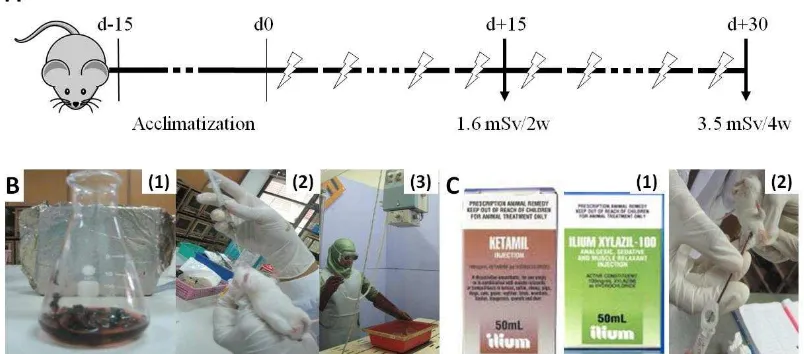
Eighteen male ddy mice 6-8 weeks age with body weight 30-40 gram were used as experimental animals. Mice were caged by plastic box covered by wire (30x40 cm) with commercial foods and ad libitum water. The mice were acclimatized for two weeks prior to ionization radiation. They were divided into 3 groups of treatment, i.e. control non-ionizing radiation group (K, n=6) 0.9% NaCl 0.2 ml orally, ionizing radiation group (P, n=6) 0.9% NaCl 0.2 ml orally and Roselle tea with ionizing radiation group (RT, n=6)
500gram roselle/100ml aquadest 0.2 ml orally.
Roselle tea was prepared by boiled system at 90oC of temperature for 15 minutes, then cooled to 30oC (Fig 1B1-2). The ionizing radiation exposure were performed by radiodiagnostic portable X-ray (VR-1020, MA Medical Corp. Japan) as tool model. Total dose of ionizing radiation for mice were 1.6 mSv/2 weeks and 3.4 mSv/4 weeks, performed every 2 days after being given saline or tea orally by 0.2 mSv/exposure radiation dose (Fig 1B3). Total Body Radiation (TBR) radio-graphy exposure setting for focal focus distance (FFD) by 100 cm, kilovoltpeak (kVp) by 80, and mili Amphere second (mAs) by 20 produced 0.2 mSv of ionizing radiation on each exposure (Noviana et al.
2010). The ionizing radiation exposures were measured by digital dosimeter MYDOSE miniTM (ALOKA CO., LTD Tokyo Japan).
Blood samples were collected from blood venous at sinus retro-orbitalis of anesthetized mice (Thrall 2004). These blood samples were examined for the numbers of erythrocyte (RBC), leucocyte (WBC), hemoglobin (Hb), hematocrit (PCV), RBC size (MCV), and Hb concen-tration (MCHC). Blood samples were col-
lected from mice which had been exposed of ionizing radiation for 2 weeks and 4 weeks period (Fig 1C1-2).
The obtained data were expressed as mean ± standard deviation of means (x±SD). A One-Way Analysis of Variance (ANOVA) was used to compare the means of the studied groups with post hoc Duncan test at 5% for those results where a significant difference was indicated. The
Statistical Package for Social Sciences (SPSS®) version 13 for Microsoft® Windows® statistical software was used.
RESULTS AND DISCUSSION
The RBC count of Roselle tea was higher than radiation and control group after receiveing 1.6 mSv total body radiation (TBR). In other hand, no
112 group (Fig 2C,D). However, RBC size on both of radiation and Roselle group were higher than control (Fig 2E).
Parameter measurement of total peripheral blood at 1.6 mSv of radiation for 2 weeks showed that there were disturbances in the blood parameters. While the total radiation was 3.5 mSv after 4 weeks of the condition in which the body had radio-adaptation response. The RBC values decreased in the radiation group after receiving 1.6 mSv total radiation exposure, indicating the signs of anemia. Anemia occurs due to the reduced number of RBC from the normal threshold values caused by exposure of ionizing radiation. The MCV increased in radiation group than control after receiving 1.6 mSv total radiation exposure. This condition was caused by low levels of vitamin B12 and folic acid in the peripheral blood. This condition occurs probably due to impaired absorption of nutrients in the digestive system and lead to macrocytic anemia. Supplementation of Roselle tea can repair this condition close to normal as the control group. Because MCH value in normal range conditions, so it is called as normochromic macrocytic anemia (NMA). The NMA is a regenerative anemia and become normal if the causative factor was eliminated (Thrall 2004).
The result showed that ionizing radiation energy has negative effect for hematopoietic process (Wambi et al.
2009). Occupational exposures of ionizing radiation from medical diagnostic imaging give 35 % contribution of about 0.8 mSv (DMP 2011). The long-term effects of low-dose exposures from repeated radiodiagnostic imaging may be real and should be given serious consideration (Tucker 2008).
Ionizing irradiation clearly induces multiple changes both within cells and in their microenvironment (Little 2000; Barcellos-Hoff et al. 2005). Irradiation also causes a high degree of in vivo hydroxy radical generation by hemolytic cleavage of body water or from the endogenous hydrogen peroxide formed by reduction of the superoxide anion. The hydroxyl radical is the major cytotoxic radical than others. The interaction of these radicals with phospholipid structures induces the peroxidation processes that increase hydroxyl radical activity in the DNA oxidative damage. Under these oxidative stress conditions, although radiation therapy is needed to patient with cancers, exogenous agents with a radical scavenging capacity need to be used to minimize the normal cell damage (Castillo
et al. 2000)
The supplementation with antioxidant vitamins and minerals may have a protective effect against X-ray-induced damage. Flavonoids compound shows 20 times more powerful antioxidant activity than vitamin in the lipoprotein oxidation model (Craig 1999). Roselle contains vitamin C more than threefold of black grape, nine fold of citrus lime, and tenfold of star fruit (Widyanto & Nelistya 2008). Phytochemical screening and structural analysis has identified a class of flavonoids called anthocyanin as responsible for antioxidant activities (Passamonti et al.
ACKNOWLEDGMENT
This study was supported by Directorate of General Higher Education (DGHE) of Indonesia in Student Creativity
Program 2010 collaborated with
Competitive Grant also from DGHE (28/I3.24.4/SPK/PD/2010) in the same year.
REFERENCES
Akindahunsi AA, Olaleye MT. 2003. Toxicological investigation of aque-ous methanolic extract of Hibscus sabdariffa L. J Ethnopharmacol.
89:161–164.
Barcellos-Hoff MH, Park C, Wright EG. 2005. Radiation and the microenv-ironment - tumorigenesis and therapy.
Nat Rev Cancer. 5:867–875.
Bouvet M, et al. 2002. Real-time optical imaging of primary tumor growth and multiple metastatic events in a pan-creatic cancer orthotopic model.
Cancer Res. 62(5):1534–1540.
Castillo J, et al. 2000. Antioxidant activity and radioprotective effects against chromosomal damage induced in vivo by X-rays of flavan-3-ols (Procyani-dins) from grape seeds (Vitis vinifera): comparative study versus other phe-nolic and organic compounds. J Agric Food Chem 48:1738-45.
Citrin D,et al. 2010. Radioprotectors and Mitigators of Radiation-Induced Nor-mal Tissue Injury. Oncologist. 15(4): perspective. Department of Mine and Petroleum, Government of Western Australia. http://www.arpansa.gov.au/ radiationprotection/basics/understand. cfm [25 September 2011]
Hou DX, Tong X, Terahara N, Luo D, Fujili M. 2005. Delphinidin -3- sambubioside, a Hibiscus anthocyan-in, induces apoptosis in human
leukemia cells through oxygen reactive species-mediated mitochon-drial pathway. Arch. Biochem. Biophys. 440(1): 101-109.
Jeong H, et al. 2003. Differential Effects of Green Tea Polyphenol in the -irradiation Induced Human Leukemic and Lymphoblastic Cell Damage. The Korean Journal of Nuclear Medicine
37(5).
Little J. B. 2000. Radiation carcinogenesis.
Carcinogenesis. 21:397–404
Mauch P, et al. 1995. Hematopoietic stem cell compartment: acute and late effects of radiation therapy and chemotherapy. Int J Radiat Oncol Biol Phys. 31:1319–1339.
McCurnin DM, Bassert JM. 2006. Clinical Textbook for Veterinary Technicians. 6th Edition. Elsevier Saunders. USA. Ningsih EM, et al. 2011. Hemogram
Characteristic and Body Weight Differences in IonizingRadiation Exposed Mice Treated with Radio-protective Roselle Simplisia by Infusion Methode. Proceedings of International Seminar and 2nd Congress of SEAVSA. 21-22 June musculus): The Preliminary Research of Radioprotective Potential of Roselle (Hibiscus sabdariffa L.) Against Radiodiagnostic Ionizing Radiation. Proceedings of the First Congress of South East Asia Veterinary School Association. IPB ICC Bogor, Indonesia 20-22 Juli 2010. pp 157
114
Passamonti SVU, Vanzo A, Mattivi F. 2003. The stomach as a site for anthocyanins absorption from food.
FEBS Lett. 544(1-3): 210-213.
Thrall DE. 2002. Textbook of Veterinary Diagnostic Radiology. 4th edition London: W. B. Saunders Company. Thrall MA. 2004. Veterinary Hematology
And Clinical Chemistry. Lippincott Williams & Wilkins: USA
Tucker JD. 2008. Low-dose ionizing radiation and chromosome transloca-tions: A review of the major consideration for human biological dosimetry. Mutat Res. 659:211–220.
Wambi CO, et al. 2009. Protective Effects of Dietary Antioxidants on Proton Total-Body Irradiation-Mediated He-matopoietic Cell and Animal Survival.
Radiat Res.172 (2): 175-186.
Wang CJ, et al. 2000. Protective effect of Hibiscus anthocyanins against tert-butyl hydroperoxide–induced hepatic toxicity in rats. Food Chem Toxicol. 38:411–416.