* corresponding author: [email protected]
Cytochrome P450 aromatase (CYP19)
gene expression in ovarian granulose cells
of hypothyroid rats induced by
propylthiouracil
Prihatin Broto Sukandar1, Sri Kadarsih Soejono2, Totok Utoro3
1Center for Research and Development for Iodine Deficiency Disorders, Magelang, Central Java, 2Department of Physiology, 3Department of Pathology Anatomy, Faculty of Medicine, Universitas Gadjah Mada, Yogyakarta
ABSTRACT
Thyroid hormones are proven to have a direct effect on granulose cells, luteal cells and oocytes due to their role in gonadotropin action on steroid hormone production.In vitro study showed that tiroxine (T4) on granulose cells can stimulate ovarian steroidogenesis. Moreover, high concentration of triiodothyronine (T3) increases the estradiol secretion and aromatase mRNA expression. Hypothyroidism influences the cytochrome P450 aromatase (CYP19) gene expression. The aim of this study was to evaluate the expression of the CYP19 gene in granulosa cells of hypothyroid rats induced by propylthiouracil (PTU). This was quasi experimental study with post-test only control group design. Eleven female Sprague-Dawley rats were divided into two groups i.e. five rats as treated group that induced by PTU 0.1 g/L in aquadest for 30 days and control group that not induced by PTU. Blood sample was taken and then T4 blood level was measured using an enzyme-linked immunosorbent assay (ELISA). Whereas, CYP19 gene expression in ovarian granulose cells was measured using immunohistochemistry. Unpaired t test was used to compare the data obtained from treated and control groups. The results showed that T4 blood level on treated group (4.02 ± 0.39 ng/dL) was significantly lower than control group (8.08 ± 1.63 ng/dL) (p = 0.000). However, CYP19 gene expression on treated group (30.84 ± 8.01%) was not significantly different compare to control group (25.06 ± 6.79%) (p = 0.227). In conclusion, the CYP19 gene expression in ovarian granulose cells of rats is not change after induction of PTU 0.1 g/L for 30 days, although the T4 blood level decreases.
ABSTRAK
dibandingkan dengan kelompok kontrol (8.08 ± 1,63 ng/dL) (p=0,000). Namun demikian, ekspresi gene CYP19 pada kelompok perlakuan (30,84 ± 8,01%) tidak berbeda nyata dibandingkan kelompok kontrol (25,06 ± 6,79%) (p = 0,227). Dapat disimpulkan, ekspresi gene CYP19 pada sel granulosa ovari tikus tidak berubah setelah induksi 0,1 g/L PTU selama 30 hari, meskipun kadar T4 darah mengalami penurunan.
Key words:CYP19 - aromatase - T4 blood level - propylthiouracil - hypothyroid
INTRODUCTION
Iodine deficiency disorder (IDD) is one of the nutrition problems in Indonesia.1It is highly
associated with infertility, stillbirths, lactation failure, and abnormalities menstruation.2 An
estimated 750 million people in the world are at risk of suffering from IDD.3 In Indonesia,
based on a survey of thyroid in children, the national Total Goitre Rate (TGR) was 9.8% in 1998 and 11.1% in 2003.4 The rates are still
above the WHO recommendation, which is less than 5%. The prevalence of hypothyroidism in the general population of reproductive age is about 2%.5 Previous study by Auchus and
Chang6showed that thyroid disease occurs five
to ten times more in women than men, and the highest incidence occurs at reproductive age. Marijata’s7 study at a hospital in Wonosari,
Yogyakarta, Indonesia found that 90% of goitre patients are female and10% are male .
Hypothyroidism affects the granulose cells, luteal cells, oocytes and eventually leads to ovarian dysfunction.8 Mild hypothyroidism
interfere ovulation and conception.9 Severe
hypothyroidism often leads to ovulatory dysfunction and infertility.10There is a causal
relationship between hypothyroidism and the development of various ovarian disorders (enlarged polycystic ovaries, cysts, and spontaneous hyperstimulation ovarian syndrome).11
Reduction of thyroid function may alter pituitary-ovarian axis. In hypothyroid condition, the pituitary is more sensitive to produce thyroid stimulating hormone (TSH) or thyroid realising
hormone (TRH). Thyrotropin releases hormone that stimulates lactotrop cells to synthesize prolactin. Prolactin interfere GnRH pulsatility and suppresses follicle stimulating hormone (FSH) and luteinizing hormone (LH), and as a consequences there is no follicle maturation. Furthermore, prolactin stimulates adrenal androgen secretion, lead to the increasing of androgen serum and follicle maturation stunts.6,12
Treatment of hypothyroidism with L-thyroxine (L-T4) can restore normal menstrual pattern and ovarian disorders.10
In addition, the effects of hypothyroidism can impair hypothalamic-pituitary axis and the function of ovaries. Hypothyroidism can reduce sex hormone binding globulin (SHBG) affinity and that increase free testosterone and estradiol due to lower clearance rate of androstenedione and estron.6 Ovarian enlargement in severe
hypothyroidism is probably due to stimulation of FSH receptor by unusually high TSH levels. TSH has a weak FSH-like activity.13Thyroid
hormones play a role in modulating LH and FSH on granulose cells function to induce steroido-genesis.14
Several studies found that one of the genes that influence the process of steroidogenesis is cytochrome P450 19 (CYP19) gene. The CYP19 gene is located in chromosome 15 on the long arm (15q21.1). It encodes the CYP aromatase enzyme and catalyzes the final step of the biosynthesis of estrogen from testosterone to estradiol and androstenedione to estron.15
ovarian follicle. Estrogen precursors such as testosterone are supplied by cells in the outer layer of the follicle (theca cells). Aromatase is a reliable marker for ovarian granulose cells in mammalian.16,17 Several cases with estrogen
deficiency reduce aromatase activity. Conversely, the increase aromatase activity can lead to gynecomastia and feminization in males.18Experiments with T4 administration on
granulose cells culture proved to stimulate ovarian steroidogenesis.6
Propylthiouracil (PTU) treated rats have been used as an animal model to study hypothyroid ovarian follicular cysts and steroidogenesis. This model can be used to investigate the biochemical changes
intra-ovarium.19 Hypothyroidism affects the
expression of CYP19 gene encoding aromatase. This study was conducted to evaluate the expression of CYP19 gene in ovarian granulose cells of hypothyroid rats induced by PTU.
MATERIALS AND METHODS
Animals and hypothyroid induction
This was a experimental study with post-test only control group design. Twelve Sprague-Dawley female rats aged 10 weeks with average of body weight of 111.08 ± 8.10 g obtained from the Integrated Research and Testing Laboratory (Laboratorium Penelitian dan Pengujian
Ter padu= LPPT), Universitas Gadjah Mada,
Yogyakarta were used in this study. The rats were housed in the Laboratory of Physiology, Faculty of Medicine, Universitas Gadjah Mada at room temperature under 12 hours cycles of dark and light. The rats were fed with AD-II pellet (PT. Japfa Comfeed Indonesia, Tbk, Sidoarjo) and provided an
access to water ad libitum. After an
adaptation period of one week, the rats were divided into two groups with six rats in each group. The animal sample size was calculated
according to the software of Ramakrishnan,20
or formula of n = 1+2 C (s/d)2described by
Dell et al.21 The first group as control was
without PTU induction. The second group as treatment group was induced by PTU 0.1 g/L in aquadest for 30 days according to the protocol described by Haponet al.22
Thyroxine blood level assay
On day 30 after PTU induction, blood samples were collected from orbital sinus and T4 blood leve was measured. Two mL blood sample was centrifuged at 3000 rpm (rounds per minuts) for 10 minutes. Serum sample was taken and kept at 2-8 oC for 24 hours until T4
serum level analyzed. The T4 serum level was then measured using ELISA. Fifty µL of serum sample was pippeted into each well of a microtiter well coated antibody to T4 on a solid phase. One hundred µL of T4 labeled with sheep peroxidase conjugate were added into each well and incubated at room temperature. After a 60 minute incubation, the microtiter well was washed with a washing solution containing surfactant in Tris beffered saline. A solution of tetramethylbenzidine (TMB) was added and incubated for 15 minutes resulting in the development of a blue color. The color development was stopped with the addition 100 µL stopping solution containing 0.25 M sulphuric acid. The resulting yellow color was measured ELISA reader at 450 nm. The intensity of the color formed was proportional to the amount of enzyme present and was inversely related to the amount of T4 in the serum sample. By reference to a series of standard processed in the same way, the concentration of T4 in the unknown serum sample was calculated.
CYP19 gen expression assay
the rat. The right ovarian tissue was then made histological preparations. The formalin-fixed and paraffin-embedded tissues were cut into serial tissue sections at a thickness of 3 µm. Each slide contains 4 tissue sections e.g. two sections as control and two sections as treatment group. Following deparaffinization, the sections were blocked with normal rabbit serum for 5 minutes. The sections were then incubated with polyclonal antibody anti CYP19 solution (1:50) purchased from Santa Cruz Biotechnology, Inc. for 60 minutes. The sections were washed with 10% phosphate buffered saline (PBS) three time for 5 minutes and incubated with diamino-benzidine (DAB) for 5 minutes. The sections were then lightly countersatined by
haemato-xylin mayer and incubated for 3 minutes at room temperature and washed in water for 10-15 minutes. The sections were then dried and coverslipped. All sections were then examined and evaluated using light microscope on 400x magnifications. The observation of CYP19 expression was performed in the Department of Pathology Anatomy at Faculty of Medicine/ Dr. Sardjito General Hospital, Universitas Gadjah Mada. The CYP19 expression in ovarian granulose cells was identified by a brown color in cytoplasma of the cell, while a blue color of a ovarian granulose cell indicated no expression of the CYP19. The CYP19 expression was observed on each sections and percentage of the CYP19 expression was calculated using a formula as follows:
Statistical analysis
Data were presented in the form of mean ± standard error of mean (SEM). Statistical analysis was performed by unpaired t test with significance level of p<0.05. If the data distribution was not normal, the Mann-Whitney test was used. This study was approved by the the Medical and Health Research Ethic Committee, Faculty of Medicine, Universitas Gadjah Mada, Yogyakarta.
RESULTS
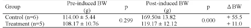
Among six rats induced by PTU in treatment group, one rats did not showed hypothyroid. Therefore only five hypothyroid rats in the treatment group were used in this study. The body weight (BW) of rats in both control (108.17±10.76 g) and treatment groups (114.00 ± 5.44 g) before induced with PTU were not significantly different (p>0.05). At the end of the study, the BW of all rats in both increased. The average BW gain of the control group rats (55.5 g) was higher than the treatment group (11.0 g) (p<0.05) (TABLE 1).
TABLE 1. The BW (mean ± SEM) of rats in both groups before and after induced with PTU 0.1 g/ L in aquadest for 30 days
The T4 blood sample after induction of PTU 0.1 g/L for 30 days in the treatment group (4.02
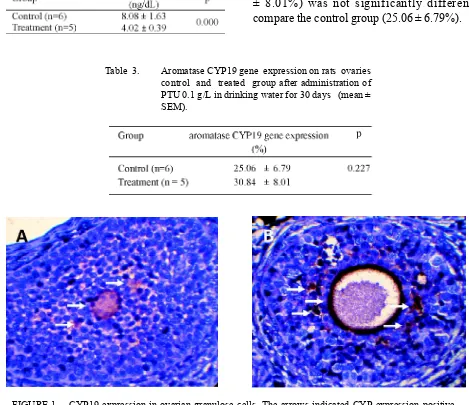
The CYP19 expression in ovarian granulose cells of rats in both control and treatment groups is presented in FIGURE 1, while the level CYP19 expression in the both groups is presented in TABLE 3. The average CYP19 expression in the treatment group (30.84 ± 8.01%) was not significantly different compare the control group (25.06 ± 6.79%). (TABLE 2). It was indicated that the PTU
induction caused hypothyroid of the rats.
TABLE 2. The T4 blood level (mean ± SEM) of rats in both groups before and after induced with PTU 0.1 g/L in aquadest for 30 days
FIGURE 1. CYP19 expression in ovarian granulose cells. The arrows indicated CYP expression positive cells. A. control group and B. treatment group. Light microscope observation on 400 x magnification.
Table 3. Aromatase CYP19 gene expression on rats ovaries control and treated group after administration of PTU 0.1 g/L in drinking water for 30 days (mean ± SEM).
DISCUSSION
The free T4 blood levels in treatment group were statistically lower than control group (p<0.05). This suggests that ingestion of PTU 0.1 g/L for 30 days was able to induce hypothyroidism.22
Weight gain was significantly higher in the control group compared to treatment group (p<0.05). It was consistent with research by Cooke et al.23which showed that
hypothyroi-virgin rats can increase CYP19, whereas T4 serum levels stay normal.
In hypothyroidism condition , gonadotropin (FSH and LH) production is not interfere by the pituitary,31while it is known that FSH is a
major trigger of aromatase activity.32
Hypo-thyroidism can inhibit the growth and development of the follicles, but the follicles are still able to produce steroid hormones. It happens because during this condition, only the number and size of corpura lutea cells are reduced.31Corpora lutea is the major source of
the hormone progesterone. Reduction of the size and the cell numbers of will reduce the production of progesteron, but it will induce the development of estrogen receptors in granulose cells. Estrogen hormone stimulation normal granulose cells development and it leads
to normal process of aromatization.33
Furthermore, ovarian enlargement in severe hypothyroidism is probably due to the stimulation of FSH receptor induced by high TSH levels. TSH has a weak FSH-like activity because it similar to the subunit of FSH and LH.13 Recent study showed that estrogen
production that induced by aromatization is not interfered by ovarian enlargement.
CONCLUSION
It can be concluded that the administration of PTU 0.1 g/L for 30 days does not influence the CYP19 gene expression in ovarian granulose cells of rats, although it can decrease the T4 blood levels. Further studies will be conducted to evaluate the effect of PTU induction in longer period. Moreover, other indicators of reproductive hormones level such as T3 will be studied.
ACKNOWLEDGEMENTS
The authors would like to thank Ms. Agustin from the Laboratory of Pathology Anatomy, dism inhibits weight gain.24Thyroid hormones
affects tissue growth, brain maturation, increases heat production and oxygen consumption due to increased activity of Na+-K+-ATPase, as well
as increased transport of glucose and amino acids.25 Thyroid hormone also stimulates the
secretion of growth hormone and stimulates the growth hormone effects (somatomedin) on protein and new structural bones synthesis.26
The results of this study showed that the expressions of CYP19 gene in granulose cells between the control group and the treatment group were similar. According to the research by Haponet al.22PTU-induced hypothyroidism
with dose 0.1 g/L given for 30 days did not reduce estradiol serum levels. However, the prolongation of PTU administration until 50 days decreases the estradiol serum levels.
In hypothyroid, free T3 may be normal. This is caused by the influence of thyroid tissue remnants that are still have normal function under the influence of increased TSH. T4 is converted to T3, and cause lower T4 level. As a consequence of this mechanism, free T3 levels remain within normal limits.27The active form
of thyroid hormone (T3) regulates development and physiological functions at the cellular level, control metabolism, proliferation, differentiati-on, and apoptosis. T3 mostly affects transcripti-on gene by binding to the thyroid hormtranscripti-one receptor found in nucleus.28However, no other
studies correspond with the results of this study. CYP19 gene expression will be decreased when levels of thyroid hormone (T3) is low, thus affecting gene transcription.28 Free T3
levels in this study were not measured. Research by Hatsutaet al.29proved that the addition of
T3 with normal dosage did not affect the secretion of estradiol. However, low dosage of estradiol reduce the secretion of estradiol, presumably through the reduction of CYP19 mRNA expression in granulose cells. Another study by Hapon et al30 stated that the
Universitas Gadjah Mada/Dr. Sardjito General Hospital for her valuable assistance in immuno-histochemistry staining techniques and Mr. Wakidi Parno from Laboratory of Physiology, Faculty of Medicine, Universitas Gadjah Mada for his assistance in animal handling and treatment.
REFERENCES
1. Astawan M. Iodium cegah lost generation. [cited 2011 April 11]. Available from: www.gizi.net/cgi-bin/berita/fullnews.cgi?newsid1043213364, 24317
2. Thomas R, Reid RL. Thyroid disease and
reproductive dysfunction: a review. Obstet Gynecol. 1987; 70(5):789-98.
3. Anonim. Assessment of the iodine deficiency disorders and monitoring their elimination. Geneva: World Health Organization, 2001. 4. Anonim. Technical assistanc for evaluation on
intensified iodine deficiency control project. Jakarta: Directorate General of Community Health, Directorate of Community Nutrition, 2003.
5. Bjoro T, Holmen J, Kruger O, Midthjell K, Hunstad K, Schreiner T, et al. Prevalence of thyroid disease, thyroid dysfunction and thyroid peroxidase antibodies in a large, unselected population. The Health Study of Nord-Trondelag (HUNT). Eur J Endocrinol 2000; 143(5):639-47.
6. Chang AY, Auchus RJ, Endocrine disturbances affecting reproduction. In: Yen, SSC, RB Jafee RB, editors. Reproductive endocrinology physiology, pathophysiology and clinical management. Philadelphia: Saunders. 2009. pp: 561-75.
7. Marijata. Pola distribusi penderita benjolan tiroid di RSU Wonosari Gunung Kidul. BKM VII 1991; 2: 88-93.
8. Wakim AN, Polizotto SL, Buffo MJ, Marrero MA, Burholt DR. Thyroid hormones in human follicular fluid and thyroid hormone receptors in human granulosa cells. Fertil Steril 1993; 59(6):1187-90.
9. Davis LE, Leveno KJ, Cunningham FG.
Hypothyroidism complicating pregnancy. Obstet. Gynecol 1988; 72(1):108-12.
10. Krassas GE, Pontikides N, Kaltsas T,
Papadopoulou P, Paunkovic J, Paunkovic N,et al.
Disturbances of menstruation in hypothyroidism. Clin Endocrinol (Oxf) 1999; 50(5):655-9.
11. Rohatgi T, Rohatgi N, Buckshee K. Recurring acute abdomen, ovarian cyst and hypothyroidism. JK Science 2007; 9(4):197-9.
12. Jacoeb TZ. Endokrinologi reproduksi pada wanita. In: H Wiknjosastro, AB Saifuddin, T Rachimhadhi. (Editor): Ilmu Kandungan. Jakarta: Gramedia 1997. pp: 43-96.
13. Mahendru RR, Mittal A, Gaba G. Is hypothyroidism a cause of ovarian cysts? This unusual case depicts so. Webmed Central 2011; 2(3):1-6.
14. Raber W, Nowotny P, Binstorfer EV, Vierhapper, H. Thyroxine treatment modified in infertile women according to thyroxine-releasing hormone testing: 5 year follow-up of 283 women referred after exclusion of absolute causes of infertility. Hum Reprod 2003; 18(4):707-14.
15. Jin JL, Sun J, Ge HJ, Cao YX, Wu XK, Liang FJ,
et al. Association between CYP19gene SNP rs2414096 polymorphism and polycystic ovary syndrome in Chinese women. BMC Med Genet 2009; 10:16:139.
16. Leung PC, Armstrong DT. Interactions of steroids
and gonadotropins in the control of
steroidogenesis in the ovarian follicle. Annu Rev Physiol 1980; 42:71-82.
17. Nakamura S, Kurokawa H, Asakawa S, Shimizu N, Tanaka M. Two distinct types of theca cells in the medaka gonad: germ cell-dependent maintenance of cyp19a1-expressing theca cells. Dev Dyn 2009; 238(10): 2652–7.
18. Strauss III JF. The synthesis and metabolism of steroid hormones. In: Yen SSC, Jafee RB, editors.
Reproductive endocrinology physiology,
pathophysiology and clinical management. Philadelphia: Saunders 2008, pp: 79-104. 19. Bagavandoss P, England B, Asirvatham A, Bruot
BC. Transient induction of polycystic ovary-like syndrome in immature hypothyroid rats. Proc Soc Exp Biol Med 1998; 219(1):77-84.
21. Dell RB, Holleran S, Ramakrishnan R. Sample size determination. ILAR J 2002; 43(4):207-13.
22. Hapon MB, Simoncini M, Via G, Jahn GA. Effect of hypothyroidism on hormone profiles in virgin, pregnant and lactating rats, and on lactation. Reproduction 2003; 126(3):371-82.
23. Cooke PS, Kirby JD, Porcelli J. Increased testis growth and sperm production in adult rats following transient neonatal goitrogen treatment: optimization of the propylthiouracil dose and effects of methimazole. J Reprod and fertil 1993; 97(2):493-9.
24. Kanz MF, Taj Z, Moslen MT. 1,1-dichloroethelyne hepatotoxicity: hypothyroidism decreases metabolism and covalent binding but not injury in the rat. Toxicology 1991; 70(2):213-29.
25. Greenspan FS. Kelenjar tiroid. In: Greenspan FS, Baxter JD, editors. Endokrinologi dasar dan klinik. Jakarta: EGC, 2000. pp:206-89.
26. Sherwood L. Human physiology: from cells to systems. Jakarta: EGC, 2001.
27. Pranoto A. Management hyperthyroid and hypothyroid. Article presented on Surabaya Thyroid Workshop-3, August 10, Surabaya, 2008.
28. Bilesimo P, Jolivet P, Alfama G, Buisine N, Le Mevel S, Havis E,et al.Specific histone lysine 4 methylation patterns define TR-binding capacity and differentiate direct T3 responses. Mol Endocrinol 2011; 25(2):225-37.
29. Hatsuta M, Tamura K, Shimizu Y, Toda K, Kogo H. Effect of thyroid hormone on CYP19 expressi-on in ovarian granulosa cells from gexpressi-onadotropin- gonadotropin-treated immature rats. J Pharmacol Sci 2004; 94(4):420-5.
30. Hapon MB, Gamarra-Luques C, Jahn GA. Short term hypothyroidism affects ovarian function in the cycling rat. Reprod Biol Endocrinol 2010; 11:8-14.
31. Armada-Dias L, Carvalho JJ, Breitenbach MM, Franci CR, Moura EG. Is the infertility in hypo-thyroidism mainly due to ovarian or pituitary functional changes? Braz J Med Biol Res 2001; 34(9):1209-15.
32. Stocco C. Aromatase expression in the ovary: hormonal and molecular regulation. Steroid 2008; 73(5):473–87.