Acoustic Range Electric-Magnetic Field Exposure Decreases
Cortical Excitability in Genetically Epilepsy-Prone Rats
Nato B*, Giorgi K
,
Marina B, Lamara M and Militsa S
Imvane Beritashvili Center of Experimental Biomedicine, Georgia
*Corresponding author: Bukia Nato, Doctor of Biological Sciences, Ivane
Beritashvili Center of Experimental Biomedicine, Georgia, E-mail: [email protected]
Abstract
In the present study, we examined the effect of acoustic range of electric-magnetic field (EMF) on the behavioral
manifestation of seizure in genetically epilepsy-prone rats (GEPRs) of Krushinsky-Molodkina strain. A 5-days exposure to
EMF (10000 -15000 Hz frequency, 1.5 m/Tesla, during 20 min) resulted in partial or complete suppression of behavior
seizure activity in GEPRs. Besides, on the background of EMF the latency period of first wild run was increased. On the
same conditions, duration of wild run decreased.
Audiogenic stimuli obtained in GEPRs changed the ECoG activity of sensomotoral cortex only in audiogenally-kindled
animals. Findings of this study suggest that the regulation of the behavioral manifestation of seizure in GEPRs does not
involve the cortex; it is mainly regulated by brainstem structures. We concluded that acoustic range of EMF could apply
for suppression of behavioral manifestation of seizure. Audiogenic stimuli obtained in GEPRs changed the ECoG activity
of sensomotoral cortex only in audiogenally-kindled animals. Findings of this study suggest that the regulation of the
behavioral manifestation of seizure in GEPRs does not involve the cortex; it is mainly regulated by brainstem structures.
Rostral part of cortex has significant role in manifestation of seizure activity in audiogenly kindling GEPRs. We concluded
that acoustic range of EMF could apply for suppression of behavioral manifestation of seizure.
Keywords:
Electric-Magnetic Field; Seizure; Rats; ECoGIntroduction
Epilepsy is one of the most common disorders. According to the WHO (World health Organization) fact sheet, epilepsy affects about 50 million people of all ages worldwide. Despite the fact that it is possible to treat epilepsy using pharmacological substances, about 30-40% of the patients are resistant to such treatment (WHO, media Center 2016). In addition, a long-term
consumption of the drugs negatively affects the patients’
cognitive functions [1-3]. The effectiveness of pharmacological agents, used for epilepsy treatment is
limited due to incomplete understanding of the mechanisms involved in the pathogenesis of the disease. Therefore, a goal of modern psychiatric research is to find the new ways for anti epilepsy therapy, which will give the opportunity to cure this group of patients.
The EMF exposure is a noninvasive treatment method; it used as a complementary to the drugs, for treating different neurodegenerative diseases (Parkinson's disease, schizophrenia, depression, tinnitus, etc.) [4,5]. EMF exposure can also be used as a separate treatment therapy [6]. Furthermore, repetitive TMS Transcranial
Research Article
Volume 2 Issue 1
Received Date: January 27, 2017
Magnetic Stimulation) is seen as a safe treatment method, without enduring side effects: no long-term neurological, cognitive, or cardiovascular side effects are reported.
The EMF appears to be biologically active, penetrating into the living tissue without any impediments [7,6]. However, it is unclear how the low-frequency EMF can block seizure activity.
One of the basic provisions underlying the given research is the assumption that the macromolecules which constitute the living organism are subjected to the conformational fluctuations under the conditions of EMF impact. In our opinion the artificially created EMF may change the tonic activity of the cells located in the local area of its action and because of it the mode of neural impulses formation. Because of it, the tonic activity is also changed in the central nuclei, through which the various sensory afferent ways coming from the various parts of the body are passing. On the other hand, oscillations of acoustic range (which belong to low frequency oscillations) due to their activity can cause the intensification of chemical processes. Then, oscillations of this range can be used both for process stimulation, intensification and optimization and for their attenuation. It will depend on impact mode and selection of duration. Both a change in membrane ion channel permeability and oriented redistribution of radicals, liquid-crystalline macromolecular structures, metalloproteins (hemoglobin, vitamins) and molecular water fragments may occur under influence of low frequency magnetic fields [8,9].
The goal of this study was to explore the potential mechanisms underlying the impact of EMF exposure on the epilepsy. In GEPRs and inbred white rats, we examined the effect of EMF exposure on ECoG activity in sensomotoral cortex. We attempted identify the optimal parameters of repeated EMF exposure, which fully or partially depress the ECoG and behavioral seizure manifestations.
Methods
Subjects and Surgical Procedures
Experiments were conducted on male white, inbred rats and GEP of Krushinsky-Molodkina (KM) strain rats (n=14). This strain of rats manifests short lasting seizure activity in neonatal age and fully-fledged seizure activity after hierarchical implication of brainstem structures [10]. In response to a strong sound (the bell -90-dB, during 60 sec), GEPRsdisplay either fear reaction accompanied by facial muscle clonus (group a) or fear reactions with elevated motor act responses (wild running, jumps), which are accompanied with tonic-clonic
behavioral seizures (group b). The present study was conducted on animals of group b.
Under ketamine (5-10 mg/kg intraperitoneally) anesthesia, the rats were implanted with stainless bipolar electrodes (8IE3633SPCXE ELEC 0.05-125 MM SS Plastics One) into sensomotoral area of neocortex.
Experimental Paradigm
At the begining of experiments, both control (Inbred, white male rats n=7) and experimental (GEPRs n=14) rats were placed into coil for EMF exposurewith defined parameters of magnetic field (see below) for a 20-min session. 10 min after the session of EMF exposure, the animals were re-tested. The GEPRs(with or without exposure) were given audiogenic stimuli,for 60 secs, before and after testing in open field for monitoring behavior correlates of seizure activity.
EMF Exposure
For EMF exposure (carried for five consecutive days), we used the coil designed at Tbilisi Technical University, Georgia. Parameters of magnetic field (stimulus frequency, its intensity and train duration), which partially or fully depressed ECoG and behavior manifestation of seizure activity, were established during pilot experiments. For repetitive (a 5-days) EMF exposure, we used the following parameters: (10000-15000 Hz, 1-1.5 m/Tesla).
EEG Registration
ECoG activity of seizure was registered before, during and after EMF exposure in free movement rats.
The Open Field Test
Emotional-motivational status of the rats was tested in a chamber of 80 cm in diameter surrounded by 30 cm height walls, Open Field. The floor of the chamber was divided into 32 squares and lighted with 200 W lamp. The rats were video-recorded for initial 5 minutes after the placement into the Open Field, for three consecutive days (the same time of day). We registered the following parameters: entering the center, numbers of crossed squares, head raise, vertical stands, the frequency and duration of grooming, number of fecal boluses and urination. After each trial, the chamber was cleaned with 30 % ethanol solution.
Data Analysis
the parameters of behavioral (latency of the first wild run, duration of the first wild run, duration of pause, and duration of second wild run, duration of tonic-clonic seizure and of post-ictal activity) and ECoG seizure activity were determined as well.
Statistics
Data obtained were processed using adequate statistical program. Data reliability was assessed using parametric and non-parametric techniques, with the use of one- and two-way layout of factorial analysis.
Results and Discussion
The Effect of EMF Exposure on Behavior Manifestation of Seizure Activity in GEPRs
During experiments revealed two subgroups of GEPRs. In the first group of animals, audiogenic stimuli resulted
in wild running (right after the delivery of stimuli) followed by tonic-clonic seizure. In the second group, we observed a different response to the stimuli: after the first episode of wild running, the rats had a short pause (7 ± 2 sec) followed by a second episode of wild running (15 ± 3 sec) and tonic-clonic seizure for 21 ± 2 sec. Exposition of EMF we performed in both group of GEPRs.
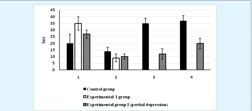
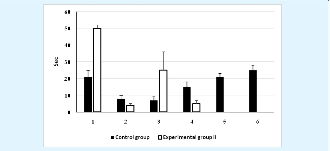
A low frequence EMF exposure (10000-15000 Hz, 1-1.5 m/Tesla) resulted in full or partial depression of general behavior in both groups of GEPRs. The latency of first wild run significantly increased p≤0.05 from 20 ± 7 sec (in control group) to 35 ± 5 sec (in GEPRs, group I) and from 21 ± 4 to 50 ± 2 sec (in GEPRs, group II). The duration of
wild run significantly decreased p≤0.05 from 14 ± 3 sec
(in control group) to 9 ± 3 sec (in GEPRs, group I) and 8 ± 2 to 4 ± 1 sec (in GEPRs, group II) (Figure 3 and 4).
Figure 3: The effect of acoustic range EMF on behavior manifestation of seizure activity in I group of GEPRs.
Black columns – GEPRs (control group). White and Patterned columns –GEPRs with EMF exposure (experimental groups). 1. Latency period for the wild run; 2. Duration of the wild run; 3. Duration of tonic-clonic seizures; 4. Duration of post-ictal activity; 5. Duration of tonic-clonic seizure (partial depression); 6. Duration of post-ictal activity (partial depression).
Group II of GEPRs demonstrated significant increases in the pause between the first and second wild run episodes from 7 ± 2 to 25 ± 11sec and decreases in the duration of second wild run from 15 ± 3 to 5 ± 2 sec
p≤0.05 . It is noteworthy that EMF exposure resulted in
Figure 4: The effect of acoustic range EMF on behavior manifestation of seizure activity in II group of GEPRs.
Black columns – GEPRs (control group). White and Patterned columns –GEPRs with EMF exposure (experimental groups). 1. latency period for the wild run; 2, duration of the wild run; 3. duration of tonic-clonic seizures; 4. duration of post-ictal activity; 5. duration of tonic-clonic seizure (partial depression); 6. duration of post-ictal activity (partial depression).
On the background of EMF exposure Group II of GEPRs did not manifest tonic-clonic seizures. In contrast, some animals of GEPRs Group I (n=2) manifested tonic-clonic seizure activity. In these animals the durations of tonic-clonic seizures and post ictal period of depression decreased from 35 ± 4 to 12 ± 4 sec, and from 37 ± 4 to 20 ± 4 sec, respectively (Figure 3). Our results suggest that I group of GEPRs, the rats that developed only one wild run, have stronger audiogenic seizure activity, compared to Group II of GEPRs. Consequently, EMF fully suppresses seizure activity in Group II of animals. However, a suppression of seizure activity was also been observed in some animals of Group I.
EMF Exposure and ECoG Activity of
Sensomotoral Cortex
In Group II of GEPRs, we recorded ECoG activity of sensomotoral cortex with or without EMF exposure. In response to a strong sound (single audiogenic stimuli), we did not see significant changes in ECoG activity of sensomotoral cortex, but we observed clear behavioral seizure activity. Changes in the ECoG activity were
observed following repeated audiogenic stimuli. Right after presenting last sound in GEPRs, we observed ECoG desynchronization. ECoG of sensomotoral cortex was predominately presented by low frequency activity (0-0.5Hz) intermixed with delta (0.5-4 Hz) and theta (4-9 Hz) waves. During pause, the activity was changing back to the background pattern.
On the background of repeated EMF exposure (10000-15000 Hz, 1-1,5 m/Tesla) a repeated audiogenic stimuli did not result in the development of ECoG activity of sensomotoral cortex observed in audiogenly kindling GEPRs and the rats did not manifest seizure activity.
Our results indicate that a single sound presentation does not change ECoG activity of sensomotoral cortex suggesting that sensomotoral cortex is not involved in the development of audiogenic seizures. In contrast, brainstem structures play a significant role in the development of audiogenic seizures. But, the initiation of seizers requires a repeated stimulation of brainstem reticular formation. Besides, rostral part of cortex has significant role in manifestation of seizure activity in audiogenly kindling GEPRs.
Summary and Conclusion
Finding of the present study suggest that low frequency acoustic range EMF exposure is an noninvasive procedure which allows to depress (fully or partially) the seizure activity developed in response to audiogenic stimuli in Krushinsky-Molodkina strain GEPRs. We tried to understand the possible mechanisms by which EMF exposure, with a mentioned field parameters, is capable positively effect on the GEPRs.
In our experiments, brain structures are subjected not only to direct transcranial stimulation, but they also receives afferent signals from whole body. We suggest that the artificially created EMF may change the tonic activity of brain cortex. The EMF can modulate Ponto -geniculo-occipital (PGO) waves. On the other hand, PGO waves have a possible inhibitory influence on EEG seizure activity [15]. Increased number of PGO spikes in animals exposed to auditory stimulation is attributed to the anatomical proximity of the structures involved in acoustic signal processing. Besides, Acoustic stimulation could promote the release of acetylcholine in the brainstem structures involved in initiation of PGO waves. Perhaps, these influences mediated by changing in membrane ion-channel permeability, which occur under influence of low frequency EMF [8,6,16]. In addition, we suggest that EMF exposure on brain results changes in electric and current density fields, accompanied by modification of synaptic activity, modes of synchronous bursts of neuronal populations, ion dynamics and other phenomena. Finally, it will manifest on behavioral and cognitive level.
Thus, proposed biological mechanisms of antiseizure action of EMF exposure in GEPRs might be include normalization of neuroendocrine, neurotransmitter, and/or neurotrophic factors.
Acknowledgement
Research was supported by ShotaRustaveli national Funding FR /257/7-270/14
References
1. He LH, Shi HM, Liu TT, Xu YC, Ye KP (2011) Effects of extremely low frequency magnetic field on anxiety level and spatial memory of adult rats. Chin Med J (Engl) 124(20): 3362-3366.
2. Okaichi Y1, Amano S, Ihara N, Hayase Y, Tazumi T (2006) Open-field behaviors and water-maze learning in the F substrain of Ihara epileptic rats. Epilepsia 47(1): 55-63.
3. Thompson PJ, Conn H, Baxendale SA, Donnachie E, Mc Grath K, et al. (2016) Optimizing memory functions in temporal lobe epilepsy. Seizure 38: 68-74.
4. Cohen OS, Orlev Y, Yahalom G, Amiaz R, Nitsan Z, et al. (2016) Repetitive deep transcranial magnetic stimulation for motor symptoms in Parkinson's disease: A feasibility study. Clin Neurol Neurosurg 140: 73-78.
5. Kim JW, Bae KY, Kim SW, Kang HJ, Shin IS, et al. (2016) Treatment-Resistant Depression Entering Remission Following a Seizure during the Course of Repetitive Transcranial Magnetic Stimulation. Psychiatry Investig 13(4): 468-471.
6. Pereira LS, Müller VT, da Mota Gomes M, Rotenberg A, Fregni F (2016) Safety of repetitive transcranial magnetic stimulation in patients with epilepsy: A systematic review. Epilepsy Behav 57(A): 167-176.
7. David Eilam (2003) Open-field behavior withstands drastic changes in arena size. Behavioural Brain Research 142: 53-62,
8. Adey WR (1992) Collective properties of cell membranes. In: Norden B, Ramel K (eds.) Interaction Mechanisms of Low-Level Electromagnetic Fields in Living Systems. Oxford: Oxford University Press, 47-77.
9. Mesquita RC, Faseyitan OK, Turkeltaub PE, Buckley EM, Thomas A, et al. (2013) Blood flow and oxygenation changes due to low-frequency repetitive transcranial magnetic stimulation of the cerebral cortex. J Biomed Opt 18(6).
11. Garcia-Cairasco NA (2002) critical review on the participation of inferior colliculus in acoustic-motor and acoustic-limbic networks involved in the expression of acute and kindled audiogenic seizures. Hear Res 168(1-2): 208-222.
12. Marescaux C, Vergnes M, Kiesmann M, Depaulis A, Micheletti G et al. (1987) Kindling of audiogenic seizures in Wistar rats: an EEG study. Exp Neurol 97(1): 160-168.
13. Carballosa-Gonzalez MM, Muñoz LJ, López-Alburquerque T, Pardal-Fernández JM, Nava E, et al. (2013) EEG Characterization of audiogenic Seizures in The hamster Strain GASH: Sal. Epilepsy research 106(3): 318-325.
14. Vataev SI, Malgina NA, Oganesian GA (2014) Inferior colliculus stimulation effects in KrushinskiI-Molodkina strain rats. Ross Fiziol Zh Im I M Sechenova 100(6): 699-709.
15. Van Rijckevorsel k (2006) Cognitive problems related to epilepsy syndromes, especially malignant epilepsies seizure. Seizure 15(4): 227-234.