The effect of curcumin as an antioxidant on cochlea fibroblasts in ototoxic
rat models
Tengku Siti Hajar Haryuna
1*, Agustinus Hamonangan Winston Purba
1, Farhat Farhat
1,
Soehartono Taat Putra
2
1. Department of Otorhinolaryngology, Faculty of Medicine,Universitas Sumatera Utara, Medan 20155, Indonesia
2. Pathobiology Division, Department of Anatomic Pathology, Faculty of Medicine, Universitas Airlangga, Surabaya 60131, Indonesia
Abstract: Aminoglycosides (e.g. Gentamicin) are ototoxic drugs and widely prescribed due to their effective antimicrobial actions and affordable prices. This study focused on determining protective effect of curcumin against the damage caused by aminoglycosides. We aimed to demonstrate the potential of curcumin as an antioxidant to increase the expressions of superoxide dismutase (SOD) in fibroblasts of cochlea lateral wall in ototoxic rat models. The experiment was conducted with randomized post test-only control group design by using 32 male Rattusnorvegicus adults which received a combination of gentamicin and curcumin with different durations and doses. Then, the rats underwent terminations and immunohistochemical assay to determine the expression of SOD. The rats receiving gentamicin injection showed significantly decreased expression of SOD (P<0.05), and the administration of curcumin before and after the gentamicin injection showed significantly increased expression of SOD (P<0.05). Collectively, we showed that curcumin was an antioxidant against oxidative stress due to ototoxicity evidenced by the expression of SOD.
Keywords: Curcumin; Antioxidant; Gentamicin; Superoxide dismutase; Preventive; Rat; Experimental CLC number: R965 Document code: A Article ID: 1003–1057(2018)12–847–08
Received: 2018-09-27, Revised: 2018-10-15, Accepted: 2018-11-05.
*Corresponding author. Tel.: +62-8126061694
E-mail: [email protected] http://dx.doi.org/10.5246/jcps.2018.12.086 1. Introduction
The integrity and functional wall of the cochlea lateral play a role in the formation of potential endo-coccal and ion homeostatic arrangements in the inner ears[1]. The research on the lateral wall of the cochlea is still very view, and this becomes the motivation for the researchers to conduct a research about lateral wall of the cochlea.
The sense of hearing has an important role in social relationships, and one of the causes of hearing loss is ototoxic medication. One type of drugs that can cause ototoxicity is gentamicin, an aminoglycoside antibiotic[2]. The incidence of cochlea damage caused by aminoglycoside is equal to 7%–90%, and the
incidence rate is varied greatly due to the differences in study designs and methodologies[3].
Aminoglycosides are ototoxic drugs, which are widely prescribed due to their effective antimicrobial action and affordable prices[4]. The local administration of gentamicin may cause ototoxicity which is not selective in the cochlea and vestibular hair cells[5]. Aminoglycosides, such as gentamicin, are classified as basic drugs used to eliminate Gram-negative bacterial infections. However, gentamicin itself can cause permanent damage to the sensory hair cells in humans and mammals[6].
Aminoglycosides possess the ability to form an active metabolite that can catalyse the production of reactive oxygen species (ROS), a highly reactive compound. Human body has a defence system against ROS in the form of antioxidant agents, namely superoxide dismutase, glutathione and catalase[7].
Curcuma longa L. is a plant originally grows in Asia, and its rhizome has anti-inflammatory properties[8]. Moreover, it is commonly used as a cooking spice and food dye[9]. A dose of curcumin up to 8 g/d is known to have no side effects in the first phase of clinical trials[10]. Other benefits exhibited by curcumin include antioxidant, antihepatotoxic, antitumor and antirheumatic activities[11]. Curcumin scavenges the oxygen to free radicals and inhibits lipid peroxidation, as well as protects cellular macromolecules, including DNA, against oxidative damage[12].
This study aimed to demonstrate the potential of curcumin as an antioxidant to increase the expressions of SOD (superoxide dismutase) on fibroblasts of cochlea lateral wall in ototoxic rat models, which could be used as the basis for further clinical practice.
2. Material and methods
A total of 32 male rats, Rattusnorvegicus type, weighing 150–250 g, were used in the present study. The rats were evenly divided into eight groups. The curcumin given to the rats was the extracted from Curcuma longa L (Turmeric, certificate number 0632/SA/V/2016, certified by Dr. rer. Nat. M. Yuwono, MS. in Pharmacy Faculty Universitas Airlangga). The level of curcumin was 16.62%±0.14% b/b counted through TLC-densitometric method. The curcumin dose was 100 mg/kg[13], which was suspended in CMC/carboxy methyl cellulose 0.5% and given through nasogastric tube. There were groups given 200 mg/kg of curcumin to analyze the effect of twice doses of the curcumin. The rats were first anesthetized by using 10 mg/kg of xylazine and 90 mg/kg of ketamine through intraperitoneal injection[13]. After the rats calmed down, they were injected with
0.03–0.05 cc of 40 mg/mL of gentamicin[14], and in
this study, the dose of 0.1 cc was injected to the anterosuperior tympanic membrane of the rats with the help of microscope. The rats were terminated for 18 h after the gentamicin injection[15].
The samples drawn in this study were rats which were in the same strain, homogeneous in regards to their gender and ages, and rats were bred in the laboratory of Biochemistry, Faculty of Medicine, Airlangga University. The meals for the rats were provided ad libitum, the room temperatures for the rats in the laboratory were between 20 ºC to 26 ºC, the lighting inside the cages during the light phase was maintained at the exposure below the reluctance threshold for the rats, and the relative ambient humidity was 55%±15%.
The ethical permission to conduct this research was approved from the Health Research Ethical Committee, Medical Faculty of Universitas Sumatera Utara/H. Adam Malik General Hospital, Medan, Indonesia, with the code number 433/KOMET/FK USU/2015.
The rats studied in Group 7 were given 20 mg of curcumin per day for 3 d and injected with gentamicin at 18 h after the last curcumin dose, and rats were sacrificed 18 h later. The rats studied in Group 8 were given 40 mg of curcumin per day for 3 d and given gentamicin injection at 18 h after the last curcumin dose, and rats were sacrificed 18 h later.
The rats were terminated and examined through necropsies, and the temporal bone tissues from their heads were collected. The samples of the tissues were fixed with 10% of formalin buffer solution, decalcified with EDTA for 4 weeks and tested in the laboratory to assess the apoptotic index on the lateral wall of cochlear fibroblasts.
2.1. Immunohistochemical assay
All samples were immunohistochemically examined for the expression of SOD in cochlear fibroblasts using a primary antibody (polyclonal anti-SOD1 antibodies (Boster Biological Technology Co. Ltd. Cat #: 1345)). The expression of SOD was investigated by using Olympus XC 10 microscope under 100× magnifications, marked by brown-stained cytoplasm. The countings were performed by two investigators, a researcher and a pathologist, with double blind method. The expression of SOD was evaluated in width (P) and intensity (I) of brown staining in cytoplasm. Intensity score: 1–3, width score 0: 0%; 1, <10%; 2, 10%–50%;
3, >50% and immune-reactive score P×I, resulted
0–9[16].
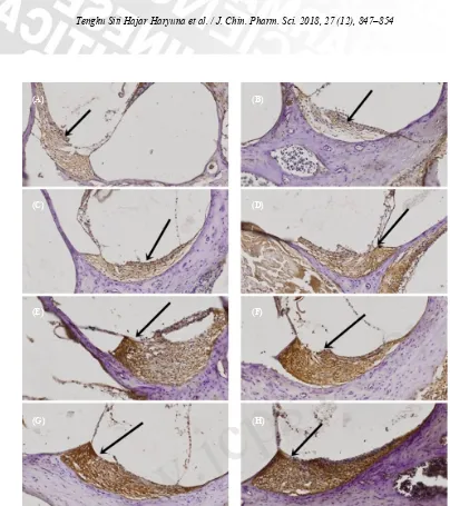
The effects of curcumin on cochlear lateral walls of ototoxic rat models were assessed by IHC. We assessed the expression of SOD by width and intensity of the brown-stained cytoplasms (immune-reactive score) (Table 1). Group given gentamicin without curcumin (Fig. 1B) showed the weakest expression of SOD compared with other group given curcumin (Fig. 1C–H). Prevention group (Fig. 1G–H) showed the strongest expression of SOD compared with group given gentamicin and curcumin (Fig. 1C–F).
Group Mean difference±Standard deviation P value
1
Table 1. ANOVA test results in regards of the expression of SOD.
*Denotes statistically significant.
4. Discussion
In this study, we analyzed the antioxidant activity of curcumin and assessed the expression of SOD in fibroblasts of ototoxic rat models.
The results of this study showed that the comparison between Groups 1 and 2 (positive control group) exhibited the statistically significant result, implying that gentamicin decreased the expression of SOD in fibroblasts of cochlea lateral wall. The similar changes were observed in melanocyte cells exposed to gentamicin, leading to significant changes in the cellular antioxidant enzymes: SOD, CAT and GPx, indicating the depletion of antioxidant
defense system[17]. Gentamicin used was 40 mg/mL, and the same gentamicin dose was used as the ototoxic control in the middle ears of Wistar Albino rats[14]. The rats were terminated at 18 h after they were given gentamicin injection in accordance with the results conducted on the Hartley strain guinea pig, in which the administration of gelatin sponge soaked in gentamicin at the round window membrane after 18 h shows the highest cell death markers[15].
The curcumin is given to the rats once per day for 7 d, which can effectively increase the SOD expression in rats[13]. Therefore, we implemented the same method.
Figure 1. The expression of SOD (100× zoom): (A) Group 1; (B) Group 2; (C) Group 3; (D) Group 4; (E) Group 5; (F) Group 6; (G) Group 7; (H) Group 8. The black arrow indicates the expression of SOD in cochlear fibroblasts marked by the brown-stained cytoplasm.
(A) (B)
(C) (D)
(E) (F)
(G) (H)
The results showed that the administration of curcumin enhanced the expression of SOD in fibroblasts of cochlea lateral wall in ototoxic rat models as seen in the comparison between Group 2 and Groups 3, 4, 5, 6. The similar result has been found in a study of Drosophila where the increased activity of SOD is observed on Drosophila receiving curcumin compared with those without curcumin administration[18]. The increased activity of SOD is also observed in a study in brain regions in rats receiving lead and curcumin compared with the group receiving lead only[19]. The curcumin dose is 100 mg/kg and this curcumin dose has been proven to effectively reduce cell death markers in rats undergoing hepatectomy[13]. A single dose of curcumin is enough to show the antioxidative effect[20]. In this study, the curcumin was given for 3 d.
The results showed that curcumin had a preventive effect as shown in Groups 7 and 8. The similar result has been found in a study conducted on rats, where SOD activity is higher in the group receiving curcumin before the administration of acetaminophen compared with the acetaminophen-administered group only[21].
Aminoglycosides, including gentamicin, are antibiotics widely used to eliminate Gram-negative bacterial infections, which have become popular again due to increased prevalence of antibiotic resistance in other classes[22]. There is no safe dose of aminoglycoside through any route of administrations (intravenous, intratympanic, oral and intrathecal)[23].
Curcumin extracted from Curcuma longa L. is widely available in Asia as a food seasoning, coloring and flavoring[12]. Curcumin has many pharmacological benefits, including antioxidant, anti-inflammatory, and anticancer activities[23].
Aminoglycosides-induced ototoxicity is developed from low frequency to high frequency and associated
with oxidative stress. Aminoglycosides, e.g. gentamicin, can react with iron to form ROS in the inner ear, inflicting permanent damage to hair cells and neurons. Excessive ROS levels trigger the apoptotic pathway, which then produces cell death caused by aminoglycoside -induced ototoxicity. Although aminoglycoside-induced
ototoxicity is well-documented, its molecular
mechanisms still have not been precisely determined[17]. The transtympanic route shows ototoxic damage in many species[24].
A general mechanism for the generation of ROS is the Fenton reaction: Fe2+ + H
2O2 → Fe3+ + HO· + HO–. When gentamicin reacts with iron salts, the gentamicin -iron complex enhances -iron-catalyzed oxidations and directly promotes the generation of ROS. In this process, unsaturated fatty acid acts as an electron donor. In turn, fatty acids, predominantly arachidonic acid, are oxidized to lipid peroxides. Since arachidonic acid is an essential fatty acid in cellular membranes, ROS can affect membrane fluidity and permeability. Through lipid peroxidation, ROS can influence nucleid proteins and acids, thereby disrupting the activity of enzymes, ion channels and receptors. If the formation of ROS exceeds the capacity of the repair and intrinsic protective systems, the cells undergo apoptosis[25].
The identification of the sensorial cell and organ of Corti endogenous defense mechanisms is in the form of antioxidant and detoxification enzymes SOD, catalase, glutathione peroxidase (GPx), reductase and glutathione S-transferase[26]. SOD converts superoxide anion radical (O2·–) into hydrogen peroxide (H2O2), and then hydrogen peroxide into highly reactive radical hydroxil (OH·–), or can be converted into water by the enzyme catalase or GPx. The use of gentamicin at high concentration may induce the decrease of antioxidant defense system[17].
Curcumin is a potent inhibitor toward the formation of ROS and also a potent scavenger against a variety of ROS, including superoxide anion radicals (O2·–)
and hydroxil radical (OH·–). Curcumin enhances the
activity of antioxidant enzyme SOD by nuclear factor erythroid-derived 2 (Nrf2)[27]. Curcumin has the role of ROS scavenger against superoxide anions (O2·–) and hydrogen peroxide (H2O2)[28] and it can increase the antioxidant activity of SOD[28–30], catalase, glutathione peroxydase, heme oxygenase-1 and glutathione transferase[28].
Curcumin is as great antioxidant agent only in higher dose[31] and as anti
-inflammatory agent being highlighted lately because it will reach great result if prepared in specific form[32].
5. Conclusions
This study showed curcumin’s ability for therapeutic and prevention against gentamicin ototoxicity in fibro-blast within the cochlear supporting tissues and lateral wall. Furthermore, this study exhibited the mechanism of curcumin underlying the increased expression of SOD and also scientific base for treatment and prevention of ototoxicity.
Competing Interests
The authors declares that there is no conflict of interest regarding the publication of this paper.
Acknowledgements
The authors thanked to DIPA Direktorat Penelitian Pengabdian kepada Masyarakat 2015, based on Surat Perjanjian Penugasan Pelaksanaan Hibah Penelitian Bagi Dosen Perguruan Tinggi Batch I Universitas Sumatera
Utara No: 120/SP2H/PL/Dit. Litabmas/II/2015, on February 5 2015 for the financial support. The authors also would like to thank Biochemisty Laboratory, Faculty of Medicine, Universitas Airlangga Surabaya; Anatomic Pathology Laboratory, Faculty of Medicine, Dr. Soetomo General Hospital Surabaya; Biochemistry Laboratory, Faculty of Medicine, Universitas Brawijaya Malang, for providing equipment and scientific apparatus.
References
[1] Hao, X.; Xing, Y.; Moore, M.W.; Zhang, J.; Han, D.; Schulte, B.A.; Dubno, J.; Lang, H. Sox10 Expressing Cells in the Lateral Wall of the Aged Mouse and Human Cochlea. PLoS ONE. 2014, 9, e97389.
[2] Baggio, C.L.; Silveira, A.F.; Hyppolito, M.A.; Salata, F.F.; Rossato, M. A functional study on gentamicin-related cochleotoxicity in its conventional dose in newborns. Braz. J. Otorhinolaryngol. 2010, 76, 91–95.
[3] Petersen, L.; Rogers, C. Aminoglycoside-induced hearing deficits of cochlear ototoxicity. S. Afr. Fam. Pract. 2015, 57, 77–82.
[4] De Oliveira, J.A.A.; Canedo, D.M.; Rossato, M.; De Andrade, M.H. Self-protection against aminoglycoside ototoxicity in guinea pigs. Otolaryngol. Head Neck Surg. 2004, 131, 271–279.
[5] Luo, J.; Xu, L. Distribution of Gentamicin in Inner Ear After Local Administration Via a Chitosan Glycerophosphate Hydrogel Delivery System. Ann. Otol. Rhinol. Laryngol. 2012, 3, 208–216.
[7] Oliveira, A.A.; Campos, M.S.; Murashima, A.A.B.; Rossato, M.; Hyppolito, M.A.; Oliveira, J.A.A. Persistence of otoprotective effect. How long does otoprotection against amikacin lasts?. Braz. J. Otorhinolaryngol. 2012, 78, 47–50.
[8] Perkins, K.; Sahy, W.; Beckett, R.D. Efficacy of Curcuma for Treatment of Osteoarthritis. J. Evid. Base. Complement. Altern. Med. 2017, 22, 156–165.
[9] Savcun, G.Y.; Ozkan, E.; Dulundu, E.; Topaloglu, U.; Sehirli, A.O.; Tok, O.E.; Ercan, F.; Sener, G. Antioxidant and anti-inflammatory effects of curcumin against hepatorenal oxidative injury in an experimental sepsis model in rats. Ulus Travma Acil. Cerr. Derg. 2013, 19, 507–515.
[10] Funk, J. L.; Oyarzo, J.N.; Frye, J.B.; Chen, G.; Lantz, R.C.; Jolad, S.D.; Solyom, A.M.; Timmermann, B.N. Turmeric Extracts Containing Curcuminoids Prevent Experimental Rheumatoid Arthritis. J. Nat. Prod. 2006, 69, 351–355.
[11] Singh, P.P.; Devi, K.R.; Devi, M.M.; Thokchom, D.S.; Sharma, G.J. Protection of Low Let Radiation-Induced DNA Damage in Rat Bone Marrow Cells by Free Radical Scavenger Curcumin. IJPSR. 2016, 7, 1168– 1178.
[12] Aktas, C.; Kanter, M.; Erboga, M.; Ozturk, S. Anti-apoptotic effects of curcumin on cadmium-induced apoptosis in rat testes. Toxicol. Ind. Health. 2012, 28, 122–130.
[13] Toydemir, T.; Kanter, M.; Erboga, M.; Oguz, S.; Erenoglu, C. Antioxidative, antiapoptotic and proliferative effect of curcumin on liver regeneration after partial hepatectomy in rats. Toxicol. Ind. Health. 2015, 31, 162–172.
[14] Sagit, M.; Somdas, M.A.; Korkmaz, F.; Akcadag, A. The ototoxic effect of intratympanic terbinafine applied in middle ear rats. J. Otolaryngol. Head Neck Surg. 2013, 42, 13.
[15] Suzuki, M.; Ushio, M.; Yamasoba, T. Time course of apoptotic cell death in guinea pig cochlea following intratympanic gentamicin application. Acta Otolaryngol. 2008, 128, 724–731.
[16] Tan, K.B.; Putti, T.C. Cyclooxygenase 2 expression in nasopharyngeal carcinoma: immunohistochemical findings and potential implications. J. Clin. Pathol. 2004, 58, 535–538.
[17] Wrzesniok, D.; Beberok, A.; Otreba, M.; Buszman, E. Impact of Gentamicin on Antioxidant Enzymes Activity in HEMn-DP Cells. Acta Pol. Pharm. 2015, 72, 447–453.
[18] Shen, L.R.; Xiao, F.; Yuan, P.; Chen, Y.; Gao, Q.K.; Parnell, L.D.; Meydani, M.; Ordovas, J.M.; Li, D.; Lai, C.Q. Curcumin-supplemented diets increase superoxide dismutase activity and mean lifespan in Drosophila. AGE (Dordr). 2013, 35, 1133–1142.
[19] Shukla, P.K.; Khanna, V.K.; Khan, M.Y.; Srimal, R.C. Protective effect of curcumin against lead neurotoxicity in rat. Hum. Exp. Toxicol. 2003, 22, 653–658.
[20] Morsy, M.A.; El-Moselhy, M.A. Mechanisms of the Protective Effects of Curcumin againsts Indomethacin -Induced Gastric Ulcer in Rats. Pharmacology. 2013, 91, 267–274.
[21] Bulku, E.; Stohns, S.J.; Cicero, L.; Brooks, T.; Halley, H.; Ray, S.D. Curcumin Exposure Modulates Multiple Pro-Apoptotic dan Anti-Apoptotic Signaling Pathways to Antagonize Acetaminophen-Induced Toxicity. Curr. Neurovasc. Res. 2012, 9, 58–71.
[22] Denamur, S.; Tyteca, T.; Marchand-Brynaert, J.; Bambeke, F.V.; Tulkens, P.M.; Courtoy, P.J.; Mingeot-Leclercq, M.P. Role of oxidative stress in lysosomal membrane permeabilization and apoptosis induced by gentamicin, an aminoglycoside antibiotic. Redox. Biol. 2011, 51, 1656–1665.
[23] Cianfrone, G.; Pentangelo, P.; Cianfrone, F.; Mazzei, F.; Turchetta, R.; Orlando, M.P.; Altissimi, G. Pharmacological drugs inducing ototoxicity, vestibular symptoms and tinitus: a reasoned and updated guide. Eur. Rev. Med. Pharmacol. Sci. 2011, 15, 601–636.
[24] Kalkandelen, S.; Selimoglu, E.; Erdogan, F.; Ucuncu, H.; Altas, E. Comparative Cochlear Toxicities of Streptomycin, Gentamicin, Amikacin and Netilmicin in Guinea-pigs. J. Int. Med. Res. 2002, 30, 406–412.
[25] Huth, M.E.; Ricci, A.J.; Cheng, A.G. Mechanisms of Aminoglycoside Ototoxicity and Targets of Hair Cell Protection. Int. J. Otolaryngol. 2011, 937861.
[26] Aquino, T.J.M.; Oliveira, J.A.A.; Rossato, M. Ototoxicity and otoprotection in the inner ear of guinea pigs using gentamicin and amikacin: ultrastructural and functional aspects. Rev. Bras. Otorrinolaringol. 2008, 74, 843–852.
[27] Attia, A.M.M.; Ibrahim, F.A.A.; El-Latif, N.A.A.;
Aziz, S.W. Antioxidant effects of curcumin againts cadmium chloride-induced oxidative stress in the blood of rats. J. Pharmacognosy Phytother. 2014, 6, 33–40.
[28] Pulido-Moran, M.; Moreno-Fernandez, J.; Ramirez-Tortosa, C.; Ramirez-Tortosa M.C. Curcumin and Health. Molecules. 2016, 21, 264.
[29] Hussein, S.A.; Azab, M.; El-Shall, S.K. Protective Effect of Curcumin on Antioxidant Defense System and Oxidative Stress in Liver Tissue of Iron Overloading Rats. Asian J. Clin. Nutrition. 2014, 6, 1–17.
[30] Motaghinejad, M.; Karimian, M.; Motaghinejad, O.; Shabab, B.; Yazdani, I.; Fatima, S. Protective effect of various dosage of Curcumin against morphine induced apoptosis and oxidative stress in rat isolated hippocampus. Pharmacol. Rep. 2015, 67, 230–235.
[31] Szaroma, W.; Lach, H.; Dziubek, K.; Goc, Z. Effect of The Single Doses of Curcumin and Kainic Acid on Changes in the Amount of Reduced Glutathione in Selected Organs of Mice. J. Microbiol. Biotechn. Food Sci. 2016, 5, 341–344.
[32] Toden, S.; Theiss, A.L.; Wang, X.; Goel, A. Essential Turmeric Oils Enhance Anti-Inflammatory efficacy of Curcumin in Dextran Sulfate Sodium-Induced Colitis. Sci. Rep. 2017, 7, 814.