TROPICAL AGRICULTURAL SCIENCE
Journal homepage: http://www.pertanika.upm.edu.my/
Article history:
Received: 18 September 2017 Accepted: 25 June 2018 Published: 29 August 2018 ARTICLE INFO
E-mail addresses:
ruth_c @staff.ubaya.ac.id (Ruth Chrisnasari) [email protected] (Devi Verina)
[email protected] (Aime Clorinda Tapatfeto) [email protected] (Stefan Pranata)
[email protected] (Tjandra Patjajani)
[email protected] (Mariana Wahjudi) [email protected] (Maria Goretti Marianti Purwanto) * Corresponding author
Isolating and Characterising Chitinolytic Thermophilic Bacteria
from Cangar Hot Spring, East Java
Ruth Chrisnasari*, Devi Verina, Aime Clorinda Tapatfeto, Stefan Pranata, Tjandra Patjajani, Mariana Wahjudi and Maria Goretti Marianti Purwanto Department of Biology, Faculty of Biotechnology, University of Surabaya, Jalan Raya Kalirungkut, Surabaya 60293, Indonesia
ABSTRACT
In the present study, chitinolytic thermophilic bacteria were collected from Cangar hot spring, East Java, Indonesia and screened. The 16S rRNA gene sequencing was used to
identify the isolated bacterium which showed highest chitinolytic activity. The identified
isolate was then characterised based on morphological and physiological analyses. The results showed the isolated bacterium belonged to Bacillus licheniformis. This isolate produced large amounts of chitinase on 0.9% (w/v) colloidal chitin (pH 7.0) at 52°C in a very short time (24 hours). Two pairs of primer were designed to detect the presence of glycosyl hydrolase (GH) 18 chitin domain sequences in the isolated bacterium. Two amplicons sized ~250 bp and ~1000 bp were obtained from PCR process. Then the amplicons were sequenced and analysed. The sequencing results showed the isolated Bacillus licheniformis was proven to have genes encoding ChiA and ChiC domain.
Keywords: Bacillus licheniformis, ChiA, ChiC, thermophilic bacteria, thermostable chitinase
INTRODUCTION
Chitinases (EC 3.2.1.14) are grouped into either Family 18 or Family 19 under glycosyl hydrolases superfamily which is capable of degrading chitin into its derivates
by hydrolysing the β-1,4-glycosidic bonds
Tjandra Patjajani, Mariana Wahjudi and Maria Goretti Marianti Purwanto
N-acetylD-glucosamine (Ramirez-Coutino, Marin-Cervantes, Huerta, Revah, & Shirai, 2006). Chitooligosaccharides produced by enzymatic hydrolysis of chitin has been
especially used in pharmaceuticals fields
as antioxidant, immunostimulant (Shahidi, Arachchi, & Jeon, 1999), antihypertensive, antibacterial, antifungal, and as a food quality enhancer (Bhattacharya, Nagpure, & Gupta, 2007).
Chitinases are produced by various microbes and recognised as extracellular inducible enzymes. Most bacteria secrete Family 18 chitinases to degrade chitin and
utilise it as an energy source (Hart, Pfluger,
Monzingo, Hoihi, & Robertus, 1995). The superiority of chitinase-producing bacteria is one of the key factors in the enzyme production. The high biodiversity in Indonesia presents a great opportunity to get potential bacteria with special characteristic to be used as enzymes producer. Therefore, the exploration of the chitinase-producing bacteria is vital Indonesia. Chitinolytic thermophilic bacteria can be isolated from both soil and aquatic thermophile habitats i.e. hot spring and crater. The advantage of using thermophilic bacteria is their ability to synthesise the heat stable molecule, including enzymes. Thermostable enzymes produced by thermophilic bacteria are
very effective and beneficial for industrial
processes that need high temperature — e.g. chitin degradation in pharmaceutical industries and waste processing in seafood industry. High temperature can improve
reaction speed, increase the solubility of the reactants and non-volatile products as well as reducing mesophilic microbial contamination (Martin, Delatorre, & Camila, 2007).
The aim of this study was to isolate the most prominent local chitinolytic thermophilic bacteria from Cangar Hot Spring, East Java for thermostable chitinase production. The obtained isolate then was
identified based on molecular, morphological and physiological analyses. The identified
isolate was used to produce chitinase
under specific condition. The isolate was
then further characterised by detection of glycosyl hydrolase (GH) 18 chitin domain sequences in the isolate genome using PCR based method.
MATERIALS AND METHODS
Enrichment and Cultural Medium
Nutrient Broth (NB) (Merck) and Luria Bertani (LB) broth (Scharlou) were used as enrichment medium. Thermus colloidal chitin (TCC) broth containing 0.7% (w/v) (NH4)2SO4, 0.1% (w/v) K2HPO4, 0.1% NaCl,
0.01% (w/v) MgSO4·7H2O, 0.05% (w/v)
yeast extract, 0.1% (w/v) bactotryptone and 0.5% colloidal chitin (Yuli, Suhartono, Rukayadi, Hwang, & Pyun, 2004) was used as culture medium. The TCC agar medium for screening process was made by adding 15 g L-1bacto agar in the TCC broth medium.
Bacterial Isolation, Screening and
Identification
A total of four different soil and water mixture samples were aseptically collected from different regions of Cangar Hot Spring, East Java, Indonesia. The four samples were enriched in NB and LB broth solution respectively with sample and medium ratio 1:3. The enriched samples were incubated for 24 hours at 52°C with 150 rpm of shaking speed. Bacterial strains were isolated and screened from enriched medium following standard procedures using spread plate technique on TCC agar plates. Morphologically distinct colonies
were sub-cultured in TCC broth and purified
to single species level using streak plating repeatedly on TCC agar plates. Pure isolates were maintained by sub-culturing on TCC slants and stored at 4°C.
The pure isolates were screened for chitinase activity in TCC broth. The isolates were previously grown in LB broth at 52°C until each isolate reach 0.5 of OD600. As
much as 1 mL of each isolate taken and added to 9 mL of TCC broth and incubated for 36 hours at 52°C. The samples were then centrifuged at 4000 rpm for 3 minutes. The supernatant was used for N-acetyl D-glucosamine detection using Nelson– Somogyi assay (Nelson, 1944).
The selected isolate was identified through partial 16S rRNA gene sequencing analysis. Chromosomal DNA of the isolate was extracted from the pure culture using Fungal/ Bacterial DNA MiniPrep Kit (Zymo Research) and amplified using
a pair of 16S universal primer (Botha, Botes, Loos, Smith, & Dicks, 2012) ordered from Macrogen, Korea (Forward: 5 ’ - C A C G G AT C C A G A C T T T G AT Y M T G G C T C A G - 3 ’ a n d R e v e r s e : 5’-GTGAAGCTTACGGYTAGCTTGTTA
CGACTT-3’). The amplification
reaction mixture contained 5 μl of 16S forward primer 10 μM/μl, 5 μl of 16S reverse primer 10 μM/μl, 25 μl of GoTaq Green Master Mix 2X (Intron), 2.5 μl of DMSO, and 12.5 μl of double-distilled
water (ddH2O). The amplification was
performed with initial denaturation at 94 ºC for 5 min, 30 cycles of denaturation at 94 ºC for 45 sec, annealing at 55 ºC for 1 min, and elongation at 72 ºC for 1.5 min
followed by final elongation at 72 ºC for
5 minutes. The preparation of samples for sequencing analysis was as follows:
(1) the PCR products were purified using PCR Purification Kit (Roche), cloned into
pGEMT-Easy (Promega) and transformed to E. coli DH5α competent, (2) the transformed
cells were confirmed by colony PCR method,
(3) DNA plasmid was extracted from the transformed cells using Plasmid Isolation Kit (Roche) and analysed for sequencing (Macrogen, Korea). The homology analysis of 16S rRNA gene sequence was conducted using BLAST algorithm in GenBank (http:// blast.ncbi.nlm.nih.gov/Blast.cgi). Bacterial
confirmation and characterisation through
Tjandra Patjajani, Mariana Wahjudi and Maria Goretti Marianti Purwanto Chitinase Production
As much as 10% (v/v) of isolate was inoculated into TCC broth medium and agitated at 180 rpm (Yin Der shaker incubator). The fermentation conditions were 0.9% (w/v) of colloidal chitin concentration, pH 7.0 and a temperature of 52ºC. Sub-sample of the culture (50
mL) at initial and final fermentation was
concentrated and analysed for chitinase activity assay (Rahayu, Fredy, Maggy, Hwang, & Pyun, 1999).
Chitin Domain Sequence Detection
Chitin Domain Sequence (CDS) was detected based on PCR method using 2 pairs of primer. The first primer was designed to detect ChiA (FChiA: 5 ’ - G G Y G T C G AT V T S G A C T G G G A G T A Y C C - 3 ’ a n d R C h i A : 5 ’ - T C R T A G G T C A T R A T A T T GATCCARTC-3’). The second primer was designed to detect ChiB (FChiB: 5 ’ - C T A C G C C G G A A T A C G A A G G G A T C G G A T A - 3 ’ a n d 5’-AACTCCGCTTCCTCACCAGGTT-3’). Amplification reaction was made in 100
μl containing 100 ng chromosomal DNA, 10 μM/μl forward and reverse primers respectively, 50 μl GoTaq Green Master Mix
2X, and ddH2O. Amplification process was
performed with initial denaturation at 95°C for 5 min, 35 cycles consist of denaturation 95°C for 45 sec, gradient annealing with varied temperature of 53-66°C for 45 sec, and elongation 72°C for 1 min, followed by
final elongation 72°C for 10 minutes. PCR
product was visualised using agarose gel
electrophoresis. The remaining PCR product
was purified and prepared for sequencing
analysis.
RESULTS AND DISCUSSION
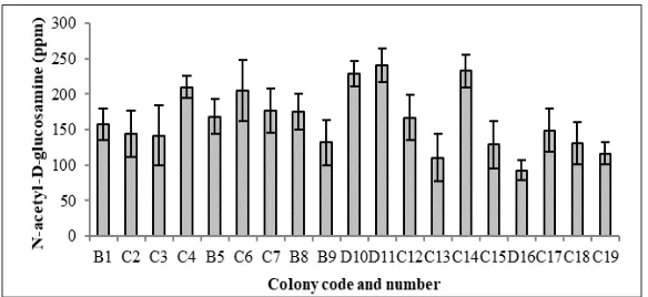
Soil and water mixture samples were taken from four different location of Cangar Hot Spring. Of the four locations (named as location “A”, “B”, “C” and “D”), 19 single colonies with chitinolytic activity were obtained, where 4 colonies obtained from location B, 12 colonies at locations C and 3 colonies at locations D. None of the colony obtained from location A. The 19 colonies then were screened for chitinolytic activity in TCC broth medium based on amount of N-acetyl D-glucosamine produced as presented at Figure 1. From the data, colony D11 showed highest chitinolytic activity compared to the other colonies, although it
is not significantly different with colony C14
and D10 (p-value > 0.05). The D11 colony
was then identified, characterised and used
for further experiments.
chitin waste degradation (Kamil, Rizk, Saleh, & Moustafa, 2007; Veith et al., 2004).
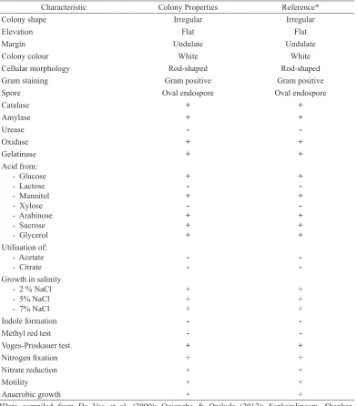
T h e c h a r a c t e r i s a t i o n a s s a y o n morphological and physiological analysis based on Bergey’s Manual of Systematic Bacteriology is presented in Table 1. Bacillus licheniformis D11 showed a positive result in the following tests: catalase, amylase, oxidase, and gelatinase production; acid production from glucose, mannitol, arabinose, sucrose and glycerol; growth in 2-7% (w/v) NaCl; Voges-Proskauer test; nitrogen fixation; nitrate reduction, motility and anaerobic growth. Bacillus licheniformis D11 showed a negative result in the following tests: acid production from lactose and xylose, hydrolysis of urea, utilization of acetate and citrate; indole formation; methyl red test and indole formation. The growth of Bacillus licheniformis D11 on TCC broth medium showed the lag (0-4 h), log (4-16 h), stationary (16-28 h) and the death phase (28-48 h) during incubation time (Figure 2).
In correlation to the cell growth curve of Figure 2, chitinase had been produced since the log phase and achieved the optimum at
the middle of stationary phase (24 h). The enzyme production was then decreased at 36-48 hours due to lack of nutrients or secretion of toxic substances which inactivated the enzymes (Saima, Roohi, & Ahmad, 2013). Bacillus licheniformis D11 achieved optimum amounts of chitinase in a very short time (Figure 3), 24 hours, compared with the other chitinase producer bacteria. Microbispora sp. (Nawani, Kapadnis, Das, Rao, & Mahajan, 2002), B. cereus, B. sphaericus and B. alvei (Wang & Hwang, 2001), as well as Aeromonas punctata and Aeromonas hydrophila (Saima et al., 2013) produced the highest chitinase after 48 h. Bacillus sp. HSA,3-1a had been reported to produce the highest chitinase at the end of the stationary phase after 72 h incubation time (Natsir, Patong, Suhartono, & Ahmad, 2010). The short production time revealed Bacillus licheniformis D11 to be one of the prominent chitinase producers.
Detecting the presence of glycosyl hydrolase (GH) 18 Chitin Domain Sequence (CDS) in Bacillus licheniformis D11 genome was done by PCR method using 2 pairs of
primer. The first primer was designed to
Tjandra Patjajani, Mariana Wahjudi and Maria Goretti Marianti Purwanto
Characteristic Colony Properties Reference*
Colony shape Irregular Irregular
Elevation Flat Flat
Margin Undulate Undulate
Colony colour White White
Cellular morphology Rod-shaped Rod-shaped
Gram staining Gram positive Gram positive
Spore Oval endospore Oval endospore
Catalase + +
Growth in salinity - 2 % NaCl
Indole formation -
-Methyl red test -
-Voges-Proskauer test + +
Nitrogen fixation + +
Nitrate reduction + +
Motility + +
Anaerobic growth + +
Table 1
Morphological and physiological characteristic of d11 isolate
*Data compiled from De Vos et al. (2009); Oziengbe & Onilude (2012); Sankaralingam, Shankar, Ramasubburayan, Prakash and Kumar (2012); Waldeck, Daum, Bisping and Meinhardt (2006).
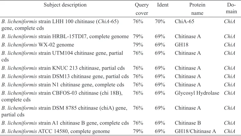
detect ChiA. Amplification using this primer by gradient thermocycler in variation of annealing temperature (Ta47-60°C) produced
one amplicon sized ~250 bp (Figure 4) which was later sequenced and analysed.
Figure 2. The growth of Bacilluslicheniformis D11in thermus colloidal chitin broth medium pH 7.0 at 52ºC for 48 hours
Figure 3. Chitinase production of Bacilluslicheniformis D11 in thermus colloidal chitin broth medium (pH 7.0) at 52ºC
Tjandra Patjajani, Mariana Wahjudi and Maria Goretti Marianti Purwanto
Subject description Query Ident Protein
Do-main
cover name
B. licheniformis strain LHH 100 chitinase (ChiA-65) gene, complete cds
76% 70% ChiA-65 ChiA
B. licheniformis strain HRBL-15TDI7, complete genome 79% 69% Chitinase A ChiA B. licheniformis WX-02 genome 79% 69% GH18 ChiA B. licheniformis strain UTM104 chitinase gene, partial
cds
76% 69% Chitinase A ChiA
B. licheniformis strain KNUC 213 chitinase, partial cds 76% 69% Chitinase A ChiA B. licheniformis strain DSM13 chitinase gene, partial cds 76% 69% Chitinase A ChiA B. licheniformis strain N1 chitinase gene, complete cds 76% 69% Chitinase A ChiA B. licheniformis strain CBFOS-03 chitinase (chi 18B),
complete cds
76% 69% Glycosyl Hydrolase ChiA
B. licheniformis strain DSM 8785 chitinase (chiA) gene, partial cds
76% 69% Chitinase A ChiA
B. licheniformis strain A1 chitinase B gene, complete cds 76% 69% Chitinase B ChiA B. licheniformis ATCC 14580, complete genome 79% 69% GH18/Chitinase A ChiA
Table 2
Sequence alignment result of ChiA amplicon using BLAST-n NCBI
licheniformis, B. cereus, B. thuringiensis, and B. pumilus. In bacteria, the function of this gene is to degrade insoluble chitin into its derivates and plays an important role in the defence mechanism against pathogens (Funkhouser & Aronson, 2007). ChiA domain sequence consists of catalytic domain (GH18), fibronectin domain III (Fn3), and chitin binding domain (CBD) (Herdyastuti, Tri, Mudasir, & Sabirin, 2009; Islam et al., 2010). Amplification using ChiB primer by gradient thermocycler in variation of annealing temperature (Ta
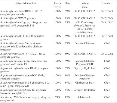
53-66°C) produced one amplicon sized ~1000 bp (Figure 5) which was sequenced and analysed. Based on sequence alignment (BLASTn) result, this sequence had high levels of similarities with ChiA and ChiC domain sequence in B. licheniformis (B. licheniformis strain HRBL-15TDI7, B.
licheniformis WX-02, dan B. licheniformis chiB gene strain F11) (Table 3). This result confirmed ChiB primer can detect the presence of ChiA and ChiC domain sequence in B. licheniformis D11 due to high level of similarity between the domains.
ChiA, ChiB, and ChiC belong to the group GH18. From the amino acid sequence, ChiC has different amino acid sequence compared with ChiA and ChiB. ChiB has
a lower specific activity than ChiA because
of the absence of fibronectin domain III.
In addition, ChiB cuts GlcNAc oligomers shorter than ChiA (Brurberg, Nesl, & Eijsink, 1996). ChiB can be found in Aspergillus fumigatus, Photorhabdus themperata, and some strains of B. licheniformis. ChiC has three functional domains, namely
N-terminal domain, fibronectin domain III,
Subject description Query Ident Protein Domain
cover name
B. licheniformis strain HRBL-15TDI7, complete genome cds
100% 99% Chi C, GH18, Chi A ChiC, ChiA
B. licheniformis WX-02 genome 100% 99% Chi C, GH18, Chi A ChiC, ChiA B. licheniformis chiB gene, chiA gene, mpr
gene and ycdF gene, strain F11
100% 99% Chi C (binding domain), Precursor
ChiB, Putative Dehidrogenase
ChiA, ChiC
B. licheniformis ATCC 14580, complete genome
100% 99% Chi C, GH18, Chi A ChiC, ChiA
B. licheniformis strain SK-1 chitinase precursor (chiB) and putative chitinase precursor
100% 99% Putative Chitinase ChiA
B. licheniformis DSM13 = ATCC 14580, complete genome
100% 99% Chi C, GH18, Chi A ChiC, ChiA
B. licheniformis chiB gene, chiA gene, mpr gene and ycdF, strain F5
100% 99% Putative Chitinase Precursor ChiB
ChiB
B. paralicheniformis strain BL-09, complete genome
100% 99% Glycosyl Hydrolase ChiA
B. paralicheniformis strain ATCC 9945a, complete genome
100% 94% Putative Chitinase Precursor
ChiA
B. licheniformis strain MS-3 chitinase A-BL3 (chiA) gene, complete cds
100% 94% Chitinase A-BL3 ChiA
B. licheniformis gh18D gene for glycoside hydrolase, complete cds
100% 94% Glycosyl Hydrolase ChiA
Bacillus sp. AV2-9 chitinase large (chiL) gene, complete cds
99% 82% Chitinase L ChiA
Table 3
Sequence alignment result of ChiB amplicon using BLAST-n NCBI
Tjandra Patjajani, Mariana Wahjudi and Maria Goretti Marianti Purwanto
ChiC is similar to the C-terminal extension of ChiA (Tsujibo et al., 1998). Chitinase gene with ChiC domain can be found in Streptomyces lividans, Paenibacillus spp., Pseudomonas sp., Serratia marcescens and Bacillus weihenstephanensis.
CONCLUSION
A total of 19 chitinolytic thermophilic bacteria were collected from Cangar hot spring, East Java, Indonesia. From the screening process, D11 isolate had the highest chitinolytic activity. The D11 isolate was identified as Bacillus licheniformis through molecular, morphological and physiological analyses. This isolate produced large amounts of chitinase (4.49x10⁻³μmol/ml. minutes) on 0.9% (w/v)
colloidal chitin (pH 7.0) at 52 °C in a very short time, 24 hours compared with other Bacillus sp. The sequence analysis showed that the isolated Bacillus licheniformis was proven to have genes encoding ChiA and ChiC domain. This isolate can be used for further application on chitinous waste degradation or chitin derivates production in pharmaceutical industries.
ACKNOWLEDGMENT
The authors thank Lembaga Penelitian dan Pengabdian Masyarakat (LPPM), University of Surabaya for its research funding through Hibah Penelitian Lanjut, ST. 087/Lit/LPPM-01/FTB/VIII/2013 and Hibah Kompetitif, ST. 007/Lit/LPPM-01/FTB/III/2016.
REFERENCES
Bhattacharya, D., Nagpure, A., & Gupta, R. K. (2007). Bacterial chitinases: Properties and potential.
Critical Reviews in Biotechnology, 27, 21–28. Botha, M., Botes, M., Loos, B., Smith, C., & Dicks, L.
M. T. (2012). Lactobacillus equigenerosi strain Le1 invades equine epithelial cells. Applied and Environmental Microbiology, 78(12), 4248-4255.
Brurberg, M. B., Nesl, I. F., & Eijsink, V. G. H. (1996). Comparative studies of chitinases A and B from Serratia marcescens. Microbiology, 142, 1581–1589.
De Vos, P., Garrity, G. M., Jones, D., Krieg, N. R., Ludwig, W., Rainey, F.A., & Whitman, W. B. (2009). Bergey’s manual of systematic bacteriology second edition: Volume 3: The
firmicutes. New York, NY: Springer.
Funkhouser, J. D., & Aronson J. (2007). Chitinase family GH18: Evolutionary insight from genomic history of a diverse protein family.
BMC Evolutionary Biology, 7(96), 1–16.
Hart, P. J., Pfluger, H. D., Monzingo, A. F., Hoihi, T., & Robertus, J. D. (1995). The refined crystal
structure of an endochitinase from Hordeum vulgare L. seeds at 1.8 Å resolution. Journal of Molecular Biology, 248, 402–413.
Herdyastuti, N., Tri, J. R., Mudasir, Sabirin, M. (2009). Kitinase dan mikroorganisme kitinolitik: isolasi, karakterisasi dan manfaatnya [Chitinase and kitinolytic microorganisms: Isolation,
characterization and its benefits]. Indonesian Journal of Chemistry, 9(1), 37–47.
Hsu, S. C., & Lockwood, J. L. (1975). Powdered chitin agar as a selective medium for enumeration of actinomycetes in water and soil. Applied Microbiology, 29(3), 422–426.
shell as a probe to detect chitin in marine shells.
Applied Microbiology and Biotechnology, 86(1), 119-129.
Kamil, Z., Rizk, M., Saleh, M., & Moustafa, S. (2007). Isolation and identification of rhizosphere soil chitinolytic bacteria and their potential in antifungal biocontrol. Global Journal of Molecular Sciences, 2(2), 57–66.
Martin, M. L. L., Delatorre, A. B. S., & Camila, R. (2007). Effect of culture conditions on the production of extracellular protease by thermophilic Bacillus sp. and some properties of the enzymatic activity. Brazilian Journal of Microbiology, 38, 253–258.
Natsir, H., Patong, A. R., Suhartono, M. T., & Ahmad, A. (2010). Production and characterization of chitinase enzymes from sulili hot spring in south Sulawesi, Bacillus sp. HSA, 3-1a. Indonesian Journal of Chemistry,10(2), 263–267.
Nawani, N. N., Kapadnis, B. P., Das, A. D., Rao,
A. S., & Mahajan, S. K. (2002). Purification
and characterization of a thermophilic and acidophilic chitinase from Microbispora sp. V2.
Journal of Applied Microbiology, 93, 965–975. Nelson, N. A. (1944). A photometric adaptation of
the somogyi method for the determination of glucose. The Journal of Biological Chemistry,
153, 375–380.
Oziengbe, E. O., & Onilude, A. A. (2012). Production
of a thermostable α-amylase and its assay using Bacillus licheniformis isolated from excavated land sites in Ibadan, Nigeria. Bajopas,5(1), 132–138.
Rahayu, S., Fredy, T., Maggy, T. S., Hwang, J. K., &
Pyun, Y. R. (1999). Eksplorasi bakteri termofilik
penghasil enzim kitinase asal Indonesia [Exploration of thermophilic bacteria producing
enzyme kitinase origin Indonesia]. Prosiding Seminar Hasil-Hasil Penelitian Bidang Ilmu Hayat (pp. 349-356). Bogor, Indonesia: Pusat Antar Universitas Ilmu Hayat IPB.
Ramirez-Coutino, L., Marin-Cervantes, M. D. C., Huerta, S., Revah, S., & Shirai, K. (2006). Enzymatic hydrolysis of chitin in the production of oligosaccha-rides using Lecanicillium fungicola chitinases. Process Biochemistry, 41, 1106–1110.
Saima, M. K., Roohi, I. Z., & Ahmad (2013). Isolation of novel chitinolytic bacteria and production optimization of extracellular chitinase. Journal of Genetic Engineering and Biotechnology, 11, 39–46.
Sankaralingam S., Shankar, T., Ramasubburayan, R., Prakash, S., & Kumar, C. (2012). Optimization of culture conditions for the production of amylase from Bacillus licheniformis on submerged fermentation. American-Eurasian Journal of Agricultural & Environmental Science,12(11), 1507–1513.
Shahidi, F., Arachchi, J. K. V., & Jeon, Y. J. (1999). Food applications of chitin and chitosan. Trends in Food Science & Technology, 10, 37–51. Shaikh, S. A. & Deshpande M. V. (1993). Chitinolytic
enzymes: Their contribution to basic and applied research. World Journal of Microbiology and Biotechnology, 9, 468–475.
Takayanagi, T., Ajisaka, K., Takiguchi, Y., & Shimahara, K. (1991). Isolation and characterization of thermostable chitinases from Bacillus licheniformis X-7u. Biochimica et Biophysica Acta (BBA) - Protein Structure and Molecular, 1078(3), 404–410.
Tantimavanich, S., Pantuwatana, S., Bhumiratana, A., & Panbangred, W. (1998). Multiple chitinase enzymes from a single gene of Bacillus licheniformis TP-1. Journal of Fermentation and Bioengineering, 85(3), 259–265.
Tjandra Patjajani, Mariana Wahjudi and Maria Goretti Marianti Purwanto
Tsujibo, H., Orikoshi, H., Shiotani, K., Hayashi, M., Umeda, J., Miyamoto, K., & Inamori, Y. (1998). Characterization of chitinase C from a marine bacterium, Alteromonas sp. strain O-7, and its corresponding gene and domain structure.
Applied and Environmental Microbiology, 64(2), 472-478.
Veith, B., Herzberg, C., Steckel, S., Feesche, J., Maurer, K. H., Ehrenreich, P., Gottschalk, G. (2004). The complete genome sequence of
Bacillus licheniformis DSM13, an organism with great industrial potential. Journal of Molecular Microbiology and Biotechnology, 7, 204–211. Waldeck J., Daum, G., Bisping, B., & Meinhardt, F.
(2006). Isolation and molecular characterization
of chitinase-deficient Bacillus licheniformis
strains capable of deproteinization of shrimp shell waste to obtain highly viscous chitin.
Applied and Environmental Microbiology,
72(12), 7879–7885.
Wang, S., & Hwang, J. (2001). Microbial reclamation of shellfish wastes for the production of chitinases. Enzyme and Microbial Technology,
28(4-5), 376–382.