Oestrogen, its receptors and function in the male reproductive
tract — a review
Rex A. Hess
a,*, David Bunick
a, Janice Bahr
b aDepartment of Veterinary Biosciences,Uni6ersity of Illinois,2001S.Lincoln,Urbana,IL61802,USA
bDepartment of Animal Sciences,Uni
6ersity of Illinois,2001S.Lincoln,Urbana,IL61802,USA
Abstract
Oestrogen is synthesized in the male reproductive system by at least three different cell types; Sertoli, Leydig and germ cells.
Although testosterone is recognized as the primary sex steroid in man, oestrogen is produced in sizable quantities in the testis, as
well as the brain and is found in extremely high concentrations in the semen of several species. The high concentration of
oestrogen in rete testis fluid of the rodent is now thought to be derived from the conversion of testosterone to estradiol by P450
aromatase in germ cells of the testis and spermatozoa traversing the reproductive tract. This new major source of oestrogen would
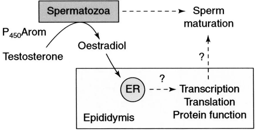
target oestrogen receptors in the male reproductive tract, in particular the efferent ductules, which contain the highest
concentration of oestrogen receptor-
a
. This recent data raises new hypotheses regarding the role of oestrogen in the function of
the male reproductive system. The oestrogen receptor-
a
knockout mouse was used to help define the function of oestrogen in the
male. It was found that oestrogen receptor-
a
is essential for fluid reabsorption in the efferent ductules and in the absence of
expression the male is infertile. © 2001 Elsevier Science Ireland Ltd. All rights reserved.
Keywords:Oestrogen receptor; Testis; Efferent ductules; Epididymis; Fluid reabsorption
www.elsevier.com/locate/mce
1. Oestrogen source and concentration
Oestrogen is synthesized in the male reproductive
system by at least three different cell types; Sertoli,
Leydig and germ cells. Although testosterone is
recog-nized as the primary sex steroid in man, oestrogen is
produced in sizable quantities in the testis, as well as
the brain (Roselli et al., 1997) and is found in extremely
high concentrations in the semen of several species
(Claus et al., 1992, 1987; Free and Jaffe, 1979; Ganjam
and Amann, 1976). Early studies of oestrogen reported
that the primary source of oestrogen in the immature
male was the Sertoli cell (van der Molen et al., 1981). In
the adult testis, Leydig cells express P450arom and
actively synthesize estradiol at a rate much greater than
that seen in the adult Sertoli cell (Payne et al., 1976;
Carreau et al., 1999; Levallet and Carreau, 1997;
Leval-let et al., 1998). Currently, a growing body of evidence
indicates that germ cells also synthesize oestrogen, and
possibly serve as the major source of this steroid in the
male reproductive tract. In 1993, in collaboration with
the laboratories of Bahr and Bunick (Nitta et al., 1993)
we reported for the first time that P450arom is present
in testicular germ cells of the adult male testis. The
enzyme was localized in the Golgi of round spermatids,
throughout the cytoplasm of elongating spermatids,
and along the flagella of late spermatids. Its presence in
these cell types was confirmed by Western blot analysis
of isolated germ cells and Northern blot analysis
demonstrated that its mRNA was present in testicular
germ cells. P450arom activity was also measured in
germ cells using the
3H
2
O water assay. Its activity was
equal to or exceeded the activity found in isolated
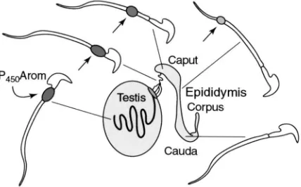
interstitial cells. Thus, it appeared from this early work
that sperm could serve as mobile endocrine units,
capa-ble of producing oestrogen that would target oestrogen
receptors (ER) downstream from the testis (Fig. 1).
The presence of P450arom in male germ cells has
now been demonstrated in several species, including
mouse, rat, brown bear and rooster (Hess et al., 1995;
Kwon et al., 1995; Nitta et al., 1993; Janulis et al.,
1998; Tsubota et al., 1993). P450arom presence in germ
cells and spermatozoa was recently confirmed and
* Corresponding author. Fax: +1-217-2441652.R.A.Hess et al./Molecular and Cellular Endocrinology 000 (2001) 000 – 000
2
shown to represent approximately 62% of the total
testicular aromatase (Carreau et al., 1999; Levallet et
al., 1998). The observation that germ cells of the testis
are capable of synthesizing oestrogen raises new and
exciting hypotheses regarding the potential for
oestro-gen to regulate functions along the epididymal tract
(Fig. 2). The protein has been immunolocalized in the
cytoplasmic droplet of the sperm tail, and the staining
becomes less intense as sperm traverse the epididymis
(Janulis et al., 1996).
The concentration of oestrogens in peripheral blood
is typically very low in the male, but ranges from 2 to
180 pg
/
ml depending upon the species. The horse is an
exception, where estrone sulfate is found as high as
2447 pg
/
ml (Setchell and Cox, 1982; Claus et al., 1992).
Oestrogen concentrations are typically higher in the
testicular vein than in general circulation (Waites and
Einer-Jensen, 1974; de Jong et al., 1973; Setchell and
Cox, 1982; Setchell et al., 1983). In the male rat
repro-ductive tract, oestrogen concentrations are quite high,
approximately 250 pg
/
ml in rete testis fluid (Free and
Jaffe, 1979), which is higher than the average serum
concentrations of estradiol in the female rat (Overpeck
et al., 1978; Robaire and Hermo, 1988). Oestrogens
have also been found to be abundant in semen (Bujan
et al., 1993; Claus et al., 1992; Eiler and Graves, 1977;
Ganjam and Amann, 1976; Claus et al., 1985).
2. Oestrogen receptors in the male tract
In 1975, Danzo suggested that oestrogen might be
capable of binding to receptors in the epididymal
ep-ithelium and serve some type of function in the male.
He found in the rabbit that cytosol-specific estradiol
(E2) binding was highest in the cauda epididymidis of
the immature animal (Toney and Danzo, 1988). During
the 1980s, autoradiographic localization of
3H-E
2
was
used to identify epithelial-positive tissues in the male
reproductive system. Schleicher et al. (1984) found
strong labeling of the efferent ductules and initial
seg-ment epididymis, with lesser binding in the distal tract.
Using immunocytochemistry (ICC), ER have been
lo-calized primarily in the epithelium of efferent ductules
(Goyal et al., 1998; Fisher et al., 1997; Goyal et al.,
1997; Kwon et al., 1997; Ergun et al., 1997; Hess et al.,
1997b; Sato et al., 1994; Iguchi et al., 1991; West and
Brenner, 1990). However, in the goat and monkey, only
non-ciliated cells of the efferent ductal epithelium
stained ER positive (Goyal et al., 1997; West and
Brenner, 1990). ER
a
localization in the epididymis has
been less clear (Hess et al., 1997b; Kwon et al., 1997;
Fisher et al., 1997; Goyal et al., 1997; West and
Bren-ner, 1990). In both rat and mouse, the epithelium of vas
deferens was negative, but the surrounding stromal cells
were positive (Hess et al., 1997b; Iguchi et al., 1991). In
contrast, rat connective tissue in the efferent ductules
and caput epididymis was positive for ER
a
(Hess et al.,
1997b), whereas these regions were negative in the
mouse (Iguchi et al., 1991).
The discovery of a second form of ER (ER
b
) has
further complicated the interpretation of earlier data.
Fig. 2. P450 aromatase (P450Arom) in sperm is noted in the cytoplasmic droplet of the rodent sperm tail (arrow). Immunostaining is stronger in testis and efferent ductules, and is reduced in size and intensity as sperm traverse the epididymis, until the cytoplasmic droplet is lost in the cauda region.
ER
b
has now been found in testis, efferent ductules,
epididymis and prostate (Kuiper et al., 1996, 1997; Hess
et al., 1997b; Saunders et al., 1997; van Pelt et al., 1999;
Krege et al., 1998; Rosenfeld et al., 1998; Saunders et
al., 1998; Prins et al., 1998). However, a function for
ER
b
in the male reproductive tract awaits further
inves-tigation, as the ER
b
knockout mouse has been shown
to be fertile and appears to have a normal testis and
epididymis (Krege et al., 1998).
3. Oestrogen function in the male reproductive tract
Oestrogen receptors are consistently present in
abun-dance in efferent ductules of every species examined,
but less so in other regions of the male tract. Therefore,
these small ducts have remained a primary focus of
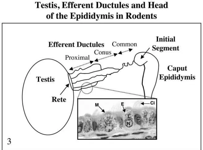
oestrogen studies. Efferent ductules are small tubule
offshoots of the rete testis, which form a series of coiled
tubules between the rete testis and epididymis (Fig. 3).
They may number between 2 and 33 depending on
species (see Ilio and Hess, 1994 for an extensive review).
Near the rete, the ductuli have a wider lumen but near
the epididymis, a coni vasculosa forms a series of highly
tortuous and narrow tubules. Within the conus, the
ductules anastomose into a common duct in the rat and
become invested with a connective tissue capsule. In
other species, some ductules merge, but others enter the
epididymis as separate tubules. As the common duct
enters the head of the epididymis it becomes smaller in
diameter and remains highly coiled. The caput
epi-didymidis in man and larger mammals is occupied
mostly by the efferent ducts that leave the testis as
parallel straight tubules, which become coiled into
lob-ules that fold over one another before emptying into
the epididymis (Yeung et al., 1991; Foley et al., 1995;
Ilio and Hess, 1994). Columnar, non-ciliated principal
cells and ciliated cells line the efferent ductule
epithe-lium (Fig. 3) with kinocilia (Ilio and Hess, 1994). The
lumen of the ductule is typically empty or contains few
spermatozoa, except in the common duct where
sper-matozoa become more (Talo, 1981).
lyso-R.A.Hess et al./Molecular and Cellular Endocrinology 000 (2001) 000 – 000
4
somes are components of an elaborate system for fluid
phase, adsorptive and receptor-mediated endocytosis
(Byers et al., 1985; Hermo and Morales, 1984; Hermo
et al., 1985; Morales and Hermo, 1983;
Veera-machaneni et al., 1990; VeeraVeera-machaneni and Amann,
1990, 1991; Ilio and Hess, 1994). Rete testis fluid is
taken up by the endocytotic apparatus from coated pits
to multivesicular bodies and then to lysosomes for
digestion by hydrolytic enzymes (Hermo and Morales,
1984). The lateral plasma membranes in the basal and
supranuclear regions of the efferent ductules form a
well-localized ‘tubular network’ (Ramos Jr. and Dym,
1977; Jones and Jurd, 1987; Robaire and Hermo, 1988;
Ilio and Hess, 1994). The intercellular spaces become
dilated when absorption is active (Pudney and Fawcett,
1984). The occurrence of these dilated intercellular
channels strongly suggests that fluid movement in this
part of the tract may be coupled to active solute
transport (Suzuki and Nagano, 1978). However, these
tubular networks show considerably less amplification
than is normally found in resorptive epithelium such as
the proximal convoluted tubules of the kidney (Ilio and
Hess, 1992).
It is now well established that efferent ductules
func-tion to reabsorb luminal fluids (Clulow et al., 1998; Ilio
and Hess, 1994). These ductules function to transport
sperm and reabsorb water, ions and proteins (Fig. 4).
The time interval required for spermatozoa to travel the
length of the ductuli efferentes is approximately 45 min
in the rat (English and Dym, 1981), but little is known
for other species. Many physiological and
micropunc-ture studies on the proximal segments of the excurrent
ducts in different species have confirmed the original
findings of Crabo (1965) that more than 90% of the
fluids secreted by the seminiferous epithelium is
reab-sorbed in the efferent ductules. The reported values, in
which the caput epididymidis is excluded, vary between
74 and 96% reabsorption (Clulow et al., 1994; Djakiew
and Jones, 1983; Jones, 1980; Jones and Jurd, 1987;
Man et al., 1997). Although the efferent ductules are
now recognized as the major site for rete testis fluid
absorption, the underlying mechanisms for absorption
remain unsettled. However, the work of several
labora-tories suggests that the primary mechanism of fluid (i.e.
water) movement in the ductules involves the coupling
of water and active ion transport (Clulow et al., 1996;
Man et al., 1997; Clulow et al., 1994, 1998; Hansen et
al., 1999; Jones and Jurd, 1987; Jones and Clulow,
1987; Ilio and Hess, 1992; Chan et al., 1995).
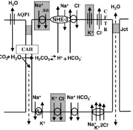
The movement of water involves several pathways,
including paracellular flow and the use of apical and
basal aquaporin (AQP) water channels for transcelluar
movement (Brown et al., 1993; Fisher et al., 1998).
However, AQP1 appears to be expressed only on the
apical surface of efferent ductules (Fisher et al., 1998),
in contrast to that reported for kidney proximal
tubules, which contain AQP1 in both apical and
baso-lateral plasma membranes (Verkman, 1998, 1999). This
Fig. 4. A physiological model representing potential sites for oestrogen regulation in the efferent ductule epithelium. Aquaporin-1 (AQP1) is a water channel that is known to exist along the apical border. Water also may move paracelluarly through the apical junctions (Jct). Carbonic anhydrase II (CAII) is abundant in efferent ductules and catalyzes the formation of carbonic acid. NHE-3 is the Na+/H+exchanger along the apical membrane that apparently co-functions with the HCO3−/Cl−exchanger. The Cl−channel, cystic fibrosis transmembrane regulator (CFTR) is present in the apical membrane. Along the basal membrane are found the Na+/K+, ATPase and the Na+/HCO
3
− and Na+/K+/2Cl− co-transporters. Channels and transporters outlined in gray are yet to be detected.
difference in the location of AQP1 in efferent ductules
may explain why males are fertile even in the absence of
AQP1 in knockout mice (Schnermann et al., 1998;
Verkman, 1999). However, other AQP molecules may
be present in the basolateral membranes of efferent
ductal cells. Alternatively, if other AQP isoforms are
not identified in the efferent ductules, the leaky cell
junctional complex that is present could provide a rapid
route for the equilibration of water across this
epithe-lium. In the kidney, it appears that AQP1 channels that
are located in both apical and basal plasma membranes
of proximal tubules (Verkman, 1999) are capable of
ensuring rapid movement of water by small differentials
in hyperosmolality from epithelial cytoplasm to
inter-cellular and connective tissue spaces. In the kidney of
the AQP1 knockout mouse, water movement is reduced
by nearly 80% (Verkman, 1999), strongly suggesting
that the transcelluar pathway is the dominant
mecha-nism for water absorption in the proximal tubules. The
paracellular pathway may prove to be more important
in the efferent ductules, because the AQP1 knockout
mouse is fertile.
The absorption of protein in the efferent ductules has
been demonstrated by the disappearance of certain
bands of proteins from the rete testis fluid between the
ductuli efferentes and the initial segment of the
epi-didymis, owing to their absorption in the ductuli and
/
or
the initial segment (Jones and Jurd, 1987; Koskimies
and Kormano, 1975; Olson and Hinton, 1985). It has
been calculated that approximately 50 – 90% of the total
protein leaving the testis was reabsorbed in the efferent
ductules (Jones and Jurd, 1987; Clulow et al., 1994;
Veeramachaneni and Amann, 1990, 1991). The capacity
of the efferent ductal epithelium to reabsorb molecules,
both through fluid-phase, adsorptive endocytosis and
receptor-mediated endocytosis has been confirmed by
several studies (Pelliniemi et al., 1981; Morales and
Hermo, 1983; Hermo and Morales, 1984; Hermo et al.,
1985; Veeramachaneni and Amann, 1991). More
re-cently it has been shown that up to 30% of inulin is
reabsorbed in the microperfused rat efferent ductules
(Clulow et al., 1998), which emphasizes the role of
endocytosis in transcellular movement of water, ions
and proteins. Endocytosis may also provide an
alterna-tive route for water in the AQP1 knockout mouse and
thus help to prevent fluid accumulation in the lumen
and subsequent infertility, as seen in the ERKO mouse
(Eddy et al., 1996; Hess et al., 1997a).
R.A.Hess et al./Molecular and Cellular Endocrinology 000 (2001) 000 – 000
6
efferent ductules contain the highest concentration of
oestrogen receptors in any organ examined to date; and
(c) efferent ductules function to reabsorb nearly 90% of
the luminal fluids. To test this hypothesis, the ER
a
gene
knockout mouse (ERKO Lubahn et al., 1993) was
evaluated for histological changes in efferent ductule
epithelium, fluid reabsorption and fluid dynamics in the
testis over time. The ERKO male is infertile, but its
testes appear normal until puberty, when unexpectedly
they begin to degenerate as early as 20 – 40 days of age
(Eddy et al., 1996). By 150 days, ERKO testes are
atrophic. Sperm from the ERKO male are abnormal
and sperm concentrations are significantly reduced in
the epididymis (Eddy et al., 1996). Rete testis in ERKO
males is dilated and protrudes into the testis (Hess et
al., 1997a). Downstream from the rete, the efferent
ductules are swollen (Hess et al., 1997a). From these
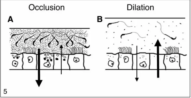
observations, two hypotheses were formed to explain
how the disruption of ER
a
could cause fluid to
accu-mulate in the ERKO testis (Fig. 5). The first hypothesis
involved excessive fluid reabsorption from the efferent
ductule lumen, which would increase the concentration
of sperm and cause luminal contents to become
com-pacted. This rapid response would induce an occlusion
of the efferent ducts, which would produce fluid
build-up in the testis and subsequent backpressure atrophy of
the testis (Cooper and Jackson, 1973; Hess et al., 1991;
Carter et al., 1987; Nakai et al., 1992). The second
hypothesis suggests that an opposite mechanism would
cause an inhibition of reabsorption and possibly a net
inward flux of water into the ductal lumen. This
exces-sive accumulation of fluid in the lumen would overload
the ductal system, because in the rodent the efferent
ductules anastomose until they form a single tubule
that enters the epididymis. This funnel-like effect of the
efferent ductules requires that luminal fluids be
reab-sorbed along the length of the tubules in order to
permit the continual movement of seminiferous tubule
fluids out of the testis (Winet, 1980). Thus, the
inhibi-tion of reabsorpinhibi-tion would also cause the accumulainhibi-tion
of fluid in the lumen, which would subsequently cause
backpressure atrophy of the testis.
In the ERKO male, it appeared that luminal fluid
was not being removed by the efferent ductules, causing
fluids to accumulate in efferent ductules, rete testis and
seminiferous tubules (Fig. 6). As predicted from these
observations, a transient increase in testis weight in
ERKO males was noted between 32 and 81 days of age
and then a continual decrease in weight out to 185 days
of age, suggesting that the long-term atrophy of testes
in the knockout mouse was caused by back pressure of
the accumulating luminal fluids (Hess et al., 1997a).
Luminal diameter was increased in ERKO ductules by
130%, 78% and 297% for the proximal, conus and
common regions, respectively and epithelial cells of the
efferent ductules in ERKO males were reduced in
height by approximately 48% (Hess et al., 2000).
The efferent ductal epithelium in ERKO tissue also
showed a loss of cellular organelles, a flattening of the
nucleus and the loss or shortening of the microvillus
border (Hess et al., 1997a, 2000). All of these changes
are consistent with a decrease in fluid reabsorption
observed in these ductules in the ERKO male (Hess et
al., 1997a). The endocytotic apparatus, including apical
vesicles and PAS
+
lysosomal granules, which are
prominent in non-ciliated cells of normal efferent
duc-tules (Ilio and Hess, 1994; Hermo and Morales, 1984;
Morales and Hermo, 1983), was greatly reduced in the
Fig. 6. (A) Wild-type efferent ductules at low magnification exhibit have narrow lumens (arrow). (B) ERKO efferent ductules have greatly dilated lumens (arrow). (C) Wild-type efferent ductule epithelium is tall, contains well-developed endocytotic vesicles (E) and numerous lysosomes (L), and has a prominent microvillus brush border (Mi). Ci, cilia. (D) ERKO efferent ductule epithelium is reduced in height, contain fewer lysosomes and endocytotic vesicles, and microvilli (Mi) are short and often absent. Ci, cilia. Bars=50mm (A, B); 10mm (C, D).
ERKO efferent ductule epithelium. Thus, the apical
surface of this absorbing epithelium appeared to be
transformed into a non-absorbing lining, when ER
a
was lacking. To test this hypothesis, the initial segment
epididymis was surgically cauterized to occlude the
terminal end of the efferent ductules in the adult male.
Testis weight 48 h post-surgery was increased 30% more
in ERKO than in wild-type males (Hess et al., 1997a).
The hypothesis was also tested in vitro, using small
segments of adult efferent ductules in organ culture.
The tubular ends were ligated with fine suture, which
prevented the inflow of culture medium, and the lumen
was observed over a 24-h period. Efferent ductules
from wild-type males were capable of rapidly
reab-sorbing the luminal fluid, resulting in a collapse of the
ductule walls; however, the luminal area of ERKO
ductules did not collapse, but instead showed a
dra-matic increase in area (Hess et al., 1997a).
Thus, the efferent ductules in ERKO mice appear to
follow the second hypothesis proposed for inducing
fluid accumulation in the male tract. Thus, the ERKO
mouse provides strong evidence that oestrogen, or more
specifically, a functional ER
a
is involved in the
regula-tion of fluid transport in the male reproductive tract,
and responsible for increasing the concentration of
sperm as they enter the epididymis. Future studies are
aimed at uncovering the biochemical and physiological
mechanisms underlying these changes in the ERKO
male and in a new animal model, in which ER are
inhibited in the adult male by treatment with the pure
anti-oestrogen, ICI 182,780 (AstraZeneca, Macclesfield,
Cheshire).
Acknowledgements
R.A.Hess et al./Molecular and Cellular Endocrinology 000 (2001) 000 – 000
8
References
Brown, D., Verbavatz, J.M., Valenti, G., Lui, B., Sabolic, I., 1993. Localization of the Chip28 water channel in reabsorptive seg-ments of the rat male reproductive tract. Eur. J. Cell. Biol. 61, 264 – 273.
Bujan, L., Mieusset, R., Audran, F., Lumbroso, S., Sultan, C., 1993. Increased oestradiol level in seminal plasma in infertile men. Hum. Reprod. 8, 74 – 77.
Byers, S.W., Musto, N.A., Dym, M., 1985. Culture of ciliated and nonciliated cells from rat ductuli efferentes. J. Androl. 6, 271 – 278.
Carreau, S., Genissel, C., Bilinska, B., Levallet, J., 1999. Sources of oestrogen in the testis and reproductive tract of the male. Int. J. Androl. 22, 211 – 223.
Carter, S.D., Hess, R.A., Laskey, J.W., 1987. The fungicide methyl 2-benzimidazole carbamate causes infertility in male Sprague – Dawley rats. Biol. Reprod. 37, 709 – 717.
Chan, H.C., Lai, K.B., Fu, W.O., Chung, Y.W., Chan, P.S., Wong, P.Y., 1995. Regional differences in bioelectrical properties and anion secretion in cultured epithelia from rat and human male excurrent ducts. Biol. Reprod. 52, 192 – 198.
Claus, R., Dimmick, M.A., Gimenez, T., Hudson, L.W., 1992. Estro-gens and prostaglandin F2a in the semen and blood plasma of stallions. Theriogenology 38, 687 – 693.
Claus, R., Hoang-Vu, C., Ellendorff, F., Meyer, H.D., Schopper, D., Weiler, U., 1987. Seminal oestrogens in the boar: origin and functions in the sow. J. Steroid. Biochem. 27, 331 – 335. Claus, R., Schopper, D., Hoang-Vu, C., 1985. Contribution of
indi-vidual compartments of the genital tract to oestrogen and testos-terone concentrations in ejaculates of the boar. Acta. Endocrinol. 109, 281 – 288.
Clulow, J., Hansen, L.A., Jones, R.C., 1996. In vivo microperfusion of the ductuli efferentes testis of the rat: flow dependence of fluid reabsorption. Exp. Physiol. 81, 633 – 644.
Clulow, J., Jones, R.C., Hansen, L.A., 1994. Micropuncture and cannulation studies of fluid composition and transport in the ductuli efferentes testis of the rat: comparisons with the ho-mologous metanephric proximal tubule. Exp. Physiol. 79, 915 – 928.
Clulow, J., Jones, R.C., Hansen, L.A., Man, S.Y., 1998. Fluid and electrolyte reabsorption in the ductuli efferentes testis. J. Reprod. Fertil. Suppl. 53, 1 – 14.
Cooper, E.R., Jackson, H., 1973. Chemically induced sperm retention cysts in the rat. J. Reprod. Fertil. 34, 445 – 449.
Crabo, B., 1965. Studies on the composition of epididymal content in bulls and boars. Acta Vet. Scand. 6, 8 – 94.
de Jong, F., Hey, A., van der Molen, H., 1973. Effect of go-nadotrophins on the secretion of oestradiol-17b and testosterone by the rat testis. J. Endocrinol. 57, 277 – 284.
Djakiew, D., Jones, R.C., 1983. Sperm maturation, fluid transport, and secretion and absorption of protein in the epididymis of the echidna,Tachyglossus aculeatus. J. Reprod. Fertil. 68, 445 – 456. Eddy, E.M., Washburn, T.F., Bunch, D.O., Goulding, E.H., Gladen,
B.C., Lubahn, D.B., et al., 1996. Targeted disruption of the oestrogen receptor gene in male mice causes alteration of sper-matogenesis and infertility. Endocrinology 137, 4796 – 4805. Eiler, H., Graves, C., 1977. Oestrogen content of semen and the effect
of exogenous oestradiol-17a on the oestrogen and androgen con-centration in semen and blood plasma of bulls. J. Reprod. Fertil. 50, 17 – 21.
English, H.F., Dym, M., 1981. The time required for materials injected into the rete testis to reach points in the caput epididymis of the rat and observations on the absorption of cationic ferritin. Ann. NY Acad. Sci. 383, 445 – 446.
Ergun, S., Ungefroren, H., Holstein, A.F., Davidoff, M.S., 1997. Estrogen and progesterone receptors and estrogen receptor-re-lated antigen (ER-D5) in human epididymis. Mol. Reprod. Dev. 47, 448 – 455.
Fisher, J.S., Millar, M.R., Majdic, G., Saunders, P.T., Fraser, H.M., Sharpe, R.M., 1997. Immunolocalisation of oestrogen receptor-al-pha within the testis and excurrent ducts of the rat and marmoset monkey from perinatal life to adulthood. J. Endocrinol. 153, 485 – 495.
Fisher, J.S., Turner, K.J., Fraser, H.M., Saunders, P.T., Brown, D., Sharpe, R.M., 1998. Immunoexpression of aquaporin-1 in the efferent ducts of the rat and marmoset monkey during develop-ment, its modulation by estrogens, and its possible role in fluid resorption. Endocrinology 139, 3935 – 3945.
Foley, G.L., Bassily, N., Hess, R.A., 1995. Intratubular spermatic granulomas of the canine efferent ductules. Toxicol. Pathol. 23, 731 – 734.
Free, M.J., Jaffe, R.A., 1979. Collection of rete testis fluid from rats without previous efferent duct ligation. Biol. Reprod. 20, 269 – 278.
Ganjam, V.K., Amann, R.P., 1976. Steroids in fluids and sperm entering and leaving the bovine epididymis, epididymal tissue, and accessory sex gland secretions. Endocrinology 99, 1618 – 1630. Goyal, H.O., Bartol, F.F., Wiley, A.A., Khalil, M.K., Chiu, J., Vig,
M.M., 1997. Immunolocalization of androgen receptor and estro-gen receptor in the developing testis and excurrent ducts of goats. Anat. Rec. 249, 54 – 62.
Goyal, H.O., Bartol, F.F., Wiley, A.A., Khalil, M.K., Williams, C.S., Vig, M.M., 1998. Regulation of androgen and estrogen receptors in male excurrent ducts of the goat: an immunohistochemical study. Anat. Rec. 250, 164 – 171.
Hansen, L.A., Clulow, J., Jones, R.C., 1999. The role of Na+– H+ exchange in fluid and solute transport in the rat efferent ducts. Exp. Physiol. 84, 521 – 527.
Hermo, L., Clermont, Y., Morales, C., 1985. Fluid-phase and adsorp-tive endocytosis in ciliated epithelial cells of the rat ductuli efferentes. Anat. Rec. 211, 285 – 294.
Hermo, L., Morales, C., 1984. Endocytosis in nonciliated epithelial cells of the ductuli efferentes in the rat. Am. J. Anat. 171, 59 – 74. Hermo, L., Spier, N., Nadler, N.J., 1988. Role of apical tubules in endocytosis in nonciliated cells of the ductuli efferentes of the rat: a kinetic analysis. Am. J. Anat. 182, 107 – 119.
Hermo, L., Wright, J., Oko, R., Morales, C., 1991. Role of epithelial cells of the male excurrent duct system of the rat in the endocyto-sis or secretion of sulfated glycoprotein-2 (clusterin). Biol. Re-prod. 44, 1113 – 1131.
Hess, R.A., Bunick, D., Bahr, J.M., 1995. Sperm, a source of estrogen. Environ. Health. Perspect. 103 (Suppl. 7), 59 – 62. Hess, R.A., Bunick, D., Lee, K.H., Bahr, J., Taylor, J.A., Korach,
K.S., et al., 1997a. A role for oestrogens in the male reproductive system. Nature 390, 509 – 512.
Hess, R.A., Bunick, D., Lubahn, D.B., Zhou, Q., Bouma, J., 2000. Morphologic changes in efferent ductules and epididymis in estro-gen receptor-alpha knockout mice. J. Androl. 21, 107 – 121. Hess, R.A., Gist, D.H., Bunick, D., Lubahn, D.B., Farrell, A., Bahr,
J., et al., 1997b. Estrogen receptor (a & b) expression in the excurrent ducts of the adult male rat reproductive tract. J. An-drol. 18, 602 – 611.
Hess, R.A., Moore, B.J., Forrer, J., Linder, R.E., Abuel-Atta, A.A., 1991. The fungicide benomyl (methyl 1-(butylcarbamoyl)-2- benz-imidazolecarbamate) causes testicular dysfunction by inducing the sloughing of germ cells and occlusion of efferent ductules. Fun-dam. Appl. Toxicol. 17, 733 – 745.
Ilio, K.Y., Hess, R.A., 1992. Localization and activity of Na+, K(+)-ATPase in the ductuli efferentes of the rat. Anat. Rec. 234, 190 – 200.
Ilio, K.Y., Hess, R.A., 1994. Structure and function of the ductuli efferentes: a review. Microsc. Res. Technol. 29, 432 – 467. Janulis, L., Bahr, J.M., Hess, R.A., Janssen, S., Osawa, Y., Bunick,
D., 1998. Rat testicular germ cells and epididymal sperm contain active P450 aromatase. J. Androl. 19, 65 – 71.
Janulis, L., Hess, R.A., Bunick, D., Nitta, H., Janssen, S., Asawa, Y., et al., 1996. Mouse epididymal sperm contain active P450 aro-matase which decreases as sperm traverse the epididymis. J. Androl. 17, 111 – 116.
Jones, R.C., 1980. Luminal composition and maturation of spermato-zoa in the genital ducts of the African elephant (Loxodonta africana). J. Reprod. Fertil. 60, 87 – 93.
Jones, R.C., Clulow, J., 1987. Regulation of the elemental composi-tion of the epididymal fluids in the tammar,Macropus eugenii. J. Reprod. Fertil. 81, 583 – 590.
Jones, R.C., Jurd, K.M., 1987. Structural differentiation and fluid reabsorption in the ductuli efferentes testis of the rat. Aust. J. Biol. Sci. 40, 79 – 90.
Koskimies, A.I., Kormano, M., 1975. Proteins in fluids from different segments of the rat epididymis. J. Reprod. Fertil. 43, 345 – 348. Krege, J.H., Hodgin, J.B., Couse, J.F., Enmark, E., Warner, M.,
Mahler, J.F., et al., 1998. Generation and reproductive pheno-types of mice lacking estrogen receptor beta. Proc. Natl. Acad. Sci. USA 95, 15677 – 15682.
Kuiper, G.G., Carlsson, B., Grandien, K., Enmark, E., Haggblad, J., Nilsson, S., et al., 1997. Comparison of the ligand binding specifi-city and transcript tissue distribution of estrogen receptors alpha and beta. Endocrinology 138, 863 – 870.
Kuiper, G.G., Enmark, E., Pelto-Huikko, M., Nilsson, S., Gustafs-son, J.A., 1996. Cloning of a novel receptor expressed in rat prostate and ovary. Proc. Natl. Acad. Sci. USA 93, 5925 – 5930. Kwon, S., Hess, R.A., Bunick, D., Kirby, J.D., Bahr, J.M., 1997.
Estrogen receptors are present in the epididymis of the rooster. J. Androl. 18, 378 – 384.
Kwon, S., Hess, R.A., Bunick, D., Nitta, H., Janulis, L., Osawa, Y., et al., 1995. Rooster testicular germ cells and epididymal sperm contain P450 aromatase. Biol. Reprod. 53, 1259 – 1264.
Levallet, J., Bilinska, B., Mittre, H., Genissel, C., Fresnel, J., Car-reau, S., 1998. Expression and immunolocalization of functional cytochrome P450 aromatase in mature rat testicular cells. Biol. Reprod. 58, 919 – 926.
Levallet, J., Carreau, S., 1997. In vitro gene expression of aromatase in rat testicular cells. CR Acad. Sci. III 320, 123 – 129.
Lubahn, D.B., Moyer, J.S., Golding, T.S., Couse, J.F., Korach, K.S., Smithies, O., 1993. Alteration of reproductive function but not prenatal sexual development after insertional disruption of the mouse estrogen receptor gene. Proc. Natl. Acad. Sci. USA 90, 11162 – 11166.
Man, S.Y., Clulow, J., Hansen, L.A., Jones, R.C., 1997. Adrenal independence of fluid and electrolyte reabsorption in the ductuli efferentes testis of the rat. Exp. Physiol. 82, 283 – 290.
Morales, C., Hermo, L., 1983. Demonstration of fluid-phase endocy-tosis in epithelial cells of the male reproductive system by means of horseradish peroxidase-colloidal gold complex. Cell. Tissue Res. 230, 503 – 510.
Nakai, M., Hess, R.A., Moore, B.J., Guttroff, R.F., Strader, L.F., Linder, R.E., 1992. Acute and long-term effects of a single dose of the fungicide carbendazim (methyl 2-benzimidazole carbamate) on the male reproductive system in the rat. J. Androl. 13, 507 – 518. Nitta, H., Bunick, D., Hess, R.A., Janulis, L., Newton, S.C., Millette, C.F., et al., 1993. Germ cells of the mouse testis express P450 aromatase. Endocrinology 132, 1396 – 1401.
Olson, G.E., Hinton, B.T., 1985. Regional differences in luminal fluid polypeptides of the rat testis and epididymis revealed by two-di-mensional gel electrophoresis. J. Androl. 6, 20 – 34.
Overpeck, J.G., Colson, S.H., Hohmann, J.R., Applestine, M.S., Reilly, J.F., 1978. Concentrations of circulating steroids in normal prepubertal and adult male and female humans, chimpanzees, rhesus monkeys, rats, mice, and hamsters: a literature survey. J. Toxicol. Environ. Health 4, 785 – 803.
Payne, A., Kelch, R., Musich, S., Halpern, M., 1976. Intratesticular site of aromatization in the human. J. Clin. Endocrinol. Metab. 42, 1081 – 1087.
Pelliniemi, L.J., Dym, M., Gunsalus, G.L., Musto, N.A., Bardin, C.W., Fawcett, D.W., 1981. Immunocytochemical localization of androgen-binding protein in the male rat reproductive tract. En-docrinology 108, 925 – 931.
Prins, G.S., Marmer, M., Woodham, C., Chang, W., Kuiper, G., Gustafsson, J.A., et al., 1998. Estrogen receptor-beta messenger ribonucleic acid ontogeny in the prostate of normal and neona-tally estrogenized rats. Endocrinology 139, 874 – 883.
Pudney, J., Fawcett, D.W., 1984. Seasonal changes in fine structure of the ductuli efferentes of the ground squirrelCitellus lateralis Say. Anat. Rec. 208, 383 – 399.
Ramos, A.S., Jr., Dym, M., 1977. Ultrastructure of the ductuli efferentes in monkeys. Biol. Reprod. 17, 339 – 349.
Robaire, B., Hermo, L., 1988. Efferent ducts, epididymis, and vas deferens: structure, functions, and their regulation. In: Knobil, E., Neill, J. (Eds.), The Physiology of Reproduction. Raven Press, New York, pp. 999 – 1080.
Roselli, C.E., Abdelgadir, S.E., Resko, J.A., 1997. Regulation of aromatase gene expression in the adult rat brain. Brain. Res. Bull. 44, 351 – 357.
Rosenfeld, C.S., Ganjam, V.K., Taylor, J.A., Yuan, X., Stiehr, J.R., Hardy, M.P., et al., 1998. Transcription and translation of estro-gen receptor-beta in the male reproductive tract of estroestro-gen receptor-alpha knock-out and wild-type mice. Endocrinology 139, 2982 – 2987.
Sato, T., Chiba, A., Hayashi, S., Okamura, H., Ohta, Y., Takasugi, N., et al., 1994. Induction of estrogen receptor and cell division in genital tracts of male mice by neonatal exposure to diethylstilbe-strol. Reprod. Toxicol. 8, 145 – 153.
Saunders, P.T., Fisher, J.S., Sharpe, R.M., Millar, M.R., 1998. Expression of oestrogen receptor beta (ER beta) occurs in multi-ple cell types, including some germ cells, in the rat testis. J. Endocrinol. 156, R13 – 17.
Saunders, P.T.K., Maguire, S.M., Gaughan, J., Millar, M.R., 1997. Expression of oestrogen receptor beta (ER-beta) in multiple rat tissues visualised by immunohistochemistry. J. Endocrinol. 154, R.13 – R.16.
Schleicher, G., Drews, U., Stumpf, W.E., Sar, M., 1984. Differential distribution of dihydrotestosterone and estradiol binding sites in the epididymis of the mouse. An autoradiographic study. Histo-chemistry 81, 139 – 147.
Schnermann, J., Chou, C.L., Ma, T., Traynor, T., Knepper, M.A., Verkman, A.S., 1998. Defective proximal tubular fluid reabsorp-tion in transgenic aquaporin-1 null mice. Proc. Natl. Acad. Sci. USA 95, 9660 – 9664.
Setchell, B.P., Cox, J.E., 1982. Secretion of free and conjugated steroids by the horse testis into lymph and venous blood. J. Reprod. Fertil. Suppl. 32, 123 – 127.
Setchell, B.P., Laurie, M.S., Flint, A.P., Heap, R.B., 1983. Transport of free and conjugated steroids from the boar testis in lymph, venous blood and rete testis fluid. J. Endocrinol. 96, 127 – 136. Suzuki, F., Nagano, T., 1978. Regional differentiation of cell
junc-tions in the excurrent duct epithelium of the rat testis as revealed by freeze-fracture. Anat. Rec. 191, 503 – 520.
Talo, A., 1981. In-vitro spontaneous electrical activity of rat efferent ductules. J. Reprod. Fertil. 63, 17 – 20.
R.A.Hess et al./Molecular and Cellular Endocrinology 000 (2001) 000 – 000
10
Tsubota, T., Nitta, H., Osawa, Y., Mason, I., Kita, I., Tiba, T., Bahr, J., 1993. Immunolocalization of steroidogenic enzymes, P450scc, 3b-HSD, P450c17, and P450arom in the Hokkaido brown bear. Gen. Comp. Endo. 92, 439 – 444.
van der Molen, H.J., Brinkmann, A.O., de Jong, F.H., Rommerts, F.F. 1981. Testicular oestrogens. J. Endocrinol. 89 (Suppl) 33 – 46. van Pelt, A.M., de Rooij, D.G., van der Burg, B., van der Saag, P.T., Gustafsson, J.A., Kuiper, G.G., 1999. Ontogeny of estrogen receptor-beta expression in rat testis. Endocrinology 140, 478 – 483.
Veeramachaneni, D.N., Amann, R.P., 1990. Oxytocin in the ovine ductuli efferentes and caput epididymidis: immunolocalization and endocytosis from the luminal fluid. Endocrinology 126, 1156 – 1164.
Veeramachaneni, D.N., Amann, R.P., 1991. Endocytosis of andro-gen-binding protein, clusterin, and transferrin in the efferent ducts and epididymis of the ram. J. Androl. 12, 288 – 294.
Veeramachaneni, D.N., Amann, R.P., Palmer, J.S., Hinton, B.T., 1990. Proteins in luminal fluid of the ram excurrent ducts: changes
in composition and evidence for differential endocytosis. J. An-drol. 11, 140 – 154.
Verkman, A.S., 1998. Role of aquaporin water channels in kidney and lung. Am. J. Med. Sci. 316, 310 – 320.
Verkman, A.S., 1999. Lessons on renal physiology from transgenic mice lacking aquaporin water channels. J. Am. Soc. Nephrol. 10, 1126 – 1135.
Waites, G.M., Einer-Jensen, N., 1974. Collection and analysis of rete testis fluid from macaque monkeys. J. Reprod. Fertil. 41, 505 – 508.
West, N.B., Brenner, R.M., 1990. Estrogen receptor in the ductuli efferentes, epididymis, and testis of rhesus and cynomolgus macaques. Biol. Reprod. 42, 533 – 538.
Winet, H., 1980. On the mechanism for flow in the efferent ducts. J. Androl. 1, 303 – 311.
Yeung, C.H., Cooper, T.G., Bergmann, M., Schulze, H., 1991. Orga-nization of tubules in the human caput epididymidis and the ultrastructure of their epithelia. Am. J. Anat. 191, 261 – 279.