22(3) : 147-153 (2016)
http://dx.doi.org/10.20307/nps.2016.22.3.147
1
Anti-inflammatory Effect of Mangosteen (
Garcinia mangostana
L.) Peel Extract
and its Compounds in LPS-induced RAW264.7 Cells
Wahyu Widowati1,*, Lusiana Darsono1, Jo Suherman1, Nurul Fauziah2,
Maesaroh Maesaroh2, and Pande Putu Erawijantari2
1Medical Research Center, Faculty of Medicine, Maranatha Christian University, Bandung 40164, West Java, Indonesia 2Biomolecular and Biomedical Research Center, Aretha Medika Utama, Bandung 40163, West Java, Indonesia
Abstract−Inflammation plays an important role in host defense against external stimuli such as infection by pathogen, endotoxin or chemical exposure by the production of the inflammatory mediators that produced by macrophage. Anti-inflammatory factor is important to treat the dangers of chronic inflammation associated with chronic disease. This research aims to analyze the anti-inflammatory effects of Garcinia mangostana L.peel extract (GMPE), α-mangostin, and γ-mangostin in LPS-induced murine macrophage cell line (RAW 264.7) by inhibiting the production of inflammatory mediators. The cytotoxic assay of G. mangostana L.extract, α-mangostin, and γ -mangostin were performed by MTS (3-(4,5-dimethylthiazol-2-yl)-5-(3-carboxymethoxyphenyl)-2-(4-sulfophenyl)-2H-tetrazolium) to determine the safe and non-toxic concentration in RAW 264.7 for the further assay. The concentration of inflammatory mediators (COX-2, IL-6, and IL-1β) were measured by the ELISA-based assay and NO by the nitrate/nitrite colorimetric assay in treated LPS-induced RAW 264.7 cells. The inhibitory activity was determined by the reducing concentration of inflammatory mediators in treated LPS-induced RAW 264.7 over the untreated cells. This research revealed that GMPE, α-mangostin, and γ-mangostin possess the anti-inflammatory effect by reducing COX-2, IL-6, IL-1β, and NO production in LPS-induces RAW 264.7 cells.
Keywords−Anti-inflammatory, GMPE, α-Mangostin, γ-Mangostin, Macrophages, Inflammatory mediator
Introduction
Natural products have over the years contributed to the development of new therapeutic drugs for a variety of human diseases.1 Many tropical plants have interesting biological activities with their therapeutic potential, including Garcinia mangostana L. (mangosteen). Man-gosteen has been used for hundreds of years around the world, mostly in Southeast Asia, as a medicine for a great variety of diseases.2
Many studies have shown that the various parts extract contain varieties of secondary metabolites such as prenylated and oxygenated xanthones. Xanthones as the major bioactive secondary metabolites were reported to have many pharmacological activities including antioxidant, antifungal, anti-bacteria, cytotoxic, anti-inflammatory, antihistamine, anti-HIV, antimalarial and other activities.3-5
The previous study confirmed that the G. mangostana peel extract (GMPE) contained α -mangostin (105 ppm), γ-mangostin (7.20 ppm), garcinone
C (3.50 ppm), and garcinone D (9.92 ppm) based on high performance liquid chromatography (HPLC) analysis.6
In this study, the anti-inflammatory potential of G.mangostana
L. peel extract (GMPE) and its compound were observed. Inflammation plays an important role in host defense that encompasses multiple processes against external stimuli such as infection by pathogen, exposure to bacterial endotoxin or chemical exposure.7
The inflammation process involves changes in blood flow, increased vascular permeability, destruction of tissue via the activation and migration of leukocytes with the synthesis of reactive oxygen derivatives (oxidative burst) and synthesis of local inflammatory mediators.8
The secretion of the inflamma-tory mediators both of proinflammainflamma-tory mediators such as interleukin (IL)-1, IL-6, tumor necrosis factor (TNF)-α, and Nitric Oxide (NO) and anti-inflammatory such as IL-10 are the primary response to inflammation in addition to leukocyte recruitment.9
Inflammation relates to various diseases such as rheumatoid arthritis, inflammatory bowel disease, artherosclerosis, Alzheimer’s, and has a role in various kinds of cancer.10 Anti-inflammatory is important to treat the danger of chronic inflammation associated *Author for correspondence
Wahyu Widowati, Faculty of Medicine, Maranatha Christian Uni-versity, Bandung 40164, Indonesia.
with chronic disease.11
Several mechanisms of action have been proposed to describe the phytochemical potential for anti-inflammatory, such as: 1) antioxidant and radical scavenging activity; 2) modulation of cellular activities of inflammation-related cells (mast cells, macrophages, lymphocytes, and neutro-phils; 3) modulation of proinflammatory enzyme activities; 4) modulation of the production of other proinflammatory molecules; and 5) modulation of proinflammatory gene expression.12
This study focused on the potential of GMPE, α-mangostin, and γ-mangostin (Fig. 1) in modu-lation of the proinflammatory molecules production by inhibiting the proinflammatory cytokines production including COX-2, IL-6, IL-1β, and NO in LPS-induced murine macrophage cell line model (RAW 264.7). The RAW 264.7 murine macrophage cell line is widely used as an inflammatory model in vitro.13
Experimental
General Experimental Procedure−Garcina mangos-tana L. was collected from Cisalak-Subang, West Java, Indonesia plantation and identified by a staff of herbarium of the Department of Biology, School of Life Sciences and Technology, Bandung Institute of Technology, Bandung, West Java, Indonesia. The murine macrophage cell line RAW 264.7 (ATCC®
TIB-71™
) was obtained from Aretha Medika Utama, Biomolecular and Biomedical Research Center, Bandung, Indonesia.
α-mangostin (95-99% purity), and γ-mangostin (95-99% purity) were bought from Biopurify Phytochemical Ltd. (Chengdu, China). The medium and its component for cell culture such as Dulbecco’s Modified Eagle Medium (DMEM), Fetal Bovine Serum (FBS), penicillin, streptomycin, and Trypsin-EDTA were purchased from Biowest. 3-(4,5-dimethylthiazol-2-yl)-5-(3-carboxymethoxy-phenyl)-2-(4-sulfophenyl)-2H-tetrazolium (MTS) used for cell viability assay was purchased from Promega (Madison, WI, USA). COX-2 ELISA kit and protocol (E-EL-M0959) was purchased from Elabscience. IL-6 and IL-1β ELISA kit and protocol (431301, and 432601) were purchased from BioLegend. Nitrate/Nitrite colorimetric assay kit (KA1342) was purchased from Abnova. MultiSkan Go (Thermo Scientific), a microplate reader, was used for viability assay, measuring COX-2, IL-6, IL-1β, and NO concentration.
Plant Extract preparation−The peels of G.
mangostana L. were collected, chopped, and kept in drier tunnel service. Extraction was performed based on the maceration method using ethanol 70% as the solvent for
collecting G. mangostana L.peel extract (GMPE).14,15 RAW 264.7 Cells Culture and Viability Assay−The murine macrophage cells were grown in DMEM supplemented with 10% FBS, 1% pennicillin-streptomycin. The cells then maintained at the 37o
C humidified atmosphere incubator with 5% CO2 until the cells were confluence. The cells were washed and harvested using Trypsin-EDTA and centrifuged at 2500 rpm for 4 minutes.16-19 Viability assay−The viability assay was performed to determine the safe and nontoxic concentration for the next assay, evaluated by MTS assay. Briefly, 100 µL cells (5× 10³cells per well) in medium (DMEM supplemented with 10% FBS and 1% pennicilin-streptomcycin) were plated in 96-well plate and incubated for 24 hours at 37oC in a humidified atmosphere incubator with 5% CO2. The medium then washed and added with 99 µL new medium and 1 µL of GMPE, α-mangostin, and γ-mangostin in different concentration and DMSO in triplicate then the plate were incubated for 24 hours. Untreated cells were served as the control. Briefly, 20 µL MTS was added to each well. The plate was incubated in 5% CO2 at 37
o C incubator for 4 hours. The absorbance was measured at 490 nm on a microplate reader.18-21
Proinflammatory activation of cells and treatment− The pro-inflammatory activation of cells was performed based on Yoon, et al. (2009) and modified method.17-19, 22 The cells were seeded in 6-well plate in density of 5×10⁵ cells per well and incubated for 24 hours at 37o
C in a humidified atmosphere and 5% CO2.18,19-23 The medium (DMEM supplemented with 10% FBS and 1% pennicilin-streptomycin) then washed and supplemented with 1,600 µL growth medium and 200 µL GMPE (20, 10, 5 µg/mL), α-mangostin, and γ-mangostin (75, 50, 25 µM). Around 1-2 hours later, the medium was added with 200 µL LPS (1 µg/mL) and incubated for 24 hours at 37oC in a humidified atmosphere and 5% CO2.
18,19 After incubation of RAW 264.7 cells with LPS for 24 hours, the quantity of COX-2, IL-6, IL-1β, and NO was accumu-lated in the cell-free supernatant. The cell-free supernatant then was taken for the next assay by centrifugation at 2,000 g for 10 minutes. The supernatant was stored at −79oC for the COX-2, IL-6, IL-1β, and NO concentration. Quantification of COX-2 concentration and inhibitory activity assay after treatment−The quantitative deter-mination of COX-2 concentration in the cell-free super-natant was performed using COX-2 ELISA Kit Elabscience (E-EL-M0959). Briefly, 100 µL of standard, blank, and sample solution was added into each well then sealed and incubated for 90 minutes at 37o
-mangostin were served as the sample. The LPS-induced cells free supernatant without extract and compounds were used as positive control. The normal cell or untreated cell was used as negative control. Subsequently, the liquid of each well removed and 100 µL of Biotinylated Detection AB working solution was added into each well, incubated for 1 hour at 37o
C. The plate then washed prior to the addition of 100 µL HRP conjugate, incubated for 30 minutes at 37o
C. The plate was washed again and 90 µL of substrate solution was added into each well, incubated for about 15 minutes at 37oC. Following that, 50 µL of stop solution was added and the absorbance measured using microplate reader at 450 nm.24 The concentration of COX-2 was determined from COX-2 standard curve, the regression equation obtained was y = 0.09543x – 0.01039 with r2
= 0.99. The percentage of inhibitory activity was calculated using following equation:
COX2 inhibitory activity (%) =
where COX2p : COX-2 concentration of positive control (ng/mL); COX2s : COX-2 concentration of samples (ng/ mL).
Quantification of IL-6 and IL-1β concentration and inhibitory activity assay after treatment−The quanti-tative determination of IL-6 and IL-1β concentration in the cell-free supernatant was performed using Mouse IL-6 ELISA MAXStandard Sets (BioLegend. 431301) and Mouse IL-1β ELISA MAX Standard Sets (BioLegend 432601), respectively. Approximately 100 µl of diluted capture antibody solution was added to each well, incubated overnight at 4o
C. Following that, the plate was washed 4 times and 200 µL of assay diluent added into each well, incubated at room temperature for 1 hour with shaking. The plate then washed again 4 times, prior to addition of the diluted standards and samples. The cell-free supernatant after treated with GMPE, α-mangostin, and γ-mangostin were served as the sample. The LPS-induced cells free supernatant without extract and compounds were used as positive control. The normal cell was used as negative control. The plate was incubated for 2 hours at room temperature with shaking, then washed for 4 times. Subsequently, 100 µL of diluted detection antibody solution was added into each well and the plate was incubated for 1 hour with shaking at room tem-perature. After the plate was washed again for 4 times, 100 µl of diluted Avidin-HRP solution was added, incubated for 30 minutes with shaking at room tem-perature. The plate was washed 5 times, then 100 µL of
TMB substrate solution was added to each well and incubated for 15 minutes in the dark. Finally, 100 µL of stop solution was added and the absorbance was read at 450 nm using microplate reader.17-19,25
The quantity of IL-6 and IL-1β were determined from the standard curve. The regression equation obtained from IL-6 standard curve was y = 0.0007x – 0.0412 with r2
= 0.98, and from IL-1β standard curve was y = 0.0013x – 0.0157 with r2= 0.99. The percentage of inhibitory activity was calculated using following equation:
Inhibitory activity (%) =
where Cp : IL-6 or IL-1β concentration of positive control (pg/mL); Cs : IL-6 or IL-1β concentration of samples (pg/ mL).
Quantification of nitrite associated with NO concentration and inhibitory activity assay after treatment−The determination of nitrite associated with NO production was performed using Nitrate/Nitrite colorimetric assay kit (Abnova KA1342).24
The cell-free supernatant after treated with GMPE, α-mangostin, and γ -mangostin were served as the sample. The LPS-induced cells without GMPE or compound free supernatant was used as positive control. The normal cell-free supernatant was used as negative control. The samples were read in 540 nm of wavelength in a microplate reader. The quantity of nitrite was determined from the sodium nitrite standard curve, the regression equation obtained was y = 0.0253x – 0.0550 with r2= 0.98. The percentage of inhibitory activity was calculated using following equation:
NO inhibitory activity (%) =
where NOp : NO concentration of positive control (µM);
NOs : NO concentration of samples (µM).
Statistical analysis−All data were derived from three independent experiments. Statistical analysis was performed using SPSS software (version 20.0). The data were pre-sented as mean±standard deviation. Significant differences between the groups were determined using the Analysis of Variance (ANOVA) followed by Duncan Post Hoc Test P < 0.05 were considered as statistical significance.18,19
Result and Discussion
Natural plant compounds are now gaining more pharmacological attention as many plant products are still unexplored and show a wide range of activities.26
Major compounds of several commonly used botanicals, including COX2p COX2s–
( )
COX2p
--- 100×
Cp–Cs
( )
Cp --- 100×
NOp–NOs
( )
mangostin have been reported to have anti-inflammatory actions. The xanthones, such as α-mangostin and γ -mangostin are major bioactive compounds found in the fruit peel of mangosteen.6,27
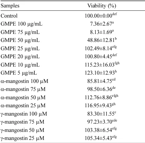
In this study, we evaluated the biological effects of GMPE, α-mangostin, and γ -mangostin on inflammatory mediators production in LPS-induced RAW 264.7 cell line as the model. The cytotoxic assay was performed to determine the safe and nontoxic concentration of GMPE and compounds for the next assay. Nontoxicity of the substrate was indicated by over 90% of cells viability by MTS assay. Viability test is an important aspect of pharmacology which deals with the adverse effect of bioactive substance on living organism prior to the use of substances as drug or chemical in clinical use.28-30
The α-mangostin and γ mangostin in the concentration of 100 µM were toxic toward RAW 264.7 cells, therefore the respective concentration was not used for the treatments and the concentration of 75, 50, and 25 was chosen instead (Table 1). In other hand, GMPE treatment showed low viability at 100, 75, 50, and 25 µg/ mL. Hence, the lower concentration of GMPE were tested. Finally, the cells in the concentration of 20, 10, and 5 µg/mL of GMPE treatment showed high viability and nontoxic to the cells (Table 1).
A stimuli of LPS can activate the macrophages that
involved in the pathological processes in several acute and chronic disorders by secreting several inflammatory mediators.7,18,19,31
The overproduction of the inflammatory mediators contributes to the pathogenesis of several diseases such as sepsis, rheumatoid arthritis, atherosclerosis, pul-monary fibrosis, and chronic hepatitis.32
Several nonsteroidal anti-inflammatory drugs were the currently available drugs to reduce the inflammation, but it possess a side effect and major problem.33
Therefore, the development of new anti-inflammatory agents from natural sources that more active and have fewer side effects become important. Based on cytotoxic result, three concentration of GMPE (20, 10, 5 µg/mL), α-mangostin (75, 50, 25 µg/ mL), and β-mangostin (75, 50, 25 µg/mL) were applied for IL-6, IL-1β, NO, and COX-2 assay. All of the anti-inflammatory assays showed that GMPE possessed potent COX-2, IL-1β, IL-6, and NO inhibitory activity. The isolated compounds from GMPE including α-mangostin and γ-mangostin also possessed the same activity. Inhibiting the synthesis of mediators that plays a role in inflammation will be useful for autoimmune diseases and inflammation treatment. COX-2 is a key regulatory enzyme of the prostaglandin/eicosanoid pathway that highly induced by pro-inflammatory cytokines in an NF-κβ-dependent manner.34
COX-2 induced several stimuli and is responsible for the pro-inflammatory cytokine at the inflammatory sites.35
COX-2 is highly induced by pro-inflammatory cytokines (IL-1β and IL-6) that serve as endogenous pyrogens that causes fever during inflammation by up-regulating the inflammatory responses and stimulating the production of acute phase reactans.36
In addition to COX-2 inhibitory activity, NO inhibitory activity may be as attractive as one of anti-inflammatory agent screening indicators. NO plays a significant role in host immune defense, vascular regulation, neurotransmission, and other system in normal condition. In human body, a family of nitric oxide synthase (NOS) enzyme is responsible for catalyzing the synthesis of NO.37 The expression of inducible NOS (iNOS) in various inflammatory and tissue cells can be induced by LPS or proinflammatory cytokines such as interleukin (IL-1).37
Overproduction of NO and iNOS are especially related to various human diseases including inflammation.16,18,19,38
GMPE and α-mangostin in the highest concentration showed the highest inhibitory activity against COX-2 production (Table 2). COX-2 with COX-1 are the key players in the inflammatory response which catalyze the conversion of arachidonic acid into pro-inflammatory prostaglandins and triggers the production of other pro-inflammatory mediators.34 IL-6 is a pleiotropic cytokine
Table 1. Effect of GMPE and mangostins in various concentrations toward RAW 264.7 cell viability
Samples Viability (%)
Control 100.00±0.00def
GMPE 100 µg/mL 557.36±2.67a GMPE 75 µg/mL 558.13±1.69a GMPE 50 µg/mL 548.86±12.81b GMPE 25 µg/mL 102.49±8.14efg GMPE 20 µg/mL 100.80±4.45def GMPE 10 µg/mL 115.23±16.03fgh GMPE 5 µg/mL 123.10±12.93h
α-mangostin 100 µM 585.81±4.75cd
α-mangostin 75 µM 598.50±6.36de
α-mangostin 50 µM 112.76±8.86efgh
α-mangostin 25 µM 116.95±9.43gh
γ-mangostin 100 µM 583.30±11.55c
γ-mangostin 75 µM 597.23±3.70cde
γ-mangostin 50 µM 103.38±6.54efg
γ-mangostin 25 µM 105.34±5.43efg
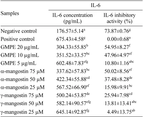
which modulates inflammatory responses.39 GMPE in the concentration of 20 µg/mL showed the highest inhibitory activity (Table 3). Inhibiting the production of IL-1β was important in finding the anti-inflammatory agent. GMPE, α-mangostin, and γ-mangostin inhibited the production of
IL-1β in a concentration-dependent manner. The highest concentration of GMPE, α-mangostin and γ-mangostin possessed the highest inhibitory activity with no significant differences observed among them (Table 4). IL-1β is a potent proinflammatory cytokine released by macrophages in systemic inflammatory responses that regulate inflam-matory reaction and immune response.40
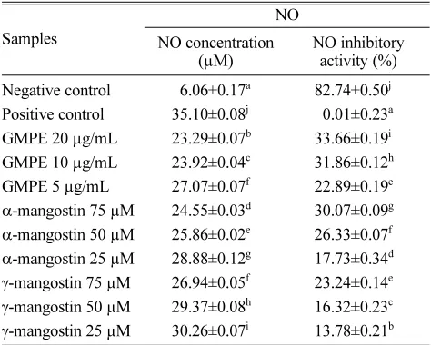
In this study, the nitrite concentration was associated with NO production. Excessive levels of NO can mediate proinflammatory and have destructive effects, thus lowering the NO concentration can be used as an anti-inflammatory action. The GMPE showed the highest NO inhibitory activity, meanwhile the positive control showed the highest concentration of NO (Table 5). This research result indicated that the LPS successfully induced the inflammation of the RAW 264.7 cells41 and GMPE had anti-inflammatory properties by reducing the NO production. The inhibition of NO pro-duction might related to suppression of iNOS expression, as NO synthesis is catalyzed by iNOS.
GMPE possessed the highest inhibitory activity against COX-2, IL-6, and NO production, but all of the tested concentration both for GMPE and its compounds have no significant differences in inhibitory activity of IL-1β. This research was consistent with previous research that G. mangostana fruit hull inhibited the inflammation-related diseases through NO and PGE2 releases.42 Likewise, α -mangostin and γ-mangostin inhibited NO and PGE2 production and COX-2 activity in LPS-induced RAW
Table 2. Effect various concentrations of GMPE and mangostins toward COX-2 concentration in RAW 264.7 cell
Samples
COX-2
COX-2 concentration (ng/mL)
COX-2 inhibitory activity (%)
Negative control 0.81±0.03a 72.93±0.97f Positive control 2.98±0.170g 50.11±5.72a GMPE 20 µg/mL 1.35±0.18b 54.59±6.09e GMPE 10 µg/mL 2.02±0.12de 32.21±3.87cd GMPE 5 µg/mL 2.16±0.07ef 27.40±2.28bc
α-mangostin 75 µM 1.43±0.08b 52.13±2.61e
α-mangostin 50 µM 1.75±0.12c 41.16±4.00d
α-mangostin 25 µM 2.06±0.05de 30.87±1.78bc
γ-mangostin 75 µM 1.89±0.03cd 36.58±0.89cd
γ-mangostin 50 µM 1.98±0.03d 33.56±1.16cd
γ-mangostin 25 µM 2.31±0.03f 22.48±0.89b The data are presented as mean ± standard deviation. Different superscript letters (a,b,bc,c,cd,d,de,e,f) in the same coloumn (among var-ious concentrations of GMPE, mangostins in COX-2 concentra-tions and inhibitory activity) are significant at p < 0.05 based on Duncan’s post-hoc comparisons (p < 0.05). The experiment was conducted in triplicate
Table 3. Effect various concentrations of GMPE and mangostins toward IL-6 concentration in RAW 264.7 cell
Samples
IL-6
IL-6 concentration (pg/mL)
IL-6 inhibitory activity (%)
Negative control 176.57±5.14a 73.87±0.76g Positive control 675.43±4.58g 50.00±0.68a GMPE 20 µg/mL 304.33±55.85b 54.95±8.27f GMPE 10 µg/mL 351.52±33.57bc 47.96±4.97ef GMPE 5 µg/mL 602.48±7.83efg 10.80±1.16abc
α-mangostin 75 µM 337.62±57.83bc 50.02±8.56ef
α-mangostin 50 µM 422.34±55.88cd 37.48±8.28de
α-mangostin 25 µM 567.52±66.90ef 15.98±9.91bc
γ-mangostin 75 µM 500.24±53.87de 25.94±7.98cd
γ-mangostin 50 µM 582.14±90.57efg 13.81±13.41abc
γ-mangostin 25 µM 645.14±92.87fg 54.49±13.75ab
The data are presented as mean ± standard deviation. Different superscript letters (a,ab,bc,abc,cd,ef,fg,efg,f,g) in the same coloumn (among various concentrations of GMPE, mangostins in IL-6 concentrations and inhibitory activity) are significant at p < 0.05 based on Duncan’s post-hoc comparisons (p < 0.05). The experi-ment was conducted in triplicate
Table 4. Effect various concentrations of GMPE and mangostins toward IL-1β concentration in RAW 264.7 cell
Samples
IL-1β
IL-1β concentration (pg/mL)
IL-1β inhibitory activity (%)
Negative control 5,841.44±18.01a 28.87±1.52c Positive control 1,183.03±35.09c 50.00±2.97a GMPE 20 µg/mL ,5894.31±77.23a 24.41±6.53c GMPE 10 µg/mL ,5950.70±115.33ab 19.64±9.75bc GMPE 5 µg/mL ,5951.72±45.98ab 19.55±3.89bc
α-mangostin 75 µM ,5877.28±35.87b 25.84±3.03c
α-mangostin 50 µM ,5910.43±79.98ab 23.04±6.76bc
α-mangostin 25 µM ,5942.64±121.95ab 20.32±10.31bc
γ-mangostin 75 µM ,5817.69±3.80a 30.88±0.32c
γ-mangostin 50 µM ,5936.41±43.20ab 20.85±3.65bc
264.7 cells according to Chen, et al. (2007) study.27 The inhibitory activity of α-mangostin and γ-mangostin from GMPE against IL-6 were also revealed by Bumrungpert,
et al. (2010) study.43 Furthermore, the α-mangostin and γ -mangostin revealed to attenuated LPS-induced inflammatory gene expression of TNF-α, IL-1β, IL-6, IL-8, MCP-1, and Toll-like receptor-2.44
The effect of G. mangostana
and its compounds against inflammation in this study also in line with Chomnawang, et al. (2007) study which reported anti-inflammatory activity of G. mangostana on inflammation caused by Propionibacterium acnes through suppression of pro-inflammatory cytokines, 45
as well as Lee, et al. (2013) study which revealed that G. mangosteen
and its compounds has great potential in the treatment and prevention of rheumatoid arthritis, a chronic inflammatory disease, showed by inhibition of TNF-α and IL-6 produc-tion in LPS-stimulated mice, reducproduc-tion of paw edema in the carrageenan-induced rats, and reduction of arthritis score in the CIA rats.46
This research revealed that GMPE, α-mangostin, and γ -mangostin possess the anti-inflammatory potential by inhibiting COX-2, IL-6, IL-1β, and NO. These extracts may have therapeutic potential for the modulation and regulation of macrophage activation, and may provide safe and effective treatment option for various inflamma-tion-mediated diseases. However, the therapeutic potential of these plant extracts will be further clear after preclinical and clinical test were conducted.
Acknowledgments
We gratefully acknowledge the financial support of Hibah Bersaing 2015 from Directorate General of Higher Education, Ministry of Research, Technology and Higher Education of the Republic of Indonesia (DIPA DIKTI No. DIPA-023-04.1.673453/2015). This study was succesfully conducted by the facilities support from Biomolecular and Biomedical Research Center, Aretha Medika Utama, Bandung, West Java, Indonesia and assisted by I Dewa Gde Sathya Deva and Balqis Balqis.
References
(1) Cragg, G. M.; Newman, D. J.; Snader, K. M. J. Nat. Prod. 1997, 60, 52-60.
(2) Ibrahim, M.; Hashim, N.; Mariod, A.; Mohan, S.; Abdulla, M.; Abdelwahab, S.; Arbab, I. Arabian J. Chem. 2016, 9,317-329.
(3) Obolskiy, D.; Pischel, I.; Siriwatanametanon, N.; Heinrich, M. Phytother. Res.2009, 23, 1047-1065.
(4) Pedraza-Chaverri, J.; Cárdenas-Rodriguez, N.; Orozco-Ibarra, M.; Pérez-Rojas, J. M. Food Chem. Toxicol.2008, 46, 3227-3239.
(5) Tjahjani, S.; Widowati, W. J. Indon. Med. Assoc. 2013, 63, 95-99. (6) Widowati, W.; Darsono, L.; Suherman, J.; Yelliantty, Y.; Maesaroh, M. Int. J. Biosci. Biochem. Bioinforma.2014, 4, 458-466.
(7) Ma li ska, D.; Gajewski, M. Folia Neuropathol.1998, 36, 199-204. (8) Shah, B. N.; Seth, A. K.; Maheshwari, K. M. Res. J. Med. Plant.
2011, 5, 101-115.
(9) Lam, D.; Harris, D.; Qin, Z. Mediators Inflamm.2013, 2013, 1-9. (10) Fang, S. C.; Hsu, C. L.; Yen, G. C. J. Agric. Food Chem. 2008, 56, 4463-4468.
(11) Naik, S. R.; Sheth, U. K. J. Postgrad. Med.1976, 22, 5-21. (12) Bellik, Y.; Boukraâ, L.; Alzahrani, H. A.; Bakhotmah, B. A.; Abdellah, F.; Hammoudi, S. M.; Iquer-Ouada, M. Molecules. 2012, 18, 322-353.
(13) Blonska, M.; Czuba, Z. P.; Krol, W. Scand. J. Immunol. 2003, 57, 162-166.
(14) Widowati, W.; Rusmana, D.; Hardiman, H.; Tiono, H.; Wargasetia, T. L.; Pujimulyani, D.; Yelliantty, Y. Eng. Tech.2013, 2013, 190-195.
(15) Darsono, L.; Hidayat, M.; Maesaroh, M.; Fauziah, N.; Widowati, W. Int. J. Med. Res. Health.Sci. 2015, 4, 566-571.
(16) Kang, C. H.; Choi, Y. H.; Choi, I. W.; Lee, J. D.; Kim, G. Y. Trop. J. Pharm. Res.2011, 10, 161-168.
(17) Yoon, W. J.; Ham, Y. M.; Kim, S. S.; Yoo, B. S.; Moon, J. Y.; Baik, J. S.; Lee, N. H.; Hyun, C. G. EurAsia J. BioSci.2009, 3, 130-143.
(18) Dewi, K.; Widyarto, B.; Erawijantari, P. P.; Widowati, W. Int. J. Res. Med. Sci. 2015, 3, 2303-2310.
(19) Rusmana, D.; Elisabeth, M.; Widowati, W.; Fauziah, N.; Maesaroh, M. Res. J. Med. Plant. 2015, 9,264-274.
s n
Table 5. Effect various concentrations of GMPE and mangostins toward NO concentration in RAW 264.7 cell
Samples
NO
NO concentration (µM)
NO inhibitory activity (%)
Negative control 56.06±0.17a 82.74±0.50j Positive control 35.10±0.08j 50.01±0.23a GMPE 20 µg/mL 23.29±0.07b 33.66±0.19i GMPE 10 µg/mL 23.92±0.04c 31.86±0.12h GMPE 5 µg/mL 27.07±0.07f 22.89±0.19e
α-mangostin 75 µM 24.55±0.03d 30.07±0.09g
α-mangostin 50 µM 25.86±0.02e 26.33±0.07f
α-mangostin 25 µM 28.88±0.12g 17.73±0.34d
γ-mangostin 75 µM 26.94±0.05f 23.24±0.14e
γ-mangostin 50 µM 29.37±0.08h 16.32±0.23c
γ-mangostin 25 µM 30.26±0.07i 13.78±0.21b The data are presented as mean ± standard deviation. Different superscript letters (a,ab,b,c,bc) in the same coloumn (among various concentrations of GMPE, mangostins in NO concentrations and inhibitory activity) are significant at p < 0.05 based on Duncan’s post-hoc comparisons (p < 0.05). The experiment was conducted in triplicate
(20) Widowati, W.; Mozef, T.; Risdian, C.; Yellianty. Y. Oxid. Antioxid. Med. Sci. 2013, 2, 137-142.
(21) Widowati, W.; Wijaya, L.; Wargasetia, T. L.; Bachtiar, I.; Yellianty.; Laksmitawati, D. R. J. Exp. Integr. Med.2013, 3, 225-230.
(22) Khan, T. Z.; Wagener, J. S.; Bost, T.; Martinez, J.; Accurso, F. J.; Riches, D. W. Am. J. Respir. Crit. Care Med.1995, 151, 1075-1082.
(23) Joo, T.; Sowndhararajan, K.; Hong, S.; Lee, J.; Park, S. Y.; Kim, S.; Jhoo, J. W. Saudi. J. Biol. Sci. 2014, 21, 427-435.
(24) Surh, J.; Yun, J. M. Prev. Nutr. Food Sci.2012, 17, 22-28. (25) Kuroishi, T.; Bando, K.; Endo, Y.; Sugawara, S. Toxicol. Sci. 2013, 135, 119-128.
(26) Verma, N.; Tripathi, S. K.; Sahu, D.; Das, H. R.; Das, R. H. Mol. Cell. Biochem. 2010, 336, 127-135.
(27) Chen, L. G.; Yang, L. L.; Wang, C. C. Food Chem. Toxicol.2008, 46, 688-693.
(28) Rajalaksmi, A.; Jayachitra, A.; Gopal, P.; Krithiga, N. Biomed. Res.
2014, 1, 1-6.
(29) Jothy, S. L.; Zakaria, Z.; Chen, Y.; Lau, Y. L.; Latha, L. Y.; Sasidharan, S. Molecules2011, 16, 5268-5282.
(30) Lalitha, P.; Shubashini, K.; Jayanthi, P. Asian J. Pharm. Clin. Res.
2012, 5, 59-61.
(31) Patel, O. V.; Wilson, W. B.; Qin, Z. Biometals. 2013, 26, 415-425. (32) Coker, R. K.; Laurent, G. J. Eur. Respir. J. 1998, 11, 1218-1221. (33) Jo, W. S.; Choi, Y. J.; Kim, H. J.; Nam, B. H.; Lee, G. A.; Seo, S. Y.; Lee, S. W.; Jeong, M. H. Toxicol. Res.2010, 26, 37-46.
(34) Nguyen, L. K.; Cavadas, M. A. S.; Kholodenko, B. N.; Frank, T. D.; Cheong, A. Cell. Mol. Life Sci.2015, 72, 2431-2443.
(35) Bak, M. J.; Truong, V. L.; Kang, H. S.; Jun, M.; Jeong, W. S. Oxid.
Med. Cell. Longev.2013, 172, 1-11.
(36) Damte, D.; Reza, M. A.; Lee, S. J.; Jo, W. S.; Park, S. C. Toxicol. Res.2011, 27, 11-14.
(37) Kornohen, R.; Lahti, A.; Kankaanranta, H.; Moilanen, E. Curr. Drug. Targets. Inflamm. Allergy. 2005, 4, 471-479.
(38) Sakurai, H; Kohsaka, H.; Liu, M. F.; Higashiyama, H.; Hirata, Y.; Kanno, K.; Saito, I.; Miyasaka, N. J. Clin. Invest. 1995, 96, 2357-2363.
(39) Kostek, M. C.; Nagaraju, K.; Pistilli, E.; Sali, A.; Lai, S. H.; Gordon, B.; Chen, Y. W. BMC Musculoskelet. Disord.2012, 13, 106
(40) Zhang, D.; Zheng, H.; Zhou, Y.; Tang, X.; Yu, B.; Li, J. BMC Cancer. 2007, 7, 45.
(41) Sarkar, D.; Fisher, P. B. Cancer Lett. 2006, 236, 13-23.
(42) Tewtrakul, S.; Wattanapiromsakul, C.; Mahabusarakam, W. J. Ethnopharmacol.2009, 121, 379-382.
(43) Bumrungpert, A.; Kalpracvidh, R. W.; Chuang, C. C.; Overman, A.; Martinez, K.; Kennedy, A.; Mclntosh, M. J. Nutr.2010, 140, 842-847. (44) Gutierrez-Orozco, F.; Failla, M. L. Nutrients2013, 5, 3163-3183. (45) Chomnawang, M. T.; Surassmo, S.; Nukoolkarn, V. S.; Gritsanapan, W. Fitoterapia.2007, 78, 401-408.
(46) Lee, L. T.; Tsai, Y. F.; Hu, N. Y.; Wang, C. W.; Huang, K. K.; Hsiao, J. K.; Shih, Y. C.; Munekazu, I. Biomed. Prev. Nutr. 2013, 3, 227-232.