Indo. J. Chem., 2009, 9 (2), 312 - 314
Fredryk Mandeyet al.
312
* Corresponding author. Tel/Fax : +62-411-586-498 Email address : [email protected]
SPECTROSCOPICAL STUDY OF AN ETHER FRACTION ISOLATE FROM
STEAM BARK OF “ JATI” (
Tectona grandis
, L)
Fredryk Mandey
1*, Raimundus Chalik and Ismail Ibrahim
2 1Department of Chemistry, Hasanuddin University, Jl. Perintis Kemerdekaan Km.10, Makassar, 90245 2
Makasar Polytechnique of Health, Jl. Baji Gau, Makassar
Received December 30, 2008; Accepted June 24, 2009
ABSTRACT
This research is aimed to primarily study the spectroscopy of ether fraction of from Jati wood (T. grandis, L). The stem bark of T.grandis, L was firstly extracted undergoes a reflux in methanol solvent. Further, the dried methanol crude extract was extracted with diethyl ether and moreover separated its chemical constituents by using of column chromatography using degraded mixtures of n-hexane – ethyl acetate from 9:1 to 7:3. After a final column chromatography from the E fraction, a white needles crystal with a melting point of 295 – 297.5 °C was resulted. This results was then confirmed undergoes a spectroscopic analysis including UV, FTIR, H-NMR, and Mass spectroscopy to get an acid of 3β-hydroxy-20(29)-lupen-28-oic or commonly known as betulinic acid.
Keywords:betulinic acid, spectroscopy, primarily study INTRODUCTION
Tectona grandis, L, commonly known as “Jati”, was an endemic plant in almost region in Indonesia and traditionally used in the industry and building materials [1]. These plants also have an ayurvedic function such as anti-bronchitis, scabbiest, diarrhoea, leucoderm, laxative, and expectorant [2]. MorphologicallyT. grandis, L, belong to the Verbenaceae family, was a typical big, tall (could reach approximately 27 – 35 meter height), cylindrical with symmetry shape. It has a brown colour bark and a typical single leaf with an elliptical form facing side by side. Jati also have small flower, about 4 – 8 mm length, white coloured and very good smell [3]. An intensive research to the part of these plant had been carried out.
Shalini and Srivastava [4], have carried out an investigation to the antifungal activities of a metanolic crude extract ofT. grandis, Land found that this crude is potentially active as antifungal agents. More recently, Majumdaret al [5] reported an intensive investigation of the wounded healing effect of the leaves of T grandis, L and discovered that leaves of T. grandis, L significantly possess an wound healing agents.
EXPERIMENTAL SECTION
Material
Samples of stem bark of T. grandis, L taken from an area of Barru Regency, South Sulawesi Province. Prior to the extraction, this sample was then cleaned, air dried, and cut into small pieces.
Within this research several chemical was used for instance : diethyl ether Merck), ethyl acetate
(E-Merck), n-hexane (E-(E-Merck), methanol (E-(E-Merck), silica gel GF 60 F245(E-Merck).
Instruments
A series of instruments was used in this investigation. Among that instruments are melting point apparatus (Electro thermal), GC-MS (Shimadzu), electric oven (Memmert), UV Spectronic 3000 (Milton Roy), FTIR Spectrophotometer (Shimadzu), HNMR Spectrophotometer (Jeol-MY 60), and analytical balance (Sartorius).
Procedure
Sample Extraction
Approximately 500 g of dried small pieces cut of stem bark ofT. grandis, Lwas extracted under reflux in methanol about three times for four hour each. Resulted methanol extract was evaporated to get resulted of dry methanol extract. This dry methanol extract was then transform into a suspension with water and further extracting with diethyl ether in a separating funnel for three times. Resulting ethereal extract was then evaporated until it dry.
Chromatographically Analysis
Indo. J. Chem., 2009, 9 (2), 312 - 314
Fredryk Mandeyet al.
313
Table 1. 2-D Chromatography Results of Fraction E Isolate
Colour No Rf values
UV lamp Sulphuric Acid 10 %
1. 0,33 - pink
2. 0,55 - pink
Table 2.FTIR Spectrum of Fraction E Isolate
Absorbtion peak (cm-1)
Functional group
883 R2C=CH2
1041,5 and 1071,1 C – O strectching of alcohol
1377,1 - CH3symmetrical and asymmetrical stretching
1454,2 Symmetrical deformation of –CH2of a cyclopentane
1639,4 -C=C vibration (R2C=CH2)
1693,4 -C=O vibration of Carboxylic acid
2857,7 -CH2vibration
2869,6 -CH3asymmetrical vibration (C – H )
2943,2 -CH3symmetrical vibration (C – H )
3440,8 OH vibration of an alcohol
Table 3.HNMR Chemical Shifting of Isolate E
Chemical shifting (δ) ppm Functional group
4,71 (m) -CH2from R2C=CH2
2,5 (t) C – H cyclic (from cyclopentane)
2,1 (m) CH2cyclic (from cyclopentane)
1,3 (s) 0
Spectroscopy Analysis
Chromatographically pure isolated sample of T. grandis, Lstem bark was then verified undergoes a two dimension chromatography and a set of qualitative analysis with a suitable chemical reagents. The results of this test were shown in Table 1. After measuring the melting point this resulting isolate was then characterized with a series of spectroscopy analysis with an instrument of UV, FTIR, HNMR, and Mass Spectrum. Results of this experiment are stated in Table 2, 3, and 4.
RESULT AND DISCUSSION
Isolation
Isolation of betulinic acid from the stem-bark of T grandis, L involving a column chromatography process
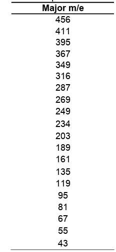
Table 4.Mass spectrum of Isolate E Major m/e
under goes a subsequently degraded polarity of the eluent. Primarily this process was done by extracting with the methanol solvent to the dried samples of T. grandis stem bark (approximately 500 grams). These methanol extract was then extracting again with diethyl ether to resulted the dried extract ether. This ether extract was then subjected to the column chromatography with an eluent of n-hexane/ethyl acetate with an arrangement of polarity from 9 : 1 to 7 : 3 ratios to give five dominant fraction of A, B, C, D, and E respectively. Among those five fractions, fraction E which forming white colour needles crystalline, physically dissolve in chloroform, diethyl ether and hot methanol. This isolate was then checked for the purity undergoes a two dimensional chromatography with an fluent of n-hexane/ethyl acetate 8 : 2 and 7 : 3 ratio to give a single spot pink colour (after spotted with 10% of sulphuric acid as a spot appearance) with an Rf of subsequently 0,22 and 0.55. These crystals also shown to have melting point range of 295 – 297.5 °C. These potentially faction E was then undergoes further for a series of spectroscopic analysis for a structural conformation.
Spectroscopic Verification
Indo. J. Chem., 2009, 9 (2), 312 - 314
Fredryk Mandeyet al.
314
Figure 1. Structure of Betulinic acid (3β -hydroxy-20(29)lupen-28-oic acid)
resulting absorption at 3440.8 cm-1which predominantly caused by an OH stretching in an alcohol. This result was also confirming by the existing of a peak in 1041.5 and 1071.1 cm-1 of the C – O vibration of an alcohol. These fragment also become one of the major fragment in MS spectrum of M-18 (M-H2O) fragment. Occurrence of asymmetric and symmetrical C – H of the methyl group clearly shown in peak of 2869.6; 2943.2; and 1377.1 cm-1respectively. A major absorption in 2857.7 cm-1 was a clear evident of –CH2- bending which conformed to a cyclopentene by an absorption in 1454.2 cm-1. These existing of –CH2- in cyclopentane were covered also by an HNMR absorption inδ2,1 ppm.
Another major functional group, carboxylic acid, is existed also in fraction E molecule. A strong absorption at 1693.4 cm-1 resulting by a stretching of C=O of carboxylic acid. A typical fragment of M-45 (M-COOH) is also a clear evident of this carboxylic acid appearance in the structure. A typical double bond of R2C=CH2pattern appeared in absorption peak at 1639.4 and 883 cm-1in FTIR spectrum and conformed also by the HNMR spectrum atδ4,7 ppm.
Gas chromatography which combined by a mass spectroscopy measurement give constant result of 11.625 retention time and a molecular ion (M+) m/e of 456.
Combining all of spectroscopy data and other physical evident it was confirmed that Fraction E is corresponding to the structure of betulinic acid (Figure 1), a potent bioactive molecules with several pharmacological activities such as anti-HIV [6,7], and
anticancer [8]. Further, this result was also confirmed with the standard spectroscopy data of betulinic acid from literature [9]. The literatures also give the same results.
CONCLUSION
In this research it was proven that Tectona grandis, L,commonly known as Jati, contain a betulinic acid, typical pentacyclic triterpenoid molecule. Further our group plans to investigate more of this betulinic acid by exploring other part of Tectona grandis to get more information of the potency of this plant.
ACKNOWLEDGEMENT
All author want give a grateful thank to Ms. Maryati, Mr Sudomo, and Mr. Wardjiman, staff of Organic Laboratory, Department of Chemistry, Faculty Mathematics and Natural Sciences, Gajah Mada University for their help and assistance in spectroscopy measurement.
REFERENCES
1. Tewari, D.N., 1992, A Monograph on Teak (Tectona grandis,L), International Books Distributor, Dehra, India.
2. Singh, J., Bhuyan, T.C., and Ahmed, A., 1996, J. Econ. Taxon. Bot, 12, 350–356.
3. Palanisamy, K. and Subramaniam, K., 2001,Silvae Genetica, 50, 5–6.
4. Shalini and Srivastava, R., 2008, The Internet Journal of Alternative Medicine, 5, 2.
5. Majumdar, M., Nayeem, N., Kamath, J.V., and Asad, M., 2007,Pak. J. Pharm.Sci., 20, 2, 120-124. 6. Pisha, E et al., 1995,Nat. Med.,1, 10, 1046–1051. 7. Huang, L., Ho, P., Lee, K.H., and Chen, C.H.,
2006,Bioorg. Med. Chem., 14, 7, 2279–2289. 8. Fulda, S., 2008,Int. J. Mol. Sci., 9, 1096–1107. 9. Pathak, N.K.R., Eogi, P., Biswas, M., Tripathi, Y.C.,