Surface Morphology of Fe(III)-Porphyrin Thin Layers as Characterized by
Atomic Force Microscopy
Utari
1,3,*, Kusumandari
1, Budi Purnama
1, Mudasir
2, and Kamsul Abraha
3 1Department of Physics, Faculty of Mathematics and Natural Sciences, Universitas Sebelas Maret Jl. Ir. Sutami 36A Kentingan Surakarta 57126, Indonesia
2
Department of Chemistry, Faculty of Mathematics and Natural Sciences, Universitas Gadjah Mada Sekip Utara PO BOX BLS 21 Yogyakarta 55281, Indonesia
3
Department of Physics, Faculty of Mathematics and Natural Sciences, Universitas Gadjah Mada Sekip Utara PO BOX BLS 21 Yogyakarta 55281, Indonesia
Received November 6, 2015; Accepted April 15, 2016
ABSTRACT
Surface morphology of Fe(III)–porphyrin thin layers was studied using atomic force microscopy. The thin layer samples used in these experiments were deposited by spin coating methods on indium–tin-oxide substrates at room temperature under atmospheric conditions. Variations of a thin layer of Fe(III)-porphyrin were done by modifying the rotational speed and the concentration of the solution. The experimental results demonstrated that the Fe(III)– porphyrin layers were observed as discrete nanomolecular islands. Both the number of nano-islands and thickness of the layer increased significantly with increasing concentration. A layer thickness of 15 nm was obtained for low concentrations of 0.00153 M and become 25 nm for dense concentrations of 0.153 M. Conversely, the higher number of islands were deposited on the surface of the substrate at a lower rotational speed.
Keywords:Fe-porphyrin; Surface morphology; Atomic Force Microscopy (AFM)
ABSTRAK
Morfologi permukaan lapisan tipis Fe(III)-porphyrin telah dipelajari dengan menggunakan atomic force microscopy (AFM). Sampel-sampel Fe(III)-porphyrin dalam penelitian ini dideposisi pada substrat indium tin oksida (ITO) dengan menggunakan metode spin-coating pada suhu kamar dibawah kondisi atmosfir. Variasi lapisan tipis Fe(III)-porphyrin dilakukan dengan memodifikasi kecepatan putar dan konsentrasi larutan. Hasil eksperimen menunjukkan bahwa lapisan Fe(III)-porphyrin membentuk pulau-pulau nano-molecular diskrit. Jumlah dan ketebalan pulau-pulau meningkat secara signifikan dengan kenaikan konsentrasi. Ketebalan sebesar 15 nm diperoleh untuk konsentrasi 0,00153 M dan menjadi 25 nm untuk konsentrasi 0,153 M. Namun demikian, jumlah nano-meter-orde pulau-pulau yang terdeposisi pada permukaan substrat lebih tinggi pada kecepatan putar rendah.
Kata Kunci:Fe-porphyrin; Morfologi permukaan; Atomic Force Microscppy (AFM)
INTRODUCTION
In the last decade, the development of organic electronics has become a widely researched field as their production involves simple, relatively low cost procedures [1]. Moreover, organic electronics can be deposited on flexible substrates such as plastics. Recently, organic electronics have been found to be complementary to conventional electronics [2]. Among organic materials, porphyrins and metalloporphyrins have been widely exploited owing to their interesting properties [3].
The arrangement of molecules on a solid surface is considered to be a key factor in the realization of a molecular device’s architecture. Moreover, nanometer
Indones. J. Chem., 2016, 16 (3), 233 - 238
234
on porphyrin-based materials has also been observed [21-27].For the detection of neurotransmitters, graphite electrodes coated by electropolymerized porphyrins have been studied since1997 [6]. Fabrication of the solid state sensor by porphyrin derivatives on glass substrates for optochemical sensing of HCl gas has also been reported in 2012 [28].
A formation of nanometer orde (10-500 nm) discrete material is a key issue for a various application technology in the future [29]. A discrete surface morphology of zinc porphyrin has been reported that can be controlled by varying the concentration as well as deposition parameters [30]. However, as far as we know, there is still very limited number of papers dealing with the parameters deposition dependence of surface morphology for porphyrin-based materials, especially Fe(III)-porphyrin. Therefore, in this work, we studied the changes in surface morphology of Fe–porphyrin thin layers as a result of the interactions between the molecules and the substrate surface. The Fe–porphyrin thin layers were assembled by spin coating at room temperature using indium–tin-oxide (ITO) coated glass substrates. The surface morphology of the Fe–porphyrin thin layers were modified by variations in the rotational speed and concentration. These changes were then characterized using atomic force microscopy (AFM).This AFM technique has been selected in this study because it is a nondestructive analytical method, requires only a very small of samples and gives valuable information about the characteristics of the materials including two and three dimension profiles, layer thickness, cross-section and distribution of islands or spot.
EXPERIMENTAL SECTION
Materials
Fe(III) mesoporphyrin IX chloride powder (100 mg) were purchased from Frontier Scientific- USA (FeM658) and used as a thin layer without further purification. Chloroform (10 mL) were purchased from Merck and used as a solvent. ITO-coated glass substrates (Aldrich-578 274) were purchased from Aldrich (Germany).
Instrumentation
Instruments used in this study consist of a magnetic stirrer, spin coater (Chemat Technology KW-4A), hot plate (IKA® C.MAG) and Atomic Force Microscopy (AFM) Park System XE-7.
Procedure
The experiments were carried out in the following three steps: (i) preparation of the Fe(III)–porphyrin
solution, (ii) deposition of the Fe(III)–porphyrin thin layer by spin coating, and (iii) characterization of the surface morphology of the Fe(III)–porphyrin film using AFM.
Preparation of Fe–porphyrin solution
Fe(III)–porphyrin powder (100 mg, Frontier Scientific-FeM658) was dissolved in chloroform (10 mL, Merck) and stirred with a magnetic stirrer for 60 min at a rotational speed of 200 rpm (solution A). To modify the concentration of the solution, 1 mL of this solution was taken and then added with chloroform until the diluted total final volume of 10 mL (solution B). Solution B was used as the basic solution for the deposition of Fe–porphyrin. The resulting layers prepared using solutions A and B are called ‘sample A’ and ‘sample B’, respectively.
Deposition of Fe–porphyrin layers
The growth of the Fe(III)–porphyrin thin layers was accomplished using a spin coater (Chemat Technology KW-4A) with ITO-coated glass substrates (Aldrich-578 274). Before use, the substrates were washed in an ultrasonic cleaner using both alcohol and acetone. The samples were deposited for 20 sec followed by post-heating at 40 °C for 60 sec. This procedure was repeated to obtain three layers. The samples were deposited with two different rotational speeds, i.e. 500 and 1000 rpm to modify the surface morphology. Fe(III)–porphyrin solutions with two different concentrations i.e. 0.153 M (solution A) and 0.00153 M (solution B).were also used to modify the surface morphology.
Characterization of Fe–porphyrin films
Surface morphology of the samples was observed using Atomic Force Microscopy (AFM) Park System XE 7. For the whole experiment was used noncontact tips
with scan area 5 μm × 5 μm and scanning frequency
are 1kHz at room temperature. Hereafter the obtained data were analyzed by using WSxM 5.0 software [31].
RESULT AND DISCUSSION
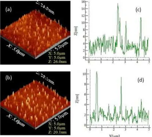
Fig 1. Surface morphology and topography of Fe–porphyrin layers (sample B) deposited by spin coating and evaluated at rotational speeds of 500 rpm (a and c) and 1000 rpm (b and d)
Fig 2. The 3D surface morphology and profiles of Fe–porphyrin layers deposited by spin coating with 0.00153 M solute
500 rpm than that found at 1000 rpm.
This phenomenon was shown more clearly in the graph of discrete islands size distribution, which resembles a Gaussian distribution for both rotational speeds but with different median size (Fig. 1 c and d). In sample B with a rotational speed of 500 rpm, the island size distribution (of the value measured topographically) widened to 20 nm, with a median size of 7.5 nm being the most abundant. In sample B, with a rotational speed
Indones. J. Chem., 2016, 16 (3), 233 - 238
236
Fig 3.Surface morphology and topography of Fe-porphyrin layers deposited by spin coating at a rotational speed of 500 rpm and evaluated at concentrations 0.00153 M (a and c) and 0.153 M(b and d)
Fig. 2 (a) and (b) show the three-dimensional (3D) surface morphology of Fe–porphyrin thin layers (sample B) on ITO-coated glass substrates with rotational speeds of 500 and 1000 rpm, respectively. If the baseline can be considered as a layer without the deposition of a molecule of Fe(III)-porphyrin, e.g. ITO-coated glass substrate surface, it can be seen that the distribution of Fe–porphyrin islands populations are spread over the surface to a certain thickness. If it is deposited in a straight line lengthwise, i.e. along the x-axis, on the surface of the sample, a surface profile would be obtained, such as those shown in Fig. 2 (c) and (d). From this profile, the thickness was approximately 13 nm
(= 15 nm of highest peak −2nm of baseline) for the rotational speed of 500 rpm and 8 nm (= 10−2) for 1000
rpm. Therefore, the results confirmed that the number of Fe(III)–porphyrin islands deposited decreases with the increasing of rotational speed.
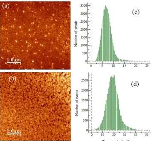
Fig. 3 (a) and (b) show the 2D AFM images and topography of the surface morphology of Fe–porphyrin thin layers on ITO-coated glass substrates with a rotational speed of 500 rpm for samples A and B. It can be seen that the amount of Fe–(III)-porphyrin islands deposited on ITO-coated glass substrates increased with increasing concentration of the solution. At higher solution concentration (sample A), more Fe(III)– porphyrin materials are deposited, which are shown in Fig. 3 (b) (sample A), where the Fe(III)–porphyrin islands cover almost the entire surface of the substrate. This result is in contrast to sample B, where the porphyrin
molecules do not cover the entire surface of the substrate.
The topographical charts clearly shows that the lateral dimension of Fe(III)–porphyrin islands forming the layers that follows a Gaussian distribution with similar shape for both samples A and B. For layers obtained from a low concentration solution of 0.00153 M (sample B), the distribution of island size extended to 20 nm (lateral dimension), with 7.5 nm being the most abundant. For layers obtained from a higher concentration solution of 0.153 M (sample A), the shape of the layer is quite similar to that of lower concentration but the lateral dimension of the Islands shifts to the higher value of 45 nm, with 22 nm being the most abundant. This finding confirms that higher concentration of Fe(III)-porphyrin leads to higher numbers of materials being deposited on the substrate surface, resulting in the increase in the number of islands with the larger size.
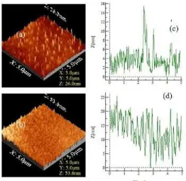
Fig 4.The 3D surface morphology and profiles of Fe–porphyrin deposited by spin coating at a rotational speed of 500 rpm and evaluated at concentrations of 0.00153 M.(a and c) and 0.153 M.(b and d)
increases the number of islands, which in turn increases both the population of Fe–porphyrin deposited on the substrate surface and the thickness of the layer. In addition, the profile becomes more crowded because the distance between the two islands is very close to each other.
CONCLUSION
We have used AFM to analyze the surface morphology of Fe(III)–porphyrin thin layers which were grown on ITO-coated glass substrates through spin coating at room temperature. Observation of the morphology indicates that a higher number of molecules were deposited on the surface of the substrate at a lower rotational speed. It is also shown that with increasing concentrations of Fe(III)–porphyrin solutions, a higher number of molecules are deposited on the substrate surface. The lateral dimensions of the island size distribution of the Fe(III)–porphyrin thin layers shows a Gaussian distribution with a sharper curve at high rotational speed and shift to the higher lateral dimension at larger concentration.
ACKNOWLEDGEMENT
This research was supported by the Scholarship for Domestic Postgraduate Program of the Directorate General of Higher Education of the Republic of Indonesia, (BPPDN DIKTI, No.1251.15/E4.4/2013) and
DIPA Universitas Sebelas Maret of Republic of Indonesia (Penelitian Unggulan Perguruan Tinggi Contract No339/UN27.11/PL/2015).
REFERENCES
1. Arai, T., Ishibashi, M., Kawasaki, M., and Shiba, T., 2008,Hitachi Rev., 57 (3), 108–115.
2. Tiwari, S., and Greenham, C.G., 2009, Opt. Quantum Electron., 41 (2), 69–89.
3. Hart, H., Craine, L.E., and Hart, D.J., 2003, Organic Chemistry: A Short Course, Houghton Mifflin Company, New York, 418–423.
4. Palmer, R., and Guo, Q., 2002, Phys. Chem. Chem. Phys., 4 (18), 4275–4284.
5. Gong, X., Milic, T., Xu, C., Batteas, J.D., and Drain, C.M., 2002,J. Am. Chem. Soc., 124 (48), 14290– 14291.
6. Duong, B., Arechabaleta, R., and Tao, N.J., 1998, J. Electroanal. Chem., 447 (1-2), 63–69.
7. Fagadar-Cosma, E., Mirica, M.C., Balcu, I., Bucovicean, C., Cretu, C., Armeanu, I., and Fagadar-Cosma, G., 2009, Molecules, 14 (4), 1370–1388.
8. Goya, M.C., Lucero, M., Orive, A.G., Marin, A., Gimeno, Y., Creus, A.H., Aguirre, M.J., Arévalo, M.C., and Armijo, F., 2011, Int. J. Electrochem. Sci., 6 (10), 4984–4998.
Indones. J. Chem., 2016, 16 (3), 233 - 238
238
Scolaro, L.M., Rowan, A.E., Elemans, J.A.A.W., and Nolte, R.J.M., 2008,Nano Lett., 8 (1), 253–259. 10. O’Sullivan, M.C., Sprafke, J.K., Kondratuk, D.V.,
Rinfray, C., Claridge, T.D.W., Saywell, A., Blunt, M.O., O’Shea, J.N., Beton, P.H., Malfois, M., and Anderson, H.L., 2009,Nature, 469 (7328), 72–75. 11. Wang, X., Wang, G.C., and Lewis, K.M., 2012,
Mater. Chem. Phys., 136 (1), 190–195.
12. Ditze, S., Röckert, M., Buchner, F., Zillner, E., Stark, M., Steinrück, H.P., and Marbach, H., 2013, Nanotechnology, 24 (11), 1–11.
13. Bernien, M., Xu, X., Miguel, J., Piantek, M., Eckhold, Ph., Luo, J., Kurde, J., Kuch, W., Baberschke, K., Wende, H., and Srivasta, P., 2007,Phys. Rev. B, 76 (21), 214406-1–214406-6.
14. Gatteschi, D., 2007,Nat. Mater., 6 (7), 471–472. 15. Wende, H., Bernien, M., Luo, J., Sorg, C.,
Ponpandian, N., Kurde, J., Miguel, J., Piantek, M., Xu, X., Eckhold, P., Kuch, W., Baberschke, K., Panchmatia, P.M., Sanyal, B., Oppeneer, P.M., and Eriksson, O., 2007,Nat. Mater., 6 (7), 516–520. 16. Bernien, M., Miguel, J., Weis, C., Ali, Md.E., Kurde,
J., Krumme, B., Panchmatia, P.M., Sanyal, B., Piantek, M., Srivastava, P., Baberschke, K., Oppeneer, P.M., Eriksson, O., Kuch, W., and Wende, H., 2009, Phys. Rev. Lett.,102 (4), 0472021–0472024.
17. Ferradás, R., García-Suárez, V.M., and Ferrer, J., 2013,J. Phys. Condens. Matter., 25 (32), 325501. 18. Danilovic, D., Lin, C.L., Yuen, T., Pan, L., and Li, J.,
2007,J. Appl. Phys., 101 (9), 09E1031–09E1033. 19. Herper, H.C., Bernien, M., Bhandary, S., Hermanns,
C.F., Krüger, A., Miguel, J., Weis, C., Schmitz-Antoniak, C., Krumme, B., Bovenschen, D., Tieg, C., Sayal, B., Weschke, E., Czekelius, C., Kuch, W.,
Wende, H., and Eriksson, O., 2013,Phys. Rev. B, 87 (17), 174425.
20. Baberschke, K., 2009,J. Phys. Conf. Ser., 190 (1), 012012-1–012012-10.
21. Podzorov, V., Menard, E., Rogers, J.A., and Gershenson, M.E., 2005,Phys. Rev. Lett., 95 (22), 226601.
22. Neidermeier, U., Baghnich, S.A., Melzer, C., Serfert, W., and Seggern, H., 2010, Synth. Met., 160 (3-4), 251–255.
23. Kozlova, N.V., Mori, N., Makarovsky, O., Eaves, L., Zhuang, Q.D., Krier, A., and Patanè, A., 2012,Nat. Commun., 3, 1–5.
24. Niedemeier, U., Vieth, M., Pätzold, R., Sarfert, W., and von Seggern, H., 2008, App. Phys. Lett., 92 (19), 193309.
25. Siles, P.F., Bufon, C.C.B., Grimm, D., Jalil, A.R., Mende, C., Lungwitz, F., Salvan, G., Dietrich, R.T.Z., Lang, H., and Schmidt, O.G., 2014, Org. Electron., 15 (7), 1432–1439.
26. Li, Z., Park, T.H., Rawson, J., Therien, M.J., and Borquet, E., 2012,Nano Lett., 12 (6), 2722–2727. 27. Yuen-Zhou, J., Saikin, S.K., Yao, N.Y., and
Aspuru-Guzik, A., 2014, Nat. Mater., 13 (11), 1026–1032.
28. Kalimuthu, P., Sivanesan, A., and John, S.A., 2012,J. Chem. Sci., 124 (6), 1315–1325.
29. Drain, C.M., Bazzan, G., Milic, T., Vinodu, M., and Goeltz, J.C., 2005,Isr. J. Chem., 45 (3), 255–269. 30. Yin, J., Guo, Q., and Palmer, R.E., 2003, J. Phys.
Chem. B, 107 (1), 209–216.
31. Horcas, I., Ferńandez, R., Gómez-Rodrigues,