Review Article
Telomere and telomerase in hematological disorders
Focusing on bone marrow failure syndromes and
hematological malignancies
Mardiah Suci Hardianti, Ibnu Purwanto, Johan Kurnianda
Division of Hematology and Medical Oncology, Department of Internal Medicine Sardjito General Hospital/Faculty of Medicine Gadjah Mada University Yogyakarta Indonesia
ABSTRACT
We review the present knowledge of telomeres and telomerase with special attention to their role in hematological disorders especially bone marrow failure syndromes including acquired aplastic anemia and myelodysplastic syndromes, as well as acute and chronic myeloid leukemia. The current understanding on the role of telomere and telomeres dysfunctions in hematological disorders leads us to a better understanding on the pathology of the diseases as well as considering some possibilities to employ the measurement of telomere length and telomere activity in disease prognostication. Several treatment options targeting telomere and telomerase being developed are also reviewed.
Keywords: telomere- telomerase- bone marrow failure syndromes- hematological malignancies
Telomeres
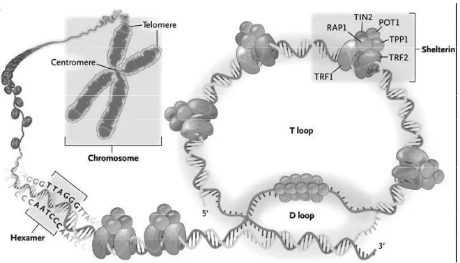
Telomeres are the non-encoding regions of DNA cappingthe ends of chromosomes, in association with various proteins, known as shelterin. The DNA that forms the telomere consists of thesequence (5′-TTAGGG-3′) n, which is referred to as a “telomeric repeat” due to its tandem repeat over 5 to15 kilobases (kb). A single-stranded 3’-hydroxyl overhang is generated by the catalytic addition of telomeric repeats to the 3’ end and by post replicative processing of the lagging strand.1 This single
stranded overhang folds back and invades the double-stranded telomeric helix, forming the T loop in order to avoid being recognized as a double-strand break and corrected by DNA repair machineries.2 Thus, telomeres function
to guard chromosomes against degradation, fusion, and rearrangements during DNA replication.3 When telomeres become very
short, they signal the arrest of cell proliferation, senescence, and apoptosis. The cells whose telomeres shorten to a “critical length” enter a stage termed replicative senescence whereby cell division is prevented.1
additional proteins can bind indirectly to telomeres, often via TRF1 and TRF2, and together these proteins function to regulate telomere homeostasis. They consist of RAP1-a binding partner of TRF2,POT1- a single-stranded DNA binding protein; and the two bridging proteins, TIN2 and TPP1.4 Not
only do these proteins function in protecting the chromosome end, they also function in telomere length regulation. Any mutations reducing their expression or impairing their binding to DNA result in telomere erosion. Telomere length is maintained within a strict range throughout cell division, suggesting a negative feedback loop involving the shelterin complex. Due to the exquisite specifi city of these DNA binding proteins, the amount of shelterin protein bound to telomeres is roughly proportional to their length.2
Telomerase
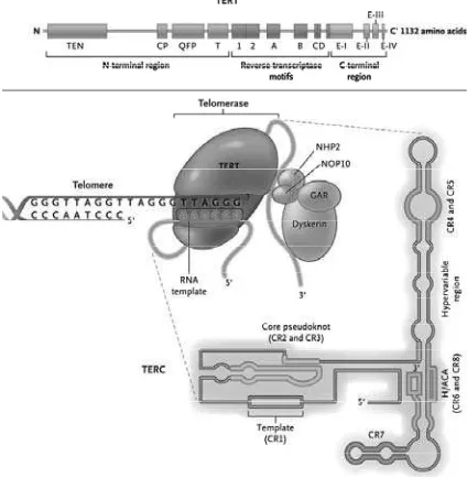
Telomerase is a DNA polymerase required to catalyze DNA synthesis to maintain telomere length. It is composed of two essential subunits, human telomerase RNA component (hTERC) and human telomerase catalytic component (hTERT). hTERT uses the telomerase RNA component (TERC) as a template to synthesize telomereDNA.1 The assembly of a functional
telomerase holoenzyme complex also requires other telomere- and/or telomerase-associated proteins (e.g., dyskerin, NOP10, GAR1, NHP2)to stabilize the complex.5 The structural
homodimerization of the protein (i.e., TERT protein-protein interaction). The functional reverse transcriptase (RT) domain is almost centrally located. The C-terminal domains of TERTs are also required for telomerase-specific enzymatic activity and/or in the telomeric nucleotide addition processivity process.5
Telomerase is regulated by a wide variety of genes or multifactorial. Removal or reduced regulation of the hTERC subunit leads to a loss of telomerase activity, erosion of telomeres and inhibition of cellular growth. Mutation of the hTERC gene has been described in distinct autosomal dominant disorder dyskeratosis congenita.6 Besides its function
as a binding site for the SP1 transcription factor, the hTERT promoter also provides other binding sites for various transcription
factors and hormone responsible elements. hTERT is repressed by retinoblastoma protein (Rb) and cyclin-dependent kinase inhibitor p21WAF1. Conversely, c-Myc activates TERT gene expression. hTERT phosphorylation is another mechanism of telomerase activity regulation. It is expressed mainly in embryonic and adult stem cells, highly proliferative cells such as mature lymphocytes, and in cancer cells, but not in most mature cells.5 While
hTERC is expressed in relatively identical amount in embryonic and somatic tissues, the expression of hTERT is precisely regulated and undetectable in many somatic cells. This means that the expression of hTERT is the limiting step in telomerase activation.6
Quantitative assays are available for telomere length and telomerase enzymatic activity. Telomeres can be visualized by fluorescent in situ hybridization (FISH) of individual cells and in-flow cytometry of specific cell populations (Young ASH). The length of telomere can be determined by a modification of Southern blotting in which the analysis of chromosome terminal restriction fragments (TRFs), as visualized with a radiolabeled telomeric repeat probe, provides the average lengths of all telomeres in a cell population. It also can be measured by quantitative polymerase chain reaction amplifi cation assays. Telomerase activity can be measured in vitro by a sensitive and effi cient polymerase chain reaction (PCR)-based detection method, also known as telomeric repeat amplifi cation protocol (TRAP).7
Telomere and on cogenesis
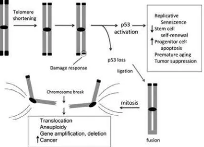
Most cancers show gross derangement in chromosome numbers. Telomere attrition has also been proposed as a mechanism for the Figure 2. The Telomerase Complex and
loss or gain of chromosomes. When telomere maintenance is disrupted in yeast, the few cells that escape senescence show chromosome abnor malities, end-to-end fusions, and consequent formation of dysenteric and circularized chromosomes. In the absence of telomerase, genetic lesions increase due to ter minal chromosome deletions and repeated cycles of break–fusion– bridge rearrangements. In “knock-out” mice that lack the RNA template component of telomerase, telomeres shorten progressively with each generation, producing chromosomal instability by end-to-end fusions.9,10 Most unstable cells
are removed by apoptosis, but they can be rescued if DNA damage is not inadequately monitored: in mTERC–/– mice that also are defi cient in the tumor suppressor gene p53, a variety of carcinomas appear associated with nonreciprocal translocations, as seen in human cancers.11
Figure 3. Telomere shortening activates p53 and drives formation of epithelial cancers through gene amplifi cation and deletion. Telomeres shorten progressively with cell division due to the end-replication problem in settings of insufficient telomerase, including in human fibroblasts, aging tissues, early cancers and diseases of high cellular turnover. Critical telomere
shortening compromises the telomere cap and results in a DNA damage response that activates the p53 tumor suppressor protein. This activation of p53 induces replicative senescence in cultured human
fi broblasts, impairs stem cell self-renewal, induces apoptosis in tissue progenitor cells, causes premature aging and strongly suppresses tumor formation. If p53 is mutated or deleted, these responses to telomere dysfunction are mitigated and chromosomal fusions are tolerated. The generation of fused chromosomes results in dysenteric chromosomes (chromosomes with two centromeres) and when these attach to opposite spindle poles, chromosome breakage occurs. These broken ends serve as potent catalysts for translocations, focal amplifi cations and focal deletions. Such CNAs drive development of carcinomas and explain the widespread gene copy number changes seen in human cancers.15
In humans, telomere length has been linked to malignant transformation— to the onset of cancer—in several diseases. When telomeres were first noted to be short in colorectal cancer, telomere loss was speculated to contribute to tumor genesis and genetic instability.12 Telomerase defi ciency has been
reported in the histologically normal mucosa of inflammatory bowel disease. Losses of chromosomes in no dysplastic tissue of ulcerative colitis patients was correlated with telomere shortening and associated with the appearance of anaphase bridges, especially in patients who progressed to cancer.13 The major
presentation is inversely proportional to the risk of later esophageal cancer, hypothesized to refl ect a genetic predisposition to repair with persistent oxidative stress.14 Telomerase
has also been reported to be up regulated in more than 90% of invasive breast cancers.15 An
epidemiology study namely the Long Island Breast Cancer Study Project conducted among 1,067 cases and 1,110 controls, provided the strongest evidence to date that breast cancer risk may be affected by telomere length among premenopausal women or women with low dietary intake of antioxidants or antioxidant supplements.16 An analysis of the relative
telomere length (RTL) of peripheral blood cells in relation to breast cancer incidence and prognosis including 265 newly diagnosed breast cancer patients and 446 female controls resulted that long RTL was a significant independent negative prognostic factor (hazards ratio, 2.92; 95% CI, 1.33–6.39; P = 0.007) in breast cancer patients with advanced disease.16
Telomere and telomerase in
hematological disorders
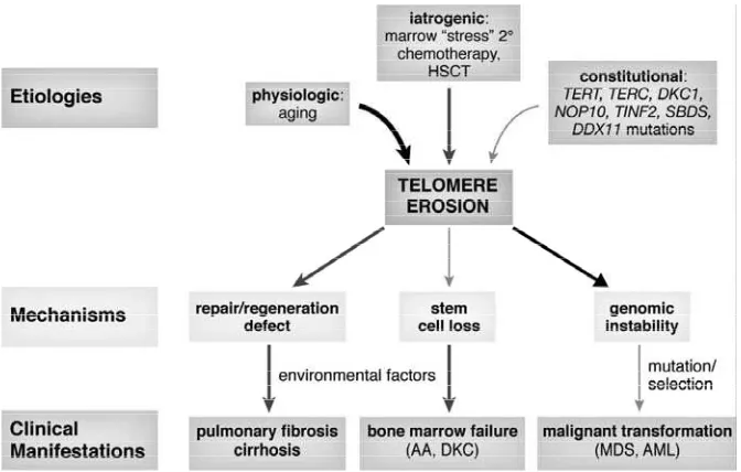
Telomere and telomerase in bone marrow failure syndromes
Dyskeratosis cong enita is a rare c o n s t i t u t i o n a l b o n e m a r r o w f a i l u r e syndrome stereotypically characterized by mucocutaneous abnormalities (nail dystrophy, hyperpigmentation, and leukoplakia) and aplastic anemia in childhood. Patients with dyskeratosis are at increased risk for malignancies, pulmonary fi brosis, and liver cirrhosis.17 The
genetic basis study of dyskeratosis congenita found X-linked dyskeratosis congenita to associate with mutations in the DKC1 gene
which produces dyskerin associated with TERC. Patients’ leukocytes showed reduced telomerase activity which explains the erosion of their chromosomes. The discovery that autosomal dominant dyskeratosis is caused by heterozygous mutations or large deletions in TERC established dyskeratosis congenita as a disease of telomerase insufficiency.18
Homozygous mutations in TERT43 and in NOP10, a telomerase-associated protein, were described in some families with autosomal recessive dyskeratosis congenita.19 More
recently, mutations in TINF2, which encodes TIN2, a shelterin protein that caps and protects the telomeres, have been identifi ed in a third of patients with dyskeratosis congenita, further implicating abnormal telomere maintenance in the pathophysiology of the disease.20
In acquired aplastic anemia, an immune pathophysiology that targets hematopoietic stem cells has been inferred in most cases of acquired aplastic anemia from the hematologic response to immunosuppressive therapies; failure to respond to such treatments might be due to a non-immune etiology or to properties of the hematopoietic compartment, a quantitatively severe loss of stem cell number or a qualitative abnormality of stem and progenitor cells.21 When some patients with
acquired aplastic anemia were found to have accelerated leukocyte telomere attrition, the cause was initially presumed to be secondary, a physiologic response to stem cell “stress.”22-24
low telomerase activity in vitro.23 TERC
mutations in either dyskeratosis congenita or acquired aplastic anemia are similar at the molecular level and produce the same effects on telomerase function, despite the diversity of clinical phenotypes (Table 1); an exception is large TERC gene deletions observed in dyskeratosis congenita families only.18 In cohort
studies, the frequency of TERC mutations was low (approximately 4% of all patients with acquired aplastic anemia).26,27 Careful study
of some families showed that healthy relatives of patients carrying TERC mutations also had short telomeres, some mild hematologic abnormalities (macrocytosis, mild anemia, thrombocytopenia, or granulocytopenia), reduced progenitor cells in peripheral blood, increased serum hematopoietic growth factors, and hypoplastic bone marrows.26 One report
that possibly explained potential clinical relevance of sometimes modest hematologic fi ndings in healthy relatives of patients carrying TERC mutations was dramatically reported in hematopoietic stem cell graft failure in one proband, whose unknowingly affected sibling donor provided only very low numbers of CD34 cells from both marrow and mobilized blood collected in multiple leukaphereses.28
A study on mutations in TERT, the gene encoding the telomerase reverse transcriptase itself, found that approximately 4% of patients with apparently acquired aplastic anemia had TERT mutations that disrupted telomerase activity, causing short telomeres of leukocytes and a hematopoietic stem cell compartment of limited proliferative capacity.29 Several
patients with TERT mutations also have a family history of blood dyscrasias, especially myelodysplastic syndrome evolving to acute myeloid leukemia, further suggesting a common genetic background for these disorders. As with TERC, apparently healthy relatives
with TERT mutations had short telomeres and reduced hematopoietic function.29 The
association between TERT mutations and aplastic anemia has been confirmed.29,30 A
few patients with marrow failure have genetic variants and specific haplotypes for genes coding for shelterin components (TERF1 and TERF2) that might contribute to disease by disrupting the telomere homeostasis.31
Despite the various mutations reported in the telomerase complex, the question whether mutations suffi cient to cause disease remains due to great variability is in the phenotype. Various clinical outcomes related with mutations in telomerase complex genes including isolated aplastic anemia, isolated pulmonary fibrosis or hepatic cirrhosis or multiorgan dyskeratosis congenita. The same mutation either in TERC or TERT in a single pedigree can associate with aplastic anemia in one patient, and pulmonary fibrosis or hepaticcirrhosis in another, suggesting that other factors contribute to organ damage. As an example, telomerase-mutant patients with pulmonary fibrosis often have a smoking history, suggesting this environmental insult as a trigger. Conversely, many relatives of patients with aplastic anemia with the same telomerase mutation have low telomerase activity, short telomeres, a hypoplastic bone marrow, and reduced hematopoietic function, but they are clinically healthy and asymptomatic.28,29
number of hematopoietic stem cells able to maintain hematopoiesis, as well as a qualitative defect by impairing hematopoietic stem cell regeneration. Higher serum interferon Ʊ and a limited T-cell receptor (TCR) Vư usage in telomerase mutant patients—similar to the skewed T-cell population typically observed in patients with immune acquired aplastic anemia32—consistent with oligoclonal T-cell
expansion and immune destruction of marrow as pathophysiologic had also been reported in these patients also.31 A telomerase mutation
does not appear to be suffi cient to determine aplastic anemia, but healthy relatives in which telomeres are short and/or a mutation need to
be further observed in long-term prospective studies.20 Since the number of acquired aplastic
anemia patients with identified mutation of either TERC or TERT genes or related genes are much fewer than those with short telomeres, it is suggested that other genetic lesions or environmental factors also may contribute to accelerated telomere erosion. In addition to aging, some environmental factors are known to cause telomere attrition. Smoking34 and even psychological stress35 have
telomere shortening has been observed in the fi rst years after allogeneic bone marrow transplantation in comparison with donor leukocytes, consistent with increased stem and progenitor turnover to replenish the bonemarrow.36 Chemotherapy for solid tumors
also causes myelotoxicity, requiring increased hematopoietic regeneration to recover blood cell counts, and premature telomere shortening has been observed following multiple cycles of cytotoxic drug therapy.37 These observations
are consistent with a contribution of “stress” hematopoiesis secondary to environmental factors to telomere shortening in aplastic anemia. However, the degree of telomere shortening produced in these clinical circumstances has been mild, less than 1 kb erosion in telomere lengths, as compared with theextreme attrition observed in individual mutations in telomerasedeficiency (usually more than 3 kb). Other bone marrow failure syndromes involving teomeres shortening including Fanconi anemia, werner syndrome, Bloom
syndrome, Nijmegen breakage syndrome, and Shwachman-Diamond syndrome.20
Telomere and telomerase in
hematologic neoplasia
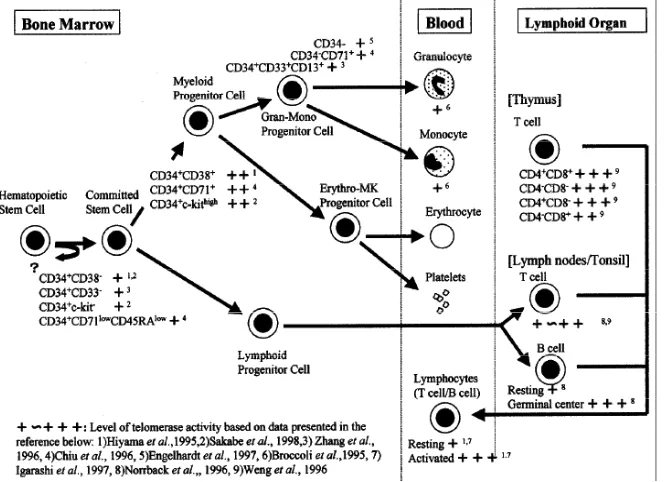
Normal hematopoietic cells express telomerase activity,however the presence of telomerase does not necessarily imply stable and thus unchanging telomere length. Therefore, before considering the telomere and telomerase status of leukemias and lymphomas we need to understand telomerase activity in normal hematopoietic cells as seen in Figure 5. The level of telomerase activity was different inhuman HSCs and their differentiate progeny. In contrast to somatic cells that lacktelomerase activity, it is clear that many hematopoieticcells are telomerase-competent. Primitive HSCs arelikely to be quiescent most of the time and thus thepopulation of cells exhibits a low level of telomerase activity. However, upon stimulation to proliferate,telomerase activity
appears to be upregulated in theirimmediate progeny and may help to slow down therate of telomere erosion. The more mature cells then become quiescent again and down-regulate telomeraseactivity. The telomeres of HSCs shorten, probably dueto inadequate levels of telomerase activity that slow but do not prevent telomere erosion. Both T cells and monocyte/B cells in peripheral blood also had low telomerase activity which was elevated when either cell types was cultured with mitogen. The highest in vivo telomerase expression in normal T cells is present in thymus, followed by T cells in the tonsil. The germinal center B-cells also show high telomerase activity. Thus, telomerase may play a permissive role inT cell and B cell development and in determining thecapacity of lymphoid cells for cell division and clonalexpansion. In summary, HSCs and lymphocytes are telomerase competent and mortal. The gradual telomere loss with aging and rapid cycling of HSCs or lymphocytes might contribute to immunosenescence, exhausted hematopoiesis, and increased likelihood of malignant transformation. Another interesting clinical aspect of these fi ndings is the future possibility of selectively upregulated telomerase and controlling telomere length in certain cell types in order to achieve a delayed induction of replicative senescence. (reviewed in ref 38).
Acute Myeloid Leukemia and
Myelodysplasia Syndrome
The overall importance of telomerase in the pathogenesis of AML has recently been confi rmed by the demonstration that hTERT is necessary for growth of primary AML cells in a mouse model.37 A number of studies have
investigated telomerase activity and telomere
length in mononuclear cells (MNC) from patients with MDS and AML (reviewed in ref.39). Calado et al examined three cohorts of AML patients who show no physical signs of DC for sequence variation in the hTERT and hTERC genes. They identifi ed three novel missense mutations in hTERT, and, while the V299M sequence change did not seem to affect telomerase enzymatic activity when tested by the TRAP assay, both the P65A and R522K mutations conferred dramatic defects. Surprisingly, they also identifi ed three AML patients who are homozygous for sequence changes previously identifi ed in a heterozygous state in AA patients and controls (A1062T and del441E). Thus, it appears that hTERT gene variants have low penetrance and are carried in patients with a wide variety of disorders. This phenomenon can be explained if short telomeres, as opposed to mutation status of telomerase, mediate disease pathogenesis, a hypothesis consistent with the fact that the median age at presentation for AML is 70.40
Telomere shortening was signifi cantly more pronounced in patients with cytogenetic alterations as compared with patients with normal karyotypes. In this study, the shortest median telomere length wasfound in the group with complex cytogenetic abnormalities. hTERT was overexpressed in patients with complex karyotypes, followed by patients with noncomplex karyotypes and patients without karyotypic changes.41 This might suggest
does not necessarily prevent cells from reaching replicative exhaustion.42
Because of the uneven distribution of telomere length on individual chromosome arms, critical shortening of telomeres on particular chromosomes could promote the formationof chromosomal aberrations and contribute to clonal evolution. This hypothesis remains relevant even if the average telomerelength remains well above the critical level of shortening.43 Distinct groups of AML
that are characterized either by aberrations that could result from telomere dysfunction (terminal deletions, gains/losses of chromosome parts, or nonreciprocaltranslocations) or by aberrations that are unlikely to result fromtelomere dysfunction (e.g., reciprocal translocations or inversions)could serve as an ideal model to study the effect oftelomere shortening and telomerase activity during tumorigenesis.44
Chronic Myeloid Leukemia
Most probably due to an increased t u r n o ve r o f t h e B C R - A B L - p o s i t i ve haematopoietic compartment, myeloid cellsfrom 123 patients with CML show accelerated telomereshortening. In Ph+
peripheralblood leukocytes, telomere length is approximately 1 kb shorter than in age-matched controls. Taking into account that roughly 100 bp (50–200 bp) are lost per celldivision in somatic cells the reduced telomerelength in CP CML cells indicates that, at a given point of time, leukaemic BCR-ABL-positive haematopoietic stemcells have undergone an excess of approximately 10 cell divisions as compared to their normal polyclonalcounterparts (HSC). Furthermore, telomere length measurements of cells
obtainedfrom CML patients suffering from different stages of disease showed signifi cantly shorter telomeres in AP and BP than in CP sd revealed by telomere fl uorescence and Southern Blot analysis (reviewed in ref 43). Successful therapy with IM was found to be associated with an increase in mean telomere length (Figure 3) refl ecting a treatment-induced shift from virtually 100% BCR-ABL+ peripheral blood cells topredominantly polyclonal, BCR-ABL haematopoiesis.46,47
Clinical implication of telomere
and telomerase roles in
hematological disorders
Bone marrow failure syndrome
A telomerase mutation does not appear to be suffi cient to determine aplastic anemia, but healthy relatives in which telomeres are short and/or a mutation need to be further observed in long-term prospective studies.20 Besides
this, protection from environmental stress should be minimized to prevent further hits for bone marrow failure development in acquired aplastic anemia. For most syndromes in which telomere shortening is known to bepathogenic, hematopoietic stem cell transplantation is the onlypotential cure. Modulation of telomerase activity may have a role in thetreatment of telomere deficiency syndromes such as dyskeratosis congenita and telomerase-mutant acquired aplastic anemia. Clinical observations suggest that androgen therapy can induce improvements in peripheral blood counts, achieving transfusion independence in as many as 60% of patients.17 Androgens stimulate
efficacy in hematologic disease.47 Androgen
therapy may cause severe adverse events, such as hepatocarcinoma andpeliosis hepatis, and liver function must be monitored. For patients with acquired aplastic anemia, hematopoietic stemcell transplantation and intensive immunosuppression with antithymocyteglobulin and cyclosporine are the main therapies.21 Immunosuppressive therapy
seems to be relatively or entirely ineffective for dyskeratosis congenita. Patients with acquired aplastic anemia and short telomeres appears to have poorer responses to immunosuppression. However, this relationship was retrospectively observed, and prospective studies of the prognostic value of telomere length are required before such measurements infl uence therapeutic decisions. Some patients with aplastic anemia with telomerase complex mutations respond hematologically to androgen treatment,48 as do
patients with dyskeratosis congenita.
Acute leukemia
Concepts of telomere and telomerase in acute leukemia are helpful for understanding the pathophysiology of the disease. Nevertheless, whatever the role of hTERT in determining the prognosis inAML, it is unlikely to surpass that of cytogenetics in discriminatingrisk groups yet.20
In primary AML samples, the transfection with dominant negative hTERT reduced the number of colony forming units (CFU) and decreased engraftment of leukaemic cells transplanted into a immunodefi cient mouse model.49
Chronic myeloid leukemia
In the BCR-ABL+ CML celline K562, 50% of the expanded clones underwent apoptosis afterdetectable telomere shortening
when transduced with thedominant negative form of hTERT.50 Competitive inhibition
of the catalytic activity of telomerase has beenattained by the small molecule inhibitor BIBR1532 as well as by the oligonucleotid GRN163L both in vitro and in vivo experiments. However, in the BCR-ABL-positive K562 cell line, BIBR1532 mediated telomerase inhibition was insuffi cient to induce telomere mediated apoptosis, while the expression of a dominant negative hTERTmutant in the respective cell line induced an increased rate of apoptosis (mostly refl ecting terminal‘crisis’ cells) and augmented radiosensitivity.50 GRN163L
produced a decreased concentration ofthe compound required for telomerase inhibition and a better biodistribution in normal and malignant tissue.51
Transcriptional inhibition of
essential telomerase components
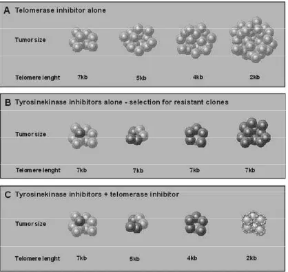
achieved earlier resulting in a mitoticcatastrophe and consequently apoptosis and necrosis. However, it needs to be emphasized that an effective treatment will require time and effects will appear with delay which may potentially lasting from months to years, as shown in fi gure 4.
Figure 4. Simultaneous inhibition of telomerase and bcr-abl tyrosine kinase may overcome emergence of resistance. (A) Telomerase inhibitor alone: while telomeres shorten in response to treatment the malignant clone continues to proliferate. Depending on initial telomere length, telomeres become critically short after a distinct amount of time leading to cell senescence and apoptosis. (B) CML Phþ cells are generally sensitive to tyrosine kinase inhibitors. However, secondary resistant subclones can emerge under therapy. (C) Combined treatment with tyrosine kinase and telomerase inhibitors represses the clonal expansion of tyrosine kinase resistant cells.44
REFERENCES
Calado RT and Young NS. Telomere 1.
diseases. N Engl J Med. 2009; 361(24):2353-65.
Carroll KA and Ly H. Telomere Dysfunction 2.
in Human Diseases: The Long and Short of It! Int J Clin Exp Pathol. 2009; 2: 528-543.
Blackburn EH. Telomeres and Telomerase: 3.
The Means to the End. Nobel Lecture, December 7, 2009. Source: http://www. nobelprize.org/nobel_prizes/medicine/ laureates/2009/blackburn-lecture.html
Elwood NJ. Telomere Biology of Human 4.
Hematopoietic Stem Cells. Cancer Control. 2004; 11(2):77-85
Ly H. Telomere dynamics in induced 5.
pluripotent stem cells: Potentials for human disease modeling. 2011; 3(10): 89-95. Gančarčíková M, Zemanová Z, Březinová 6.
J, Berková A, Včelíková S, Šmigová J, Michalová K. The Role of Telomeres and Telomerase Complex in Haematological Neoplasia: The Length of Telomeres as a Marker of Carcinogenesis and Prognosis of Disease. Prague Medical Report. 2010; 111(2): 91–105. Charles University in Prague – Karolinum Press.
Herber t BS, Shay JW, Wright WE. 7.
Analysis of Telomeres and Telomerase. Current Protocols in Cell Biology. 2003. Source: http://www.currentprotocols. com/WileyCDA/CurPro3Title/isbn-0471143030.html
Young NS. Telomere Biology and Telomere 8.
Blackburn EH, Greider CW, Szostak 9.
JW. Telomeres and telomerase: the path from maize, tetrahymena and yeast to human cancer and aging. Nat Med. 2006; 12:1133–1138.
Hackett JA, Feldser DM, Greider CW. 10.
Telomere dysfunction increases mutation rate and genomic instability. Cell. 2001; 106: 275–286.
Artandi SE, Chang S, Lee S-L, et al. Telomere 11.
dysfunction promotes non-reciprocal translocations and epithelial cancers in mice. Nature. 2000; 406:641– 645.
Artandi SE and DePinho RA. Telomeres 12.
and telomerase in cancer. Carcinogenesis. 2010;31(1):9–18.
Shen J, Gammon MD, Terry MB, Wang Q, 13.
Bradshaw P, Teitelbaum SL, Neugut AI, Santella RM. Telomere length, oxidative damage, antioxidants and breast cancer risk. Int J Cancer. 2009; 124(7):1637-43. Risques RA, Baughan TL, Li X, et al. 14.
Leukocyte telomere length predicts cancer risk in Barrett’s esophagus. Cancer Epidemiol Biomarkers Prev. 2007; 16:2649 –2655.
Artandi SE. Complex roles for telomeres 15.
and telomerase in breast carcinogenesis. Breast Cancer Res 2003, 5:37-41
O’Sullivan JN, Bronner MP, Brentnall TA, 16.
et al. Chromosomal instability in ulcerative colitis is related to telomere shortening. Nat Genet. 2002; 32:280 –284.
Vulliamy T, Dokal I. Dyskeratosis congenita. 17.
Semin Hematol. 2006; 43:157-166.
Vulliamy T, Marrone A, Goldman F, et 18.
al. The RNA component of telomerase is mutated in autosomal dominant dyskeratosis congenita. Nature. 2001; 413:432-435.
Walne AJ, Vulliamy T, Marrone A, et 19.
al. Genetic heterogeneity in autosomal recessive dyskeratosis congenita with one subtype due to mutations in the telomerase-associated protein NOP10. Hum Mol Genet. 2007; 16:1619-1629. C a l a d o RT, Yo u n g N S. Te l o m e r e 20.
maintenance and human bone marrow failure. Blood. 2008; 111(9):4446-55.
Young NS, Calado RT, Scheinberg P. 21.
Current concepts in the pathophysiology and treatment of aplastic anemia. Blood. 2006; 108:2511-2521.
Ball SE, Gibson FM, Rizzo S, et al. 22.
Progressive telomere shortening in aplastic anemia. Blood. 1998; 91:3582-3592.
Brummendorf TH, Maciejewski JP, Young 23.
NS, Lansdorp PL. Telomere length in leukocyte subpopulations of patients with aplastic anemia. Blood. 2001; 97:895-900.
Lee JJ, Kook H, Chung IJ, et al. Telomere 24.
length changes in patients with aplastic anaemia. Br J Haematol. 2001; 112:1025-1030.
Ly H, Calado RT, Allard P, et al. Functional 25.
characterization of telomerase RNA variants found in patients with hematological disorders. Blood. 2005; 105:2332-2339.
Yamaguchi H, Baerlocher GM, Lansdorp 26.
PM, et al. Mutations of the human telomerase RNA gene (TERC) in aplastic anemia and myelodysplastic syndrome. Blood. 2003; 102:916-918.
Calado RT, Pintao MC, Silva WA, Falcao 27.
RP, Zago MA. Aplastic anaemia and telomerase RNA mutations. Lancet. 2002; 360:1608.
Fogarty PF, Yamaguchi H, Wiestner A, 28.
congenita as apparently acquired aplastic anaemia due to mutations in telomerase RNA. Lancet. 2003; 362:1628-1630.
Yamaguchi H, Calado RT, Ly H, et al. 29.
Mutations in TERT, the gene for telomerase reverse transcriptase, in aplastic anemia. N Engl J Med. 2005; 352:1413-1424.
Vulliamy T, Walne A, Baskaradas A, et 30.
al. Mutations in the reverse transcriptase component of telomerase (TERT) in patients with bone marrow failure. Blood Cells Mol Dis. 2005; 34:257- 263.
Savage SA, Calado RT, Xin Z-T, et al. 31.
Genetic variation in telomeric repeat binding factor 1 and 2 in aplastic anemia. Exp Hematol. 2006; 34: 664-671.
Risitano AM, Maciejewski JP, Green S, et 32.
al. In-vivo dominant immune responses in aplastic anaemia: molecular tracking of putatively pathogenetic T-cell clones by TCR beta-CDR3 sequencing. Lancet. 2004; 364:355-364.
Calado RT, Bruno T, Wilkerson KL, 33.
Young NS. Evidence for T-cell oligoclonal expansion in aplastic anemia associated with telomerase complex mutations: pathophysiological and clinical implications [abstract]. Blood. 2005; 106: 307a. Abstract no. 1052.
Morla M, Busquets X, Pons J, et al. 34.
Telomere shortening in smokers with and without COPD. Eur Respir J. 2006; 27:525-528.
Epel ES, Blackburn EH, Lin J, et al. 35.
Accelerated telomere shortening in response to life stress. Proc Natl Acad Sci U S A. 2004; 101:17312- 17315.
Notaro R, Cimmino A, Tabarini D, Rotoli 36.
B, Luzzatto L. In vivo telomere dynamics of human hematopoietic stem cells. Proc
Natl Acad Sci U S A. 1997; 94:13782-13785.
Beeharry N, Brocolli D. Telomere dynamics 37.
in response to chemotherapy [abstract]. Curr Mol Med. 2005; 5:187-196.
Ohyashiki JH, Sashida G, Tauchi T, 38.
Ohyashiki K. Telomeres and telomerase in hematologic neoplasia. Oncogene. 2002 Jan 21; 21(4):680-7.
Roth A, Vercauteren S, Sutherland HJ et al. 39.
Telomerase is limiting the growth of acute myeloid leukemia cells. Leukemia 2003; 17: 2410 –2417.
Calado RT, Regal JA, Hills M, Yewdell 40.
WT, Dalmazzo LF, Zago MA, Lansdorp PM, Hogge D, Chanock SJ, Estey EH, Falcão RP and Young NS. Constitutional hypomorphic telomerase mutations in patients with acute myeloid leukemia. Proc Natl Acad Sci USA 2009; 106:1187-1192.
Grimwade D, Walker H, Oliver F et al. 41.
The importance of diagnostic cytogenetics on outcome in AML: Analysis of 1,612 patients entered into the MRC AML 10 trial. Blood 1998; 92:2322–2333.
Hemann MT, Strong MA, Hao LY et 42.
al. The shortest telomere, not average telomere length, is critical for cell viability and chromosome stability. Cell 2001; 107:67–77.
Swiggers SJ, Kuijpers MA, de Cort MJ 43.
et al. Critically short telomeres in acute myeloid leukemia with loss or gain of parts of chromosomes. Genes Chromosomes Cancer 2006; 45:247–256.
Keller G,Brassat U, Braig M, Heim D, 44.
Wege H and Bru¨mmendorf TH. Hematol Oncol 2009; 27: 123–129
Brummendorf TH, Ersoz I, Hartmann 45.
blood granulocytes refl ects response to treatment with imatinib in patients with chronic myeloid leukemia. Blood 2003; 101(1): 375–376.
Hartmann U, Balabanov S, Ziegler P, et al. 46.
Telomere length and telomerase activity in the BCR-ABL-transformed murine Pro-B cell line BaF3 is unaffected by treatment with imatinib. Exp Hematol 2005; 33(5): 542–549.
Calado RT, Yewdell WT, Wilkerson KL, 47.
Kajigaya S, Young NS. Sex hormones up-regulate telomerase activity of normal human hematopoietic cells and restore telomerase activity in carriers of telomerase complex mutations [abstract]. Blood 2005; 106:641a. Abstract no. 2276.
Xin ZT, Beauchamp AD, Calado RT, et 48.
al. Functional characterization of natural
telomerase mutations found in patients with hematological disorders. Blood. 2007; 109:524-532.
Roth A, Vercauteren S, Sutherland HJ, 49.
Lansdorp PM. Telomerase is limiting the growth of acute myeloid leukemia cells. Leukemia 2003; 17(12): 2410–2417.
Parsch D, Brassat U, Brummendorf TH, 50.
Fellenberg J. Consequences of telomerase inhibition by BIBR1532 on proliferation and chemosensitivity of chondrosarcoma cell lines. Cancer Invest 2008; 26(6): 590–596.
Herbert BS, Gellert GC, Hochreiter A, 51.