International Journal of Integrated Health Sciences. 2016;4(1) 43
Fetus inside Brain - Mature Cystic Teratoma in Posterior Fossa
Introduction
Meckel first reported a rare case of fetus-in-fetu in the 18th century, mostly presented in the sacrum, scrotum, mouth, liver, and cranium.1 Accounting for 0.5–5% of all intracranial tumors, intracranial teratoma represents a rare lesion with two peaks distribution before 5 years of age (10%) and between 5 and 14 years of age (48%) with males more affected than females.2–5,10 About 54% to 70% of all childhood brain tumors originated from the posterior fossa are cystic mass. Therefore, a teratoma located in the posterior fossa is uncommon. Although most of intracranial teratomas are located in the midline structures, particularly the pineal, corpus callosum and suprasellar regions, teratomas can also be identified outside the midline regions, including in the cerebellar hemisphere, basal ganglia, lateral ventricle,
fourth ventricle, sylvian fissure, cavernous sinus, sella, and temporal bone.4,6,7
Case
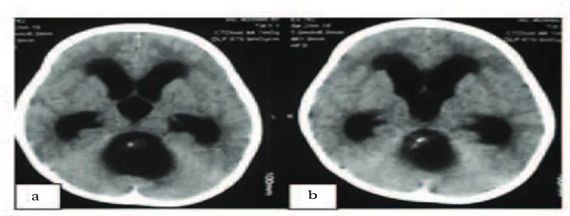
A 2 -year old boy admitted with a chief complaint of sudden decreased of consciousness. He was also presented with a tonic clonic seizure. NCCT examination revealed a well-defined extra-axial cystic mass located in the posterior fossa, 3.4×3.9×4.2 cm in size (Fig 1). Based on the NCCT image, the mass appear to be occupying the posterior fossa area and originating from the roof of the fourth ventricle. Gross examination presented cystic components and hard nodule features. Resection of the tumor was performed through the use of suboccipital craniotomy surgical technique. The mass was completely excised and sent for histopathological examination. The histopathological examination showed multiple cystic areas lined by the brain tissue admixed with islands of cartilage and salivary gland, elements and occasional intestinal type glands.
Abstract Objective: To describe a rare case of posterior fossa cystic teratoma in central
nervous system.
Methods: A 2-year old boy was admitted with a chief complaint of decreased
consciousness with tonic clonic seizure. Non-Contrast Head Computed Tomography (NCCT) scan showed acute hydrocephalus and a well-defined extra-axial cystic lesion mass, 3.4x3.9x4.2 cm in size, in the posterior
fossa.
Results: Patient underwent a total resection of the tumor. No systemic
complication was found in ten days following the surgery. There was no evidence of tumor recurrence one year after the surgery. The histopathologycal diagnosis of the tumor was cystic teratoma.
Conclusions: Mature cyst teratoma located in the posterior fossa is a rare
case. Total resection and long-term follow up is the treatment of choice for
mature teratoma.
Keywords:Hydrocephalus, posterior fossa, teratoma
IJIHS. 2016;4(1):43–6
Farid Yudoyono, Mirna Sobana, Agung Budi Sutiono, Muhammad Zafrullah Arifin
Department of Neurosurgery, Faculty of Medicine, Universitas Padjadjaran-Dr. Hasan Sadikin General Hospital
Case
Correspondence:
Farid Yudoyono, Department of Neurosurgery, Faculty of Medicine, Universitas Padjadjaran-Dr. Hasan Sadikin General Hospital
Jl. Pasteur No. 38, Bandung, Indonesia e-mail: [email protected]
44 International Journal of Integrated Health Sciences. 2016;4(1) The mass was covered with a
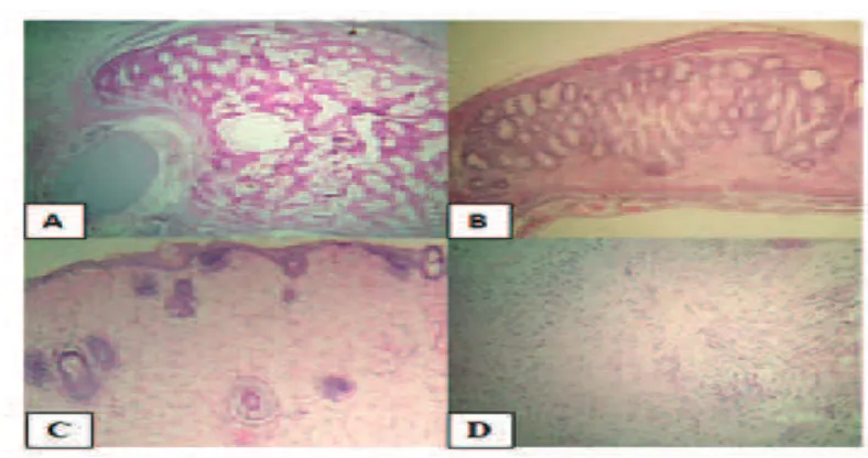
whitish-yellow membrane and located extraaxially. It was easily detached from the surrounding tissues of the brain and removed completely (Fig. 2a, b, c). During the process of debulking, gut-like structures were observed (Fig. 2d). Postoperatively, there was no neurological deficit observed. On gross examination, cystic component and hard nodule were revealed. The samples from excised tumor were fixed in 10% formalin, routinely processed and
paraffin-embedded. Five μm sections were stained using the hematoxylin and eosin (HE) method. The entire specimen was submitted for histologic examination which revealed the presence of various fully differentiated tissue elements. Mesodermal derivatives (Fig. 3a, b) including adipose and fibrous connective tissues, cartilage, bone and striated muscle were abundant. Neuroectodermal components were represented by delicate fibrillar tissue, in which mature glial elements (mainly astroglia)
:43–6
1a Non-Contrast Head Computed Tomography Imaging Revealed Acute Hydrocephalus. 1b Non-Contrast Head Computed Tomography Imaging Showed a Cyst-like Mass and Non-homogenous Hyperintense of Posterior Fossa
Fig.
a b
2a, b Intraoperative Photographs. After Suboccipital Craniotomy and the Process of Debulking. 1c, The Mass was Easily Detached from the Surrounding Brain Parenchym. 1d, Tumor Mass Presented Gut-like Structure, Brain, and Amniotic Sac
Fig.
a b
International Journal of Integrated Health Sciences. 2016;4(1) 45
Farid Yudoyono, Mirna Sobana, et al.
3a, b Histopathology H&E Stain 40x. Nodule of Moderately Cellular Cartilage, Connective Fibrous Tissue and Mature Fat Cells. 3c, d Neuroectodermal Components of Mature Teratoma, Hypocellular Fibrillar Tissue in which Glial Cells Predominate
Fig.
:43–6
predominated (Fig. 3c, d), but mature neuronal elements were also present. The tumor was moderately vascularized with predominantly smaller vessels and small foci probably injury took place during the operation. Mitotic figures were not present. The diagnosis of mature teratoma was established.
Discussion
Teratomas are nongerminomatous germ cell tumors differentiating into all three germ layers ectodermal derivatives in 100% of the tumors, mesodermal structures in 93%, and endodermal derivatives in 71% (which occurs in ovarian teratomas). The incidence intracranial teratomas 0.2% higher in children and the ration of male : female is 5:1. Mostly these tumor located are in the supratentorial midline and the pineal region.4,7
The World Health Organization classification of intracranial teratoma delineates three histological variants: mature, immature and teratoma with malignant transformation. Mature teratomas are composed of fully differentiated tissue elements of the three germ cells and have a good prognosis with
a reported 10-year survival rate of greater than 90%. The prognosis for patients with immature teratoma is less favorable, with a 5-year survival rate of up to 70%. The hallmark of immature teratomas is the presence of undifferentiated components exhibiting embryonal or fetal appearances. The immature component is frequently composed of neuroepithelial elements that can give rise to more malignant tumors. Teratomas with malignant transformation, containing a conventional malignant tumor of somatic type (i.e. sarcoma or carcinoma), are associated with poor prognosis.1,3,4
There were only a few cases of mature posterior fossa teratomas have been reported. Morelli from India published a radiographic report and Blackwell et al published the first report with histological examination.1
46 International Journal of Integrated Health Sciences. 2016;4(1)
References
1. Kim JW, Park SS, Wang KC, Cho BK, Kim SY, Ra EK
et al. Fetus-in-fetu in the cranium of a
4-month-old boy: histopathology and short tandem repeat polymorphism–based genotyping: Case report. J Neurosurg Pediatr. 2008;1(5):410–4.
2. Saada J, Delahaye S, Martinovic J, Macaleese
J And Benachi A. Early second-trimester
diagnosis of intracranial teratoma. Ultrasound
Obstet Gynecol. 2009;33(1):109–11.
3. Khan Z, Watson WJ. Congenital intracranial
teratoma prenatal diagnosis and vaginal delivery. J Ultrasound Med. 2010;29(7):1147– 9.
4. Dimov I, Stojanović I, Stojanović S, Stojanović
N, Stefanović I, Dimov D. Mature Intracranial Teratoma: Case report. Sci J Fac Med. 2013;30(2):97–102.
5. O’Grady J, Kobayter L, Kaliaperumal C, O’Sullivan M. ‘Teeth in the brain’–a case of giant intracranial mature cystic teratoma. BMJ Case
Rep. 2012; [cited 2015 Jan 10]:[about 4 p.].
Available from: http://www.ncbi.nlm.nih.gov/ pmc/articles/PMC3387452/.
6. Zhao J, Yu J, Zhong Y, Ge P. Cerebral Falx Mature
Teratoma with Rare Imaging in an Adult. Int J Med Sci. 2012;9(4):269–73.
7. Sanyal P, Barui S, Mathur S, Basak U. A case of mature cystic teratoma arising from the fourth
ventricle. Case reports in pathology. 2013; [cited 2015 Jan 10]:[about 3p.]. Available from: http://www.ncbi.nlm.nih.gov/pmc/articles/ PMC3677652/.
8. Chan LW, Leung TN, Fung TY, Lau TK. Foetal intracranial teratoma: choosing the best time and mode of delivery. Hong Kong Med J.
2007;13(4):323–6.
9. Kharosekar UK, Jasmit S, Velho V, Palande DA:
Congenital orbital teratoma up to posterior fossa. J Pediatr Neurosci. 2014;9(2):182–4.
10. Gao J, Zheng Z. Immature teratoma of the
posterior cranial fossa in a 4-month-old infant: A case report. Oncol Letters. 2013;6(1):19–22.
:43–6 mature fat cells and cartilage (mesodermal),
salivary gland tissue (endodermal), as well as intestinal type glands adjacent to brain tissue in the tumor. Total removal of the tumor is thought to be the gold standard for treatment of mature teratoma and generally shows no recurrence.7,9,10
Early ultrasonography examination during the second or third trimester of pregnancy may indicate intracranial teratoma, heterogeneous mass with hyperechogenic mixed with hypoechogenic features which also associated with polyhydramnios and hydrocephalus. A study in 2007 reviewed of 39 cases with10% were detected before 24 weeks of gestation
and 44% were detected between 28 and 32 weeks of gestation.8 Early appearance can be
observed in ultrasonograph during prenatal care and termination of pregnancy might be an option though but it is illegal in some of asian country.2,3,8 The patient underwent a total resection in April, 2012. During the follow-up period of one year, there was no evidence of tumor recurrence.
In summary, this is an unusual case of mature cyst teratoma especially for tumor located in the posterior fossa. Total surgical resection and long-term follow-up is the treatment of choice for mature teratoma.