Series Editors ALAN R. SHALITA, M.D.
Distinguished Teaching Professor and Chairman Department of Dermatology
1. Cutaneous Investigation in Health and Disease: Noninvasive Methods and Instrumentation, edited by Jean-Luc Lévêque
2. Irritant Contact Dermatitis, edited by Edward M. Jackson and Ronald Goldner 3. Fundamentals of Dermatology: A Study Guide, Franklin S. Glickman and Alan R.
Shalita
4. Aging Skin: Properties and Functional Changes, edited by Jean-Luc Lévêque and Pierre G. Agache
5. Retinoids: Progress in Research and Clinical Applications, edited by Maria A. Livrea and Lester Packer
6. Clinical Photomedicine, edited by Henry W. Lim and Nicholas A. Soter 7. Cutaneous Antifungal Agents: Selected Compounds in Clinical Prac tice and
Development, edited by John W. Rippon and Robert A. Fromtling
8. Oxidative Stress in Dermatology, edited by Jürgen Fuchs and Lester Packer 9. Connective Tissue Diseases of the Skin, edited by Charles M. Lapière and Thomas
Krieg
10. Epidermal Growth Factors and Cytokines, edited by Thomas A. Luger and Thomas Schwarz
11. Skin Changes and Diseases in Pregnancy, edited by Marwali Harahap and Robert C. Wallach
12. Fungal Disease: Biology, Immunology, and Diagnosis, edited by Paul H. Jacobs and Lexie Nall
13. Immunomodulatory and Cytotoxic Agents in Dermatology, edited by Charles J. McDonald
16. Psoriasis: Third Edition, Revised and Expanded, edited by Henry H. Roenigk, Jr., and Howard I. Maibach
17. Surgical Techniques for Cutaneous Scar Revision, edited by Marwali Harahap 18. Drug Therapy in Dermatology, edited by Larry E. Millikan
19. Scarless Wound Healing, edited by Hari G. Garg and Michael T. Longaker 20. Cosmetic Surgery: An Interdisciplinary Approach, edited by Rhoda S. Narins 21. Topical Absorption of Dermatological Products, edited by Robert L. Bronaugh and
Howard I. Maibach
22. Glycolic Acid Peels, edited by Ronald Moy, Debra Luftman, and Lenore S. Kakita 23. Innovative Techniques in Skin Surgery, edited by Marwali Harahap
24. Safe Liposuction and Fat Transfer, edited by Rhoda S. Narins
25. Pyschocutaneous Medicine, edited by John Y. M. Koo and Chai Sue Lee 26. Skin, Hair, and Nails: Structure and Function, edited Bo Forslind and Magnus
Lindberg
27. Itch: Basic Mechanisms and Therapy, edited Gil Yosipovitch, Malcolm W. Greaves, Alan B. Fleischer, and Francis McGlone
28. Photoaging, edited by Darrell S. Rigel, Robert A. Weiss, Henry W. Lim, and Jeffrey S. Dover
29. Vitiligo: Problems and Solutions, edited by Torello Lotti and Jana Hercogova 30. Photodamaged Skin, edited by David J. Goldberg
31. Ambulatory Phlebectomy, Second Edition, edited by Mitchel P. Goldman, Mihael Georgiev, and Stefano Ricci
32. Cutaneous Lymphomas, edited by Gunter Burg and Werner Kempf 33. Wound Healing, edited by Anna Falabella and Robert Kirsner
34. Phototherapy and Photochemotherapy for Skin Disease, Third Edition, Warwick L. Morison
35. Advanced Techniques in Dermatologic Surgery, edited by Mitchel P. Goldman and Robert A. Weiss
36. Tissue Augmentation in Clinical Practice, Second Edition, edited by Arnold W. Klein
edited by
Mitchel P. Goldman
University of California San Diego, California, U.S.A.and La Jolla Spa MD La Jolla, California, U.S.A.
Pier Antonio Bacci
University of SienaSiena, Italy and
Cosmetic Pathologies Center Arezzo, Italy
Gustavo Leibaschoff
University of Buenos Aires School of Medicine and
International Union of Lipoplasty Buenos Aires, Argentina
Doris Hexsel
School of Medicine University of Passo Fundo Passo Fundo, Rio Grande do Sul, BrazilFabrizio Angelini
Endocrinology Service Research Clinic Institute Ecomedica Empoli Florence, Italy
and
University of Parma Parma, Italy
Cellulite
Pathophysiology
New York, NY 10016
© 2006 by Taylor & Francis Group, LLC No claim to original U.S. Government works
Printed in the United States of America on acid-free paper 10 9 8 7 6 5 4 3 2 1
International Standard Book Number-10: 0-8247-2985-4 (Hardcover) International Standard Book Number-13: 978-0-8247-2985-1 (Hardcover) Library of Congress Card Number 2005055997
This book contains information obtained from authentic and highly regarded sources. Reprinted material is quoted with permission, and sources are indicated. A wide variety of references are listed. Reasonable efforts have been made to publish reliable data and information, but the author and the publisher cannot assume responsibility for the validity of all materials or for the consequences of their use.
No part of this book may be reprinted, reproduced, transmitted, or utilized in any form by any electronic, mechanical, or other means, now known or hereafter invented, including photocopying, microfilming, and recording, or in any information storage or retrieval system, without written permission from the publishers.
For permission to photocopy or use material electronically from this work, please access www.copyright.com (http://www.copyright.com/) or contact the Copyright Clearance Center, Inc. (CCC) 222 Rosewood Drive, Danvers, MA 01923, 978-750-8400. CCC is a not-for-profit organization that provides licenses and registration for a variety of users. For organizations that have been granted a photocopy license by the CCC, a separate system of payment has been arranged.
Trademark Notice: Product or corporate names may be trademarks or registered trademarks, and are used only for
identification and explanation without intent to infringe.
Library of Congress Cataloging-in-Publication Data
Cellulite : pathophysiology and treatment / edited by Mitchel P. Goldman … [et al.]. p. ; cm. -- (Basic and clinical dermatology ; 37)
Includes bibliographical references and index. ISBN-10: *978-0-8247-2985-1 (hardcover : alk. paper) ISBN-10: 0-8247-2985-4 (hardcover : alk. paper)
1. Cellulite--Pathophysiology. 2. Obesity--Pathophysiology. 3. Adipose
tissues--Pathophysiology. 4. Obesity--Treatment. I. Goldman, Mitchel P. II. Series. [DNLM: 1. Obesity--therapy. 2. Adipose Tissue--physiopathology. 3.
Obesity--physiopathology. WD 210 C393 2006] RC628.C42 2006
616.3'98--dc22 2005055997
Visit the Taylor & Francis Web site at http://www.taylorandfrancis.com Taylor & Francis Group
During the past 25 years, there has been a vast explosion in new information relating to the art and science of dermatology as well as fundamental cutaneous biology. Further-more, this information is no longer of interest only to the small but growing specialty of dermatology. Clinicians and scientists from a wide variety of disciplines have come to recognize both the importance of skin in fundamental biological processes and the broad implications of understanding the pathogenesis of skin disease. As a result, there is now a multidisciplinary and worldwide interest in the progress of dermatology.
With these factors in mind, we have undertaken this series of books specifically oriented to dermatology. The scope of the series is purposely broad, with books ranging from pure basic science to practical, applied clinical dermatology. Thus, while there is something for everyone, all volumes in the series will ultimately prove to be valuable addi-tions to the dermatologist’s library.
The latest addition to the series, volume 37, edited by Drs. Goldman, Bacci, Leibaschoff, Hexsel, and Angelini is both timely and pertinent. The editors are well known authorities in the field of dermatological surgery and cosmetic dermatology. We trust that this volume will be of broad interest to scientists and clinicians alike.
Alan R. Shalita, M.D. Distinguished Teaching Professor and Chairman Department of Dermatology SUNY Downstate Medical Center Brooklyn, New York, U.S.A.
What exactly is cellulite?Is it a disease or a normal finding in postpubcrtal women?Should it be treated or ignored?Is it nothing more than a convenient marketing opportunity for cos-metic manufacturers or something worthy of medical attention?These are but a few of the controversies surrounding the condition commonly known as ‘‘cellulite.’’ Perhaps the only point of agreement is that cellulite is unattractive and undesirable. It appears shortly after the initiation of menstruation in young girls on the upper outer thighs and buttocks and con-tinues to worsen with the passage of time. Cellulite seems to affect tall and short, fat and thin, asthenic and curvy females. For many women, cellulite marks the end of the idyllic youthful body and the onset of the aging, declining female shape. Certainly, there must be something that technologic medical science can offer. Even in the 1960s, cellulite treatments abounded with the vibrating belt machines designed to firm the buttock and thighs while minimizing cellulite. At the time of this writing, there are many creams, devices, and proce-dures that attempt to deal with the ubiquitous problem of cellulite, but an organized scientific treatise is lacking.
This text is the first serious evaluation of the etiology and treatment of cellulite. The editors have assembled an international panel of cellulite researchers and clinicians to share their combined knowledge on the subject. The book is nicely organized with an introduction into the social impact of cellulite, followed by a characterization of the problem through visual and noninvasive techniques, with a major focus on the various treatment modalities. Cellulite improvement through the use of topical agents, Endermologie1
, surgery, lympha-tic drainage, electroporation, and mesotherapy are investigated by practitioners of each of the arts. The editors thus provide a full critical evaluation of how each of these treatments impacts the appearance of cellulite.
Most dermatologists would agree that not a day goes by in clinical practice without a patient asking about cellulite treatments. To date, it has been difficult to find any reputable reference source on the subject. This text is a large step forward in characterizing the etiol-ogy of cellulite and evaluating worthwhile treatment approaches. The editors and their
authors should be congratulated for tackling a complex subject and organizing a text to highlight and discuss the controversies. This book is an illuminating treatise on the cloudy topic of cellulite.
Beauty has been extolled and made a cult object in all cultures and civilizations, whatever their geographic distribution, ethnic origin, or religion. In ancient Egypt, beauty was associated with a sacred nature and personified by Queen Nefertiti, a woman who had high brows, wide and well-delineated eyes, rich lips, a dignified countenance, and an upright bearing, the very image of subtle energy; the ancient Egyptians regarded beauty closely akin to ‘‘holiness.’’
The Greek aesthetic ideal was characterized by ‘‘perfect proportions’’ in the sense of the geometric relationships defining body harmony. Aphrodite, the goddess of beauty, was also worshipped as the goddess of love. Among the Etruscans, the Venus of Melos repre-sented beauty and harmony; this has remained intact and unpolluted throughout subse-quent civilizations.
An ancient depiction of the ideal female form.
Etruscan Venus of Melos.
During the Renaissance, the tall figures of Aphrodite and Venus, slim but muscular at the same time although somewhat androgynous, became impressive and important, as is evident in the works of Rembrandt and Rubens. The beauty of women was embodied in figures with abundant localized adiposity, though not obese: the faces were round and blissful and expressed a superb femininity and kindness that conveyed the idea of motherhood and protection.
After the French Revolution, the standard representation of the woman took a new turn. The feminine body started to express activity, labor, functionality, precision, and har-mony, losing some traits of Renaissance femininity. In the new society established after the Revolution, women slowly acquired new roles, carried out new activities, and achieved an unprecedented independence. As time went by, women even started to smoke cigarettes and practice sports. There were no objections to this new role as long as the exaggeration and myths of a sculptured body—such as those characteristic of the 1960s—are avoided.
&
WHY CELLULITE IS A CONCERN FOR US
The disorders characteristic of cellulite involve endocrino-metabolic alterations that affect the microcirculatory system. They also draw our attention to the functionality and the cleansing process of the whole organism. Besides, it involves hardly controllable changes in the locomotor, digestive, and endocrine system. Last, but not least, it is a cause of dis-comfort and an ill-tolerated lack of aesthetics that drives the patient to accept any type of so-called therapeutic treatments in order to solve the problem. Too frequently such ‘‘treat-ments’’ have no scientific basis.
Unfortunately, the ‘‘industrial exploitation ofpeau d’orange’’ results in permanently new offerings of therapeutic methods outside the medical sphere. Remedies for this situation are not simple, but the medical world is accountable for leaving plenty of room for other actors, perhaps in collusion or motivated by self-interest, but often because no serious scien-tific research on the physiopathology and therapeutics of cellulite syndromes is available.
Our efforts should be focused on the recovery of trophism and tissue tone, as well as on the control of endocrino-metabolic alterations that may entail irrecoverable tissue damage, not only from an aesthetic point of view. Let us recall, for example, damages resulting from hard massages on tissues affected by lipolymphedema, those derived from liposuction and vacuum applied on soft tissues, or from local, uncontrolled application of heat, as well as those arising from desperate attempts to reduce hip circumference in a few centimeters, a reduction which is often the evidence of tissue damage rather than of its improvement.
Physicians should be reminded that in their diagnostic activity as well as in therapeu-tic practherapeu-tice the Hippocratherapeu-tic Oath is still in force: ‘‘Primum, non nocere.’’ This also applies to paramedical professionals, such as physical therapists, nurses, and podiatrists. Even those who are not physicians should be highly professional and serious. Their practice should be guided by sound common sense and be aimed at prevention and health care.
Aesthetic considerations are not unbecoming for the physician and should not be deemed as such. If it comes to it, we may say they are a kind of sublimated medical attitude and therefore require still greater professionalism. We should always bear in mind that ineffective or hardly effective aesthetic treatments have three inescapable consequences: clinical damage, aesthetic injury and, more frequently, serious psychological damage.
In summary, only within the last three decades has today’s society defined the ideal female and male body as youthful and almost pre-pubertal. Well-defined muscles with very little body fat being the ideal. This recent definition of beauty has led to the development of a new medical ‘‘disease,’’ cellulite.
Cellulite can best be described as a normal physiologic state in post-adolescent women whose purpose is to maximize adipose retention to ensure adequate caloric avail-ability for pregnancy and lactation. Almost all women who are not cachectic have cellulite. The treatment of cellulite is extremely popular in Europe and Latin America. Sales of various topical therapies in those countries is a multimillion dollar business with an entire division of ROC (Johnson & Johnson) devoted to its sales and development. Unique to those countries is the purchasing and development of equipment to treat cellulite. It is estimated that the sales of cellulite equipment is over 10 million dollars each year. This supports the popularity of awareness of cellulite outside of the United States.
The time is right for a textbook on the treatment of cellulite. This subject is not taught in medical schools or in residency programs and there is no textbook in the English language on this subject. As patients go to their physicians (mostly Cosmetic, Dermatolo-gic and Plastic surgeons) to seek advice on the pathophysiology and treatment of cellulite, physicians will need to educate themselves on this subject.
To this end, this textbook represents the work of the world leaders in cellulite research. We present new ideas to challenge current medical thought on the pathophysiol-ogy of cellulite as well as a review of many different techniques for its treatment. We hope this book stimulates an interest in this underserved condition. As in many other fields of medicine and surgery, the advances in one field may be utilized in other fields. We believe that this textbook will serve this function.
&
REFERENCES
1. Curri SB. Liposclerosi e microcircolo. La dermoestetica 1990; 1:6–7.
2. Binazzi M, Papini M. Aspetti clinico istomorfologici. In: Ribuffo, Bartoletti, eds. La cellulite. Roma: Salus, 1983:7–15.
Series Introduction
Alan R. Shalita
. . . .
iii
Foreword
Zoe Diana Draelos
. . . .
v
Preface . . . .
vii
Contributors . . . .
xix
1. Social Impact of Cellulite and Its Impact on Quality of Life . . . .
1
Doris Hexsel and Camile Luisa Hexsel
Introduction . . . . 1
Aspects of Cellulite Related to QOL . . . . 2
Conclusions . . . . 4
References . . . . 5
PART I: DIAGNOSIS OF CELLULITE
2. Definition, Clinical Aspects, Associated Conditions, and Differential
Diagnosis . . . .
7
Doris Hexsel, Taciana de Oliveira Dal’Forno, and Stela Cignachi
Definition . . . . 7
Nomenclature . . . . 9
Clinical Aspects . . . . 9
Differential Diagnosis . . . . 21
References . . . . 24
Appendix . . . . 27
3. Anatomy of Cellulite and the Interstitial Matrix . . . .
29
Pier Antonio Bacci
Introduction . . . . 29
Cellulite . . . . 29
Interstitial Matrix . . . . 32
References . . . . 38
4. Pathophysiology of Cellulite . . . .
41
Pier Antonio Bacci and Gustavo Leibaschoff
What Is Cellulite
?
. . . . 41
Definition . . . . 41
Classification . . . . 43
Evolution . . . . 47
Lipedema and Lipolymphedema: Patholophysiologic
Hypotheses . . . . 50
Lipodystrophy . . . . 57
The Lymphoadipose System . . . . 58
Venous–Lymphatic Stasis . . . . 61
How Cellulite Develops . . . . 62
Manifestations of Cellulite . . . . 62
Clinical Classification . . . . 63
Why Cellulite Is a Concern . . . . 63
Liposclerosis and Localized Adiposity . . . . 63
Etiopathogenic Factors . . . . 68
The Term ‘‘Cellulite’’ . . . . 70
References . . . . 72
5. Diagnosis . . . .
75
Gustavo Leibaschoff
Patient History . . . . 75
Clinical Examination . . . . 75
Laboratory Investigations . . . . 80
ROM Test . . . . 81
Primary Instrument Examinations . . . . 82
Secondary Instrument Examinations . . . . 86
Photography . . . . 88
Diagnosis . . . . 88
Therapeutic Strategy . . . . 95
Medical History . . . . 95
References . . . . 97
Appendix A . . . . 98
Appendix B . . . . 101
6. Cellulite Characterization by High-Frequency Ultrasound and
High-Resolution Magnetic Resonance Imaging
. . . .
105
Pier Antonio Bacci and Gustavo Leibaschoff
Introduction . . . . 115
Bartoletti’s Classification . . . . 116
BIMED Classification . . . . 117
BIMED–TCD Classification . . . . 127
Pathophysiological Classification and Protocols of
BIMED–TCD . . . . 130
Edematous Cellulite . . . . 131
Adipose Cellulite . . . . 134
Interstitial Cellulite (Lipedema) . . . . 135
Fibrous Cellulite . . . . 137
Abstracts of Studies in Cellasene
1. . . . 146
New Research About the Use of Cellasene
1or Cellulase Gold
1. . . . 147
References . . . . 153
9. Theory and Working Principles of Beautytek
Òin Cosmetic Medicine . . . .
155
Valerio Genitoni
10. Topical Management of Cellulite . . . .
159
Doris Hexsel, Debora Zechmeister do Prado, Jaggi Rao,
and Mitchel P. Goldman
Introduction . . . . 159
Definition and Nature of Cellulite . . . . 160
Pathophysiologic Mechanisms of Cellulite Formation . . . . 160
Topical Management . . . . 161
Adipose Tissue and Lipoderma . . . . 176
The Superficial Fascia . . . . 177
Massage . . . . 178
12. The Use of TriActive
TMin the Treatment of Cellulite
. . . .
189
Anju Pabby and Mitchel P. Goldman
Mechanism . . . . 189
Review of Cellulite . . . . 189
Parameters . . . . 189
Initial Studies . . . . 190
Other Uses . . . . 191
Contraindications . . . . 191
Protocol . . . . 191
A: Lipoplasty, Vibro-Assisted Liposuction, Lipofilling,
and Ultrasonic Hydroliposuction . . . .
211
Gustavo Leibaschoff
Lipoplasty . . . . 211
Historical Background . . . . 212
B: VASER
Ò. . . .
220
Alberto Di Giuseppe
Definition . . . . 222
Fat Tissue Vascularization . . . . 223
Patient Selection . . . . 223
Prescriptions . . . . 228
Postoperative Scheme . . . . 229
Conclusion . . . . 230
C: Vibro-Assisted Liposuction . . . .
231
Pier Antonio Bacci, M. Scatolini, P. Belardi, S. Leonardi, and
S. Mancini
Introduction . . . . 231
Lipoplasty . . . . 232
Lipolymphosuction . . . . 232
The Vibro-Assisted Method . . . . 232
D: Lipofilling
. . . .
234
S. V. Savchenko, Marlen A. Sulamanidze, and George M. Sulamanidze
E: Ultrasonic Hydrolipoclasis (External Ultrasound)
. . . .
239
Doris Hexsel and Rosemari Mazzuco
Introduction . . . . 251
Indications and Mechanisms of Action . . . . 251
Contraindications for Subcision
1. . . . 254
Preoperative Consultation . . . . 254
Surgical Technique . . . . 254
The Postoperative Period . . . . 257
Complications . . . . 257
Conclusions . . . . 260
References . . . . 262
16. Mesotherapy in the Treatment of Cellulite . . . .
263
Gustavo Leibaschoff and Denise Steiner
A Brief History . . . . 263
The Concept . . . . 263
Action Mechanism of Mesotherapy . . . . 264
Benefits and Advantages of the Method . . . . 265
Materials and Techniques . . . . 266
General Indications for Mesotherapy . . . . 267
Mesotherapy in Cellulite . . . . 267
Uses for Mesotherapy . . . . 270
What Is Mesotherapy?
. . . . 271
Why Are Drugs Injected into the Skin
?
. . . . 271
What Drugs Are Used
?
. . . . 271
Which Drugs Should Be Used and How Should They Be
Administered
?
. . . . 272
Drugs and Products Used in Mesotherapy . . . . 277
No-Needle Mesotherapy
TM. . . . 282
Conclusion . . . . 283
References . . . . 284
17. Manual Lymphatic Drainage
. . . .
287
Gustavo Leibaschoff
18. The Role of Dermoelectroporation . . . .
291
Pier Antonio Bacci
Introduction . . . . 291
Transderm
1Method . . . . 292
Characteristic Features . . . . 292
Possible Uses . . . . 293
Dermoelectroporation Treatment . . . . 293
Clinical Study . . . . 294
Conclusions . . . . 297
References . . . . 298
19. Lipodissolve for Body Sculpting
. . . .
301
Martin Braun
Introduction . . . . 301
Phosphatidylcholine (PC) . . . . 302
Deoxycholate (Deoxycholic Acid) . . . . 305
Availability of Lipostabil
1in the United States and Canada . . . . 306
Contraindications for Lipodissolve Injections . . . . 307
Side Effects of Lipodissolve Injections . . . . 307
Dosages and Techniques for Lipodissolve Injections . . . . 309
General Considerations for Lipodissolve Injections . . . . 309
Lipodissolve Formulae for Fat Pad Dissolution . . . . 310
Procedure for Injecting Lipodissolve into Fatty Pads . . . . 312
Clinical Examples of Lipodissolve Therapy . . . . 313
Lipodissolve Summary: Simple Injections for Fat Reduction . . . . 313
References . . . . 322
Pier Antonio Bacci University of Siena, Siena, Italy and Cosmetic Pathologies Center, Arezzo, Italy
P. Belardi Department of Nuclear Medicine, Privat Hospital Santa Chiara, Florence, Italy
Martin Braun Vancouver Laser and Skin Care Center, Vancouver, British Columbia, Canada
Maurizio Ceccarelli Rome, Italy
Stela Cignachi School of Medicine, Lutheran University of Brazil (ULBRA) of Rio Grande do Sul, Canoas, Rio Grande do Sul, Brazil
Taciana de Oliveira Dal’Forno Medical Sciences, Federal University of Rio Grande do Sul, Porto Alegre, Rio Grande do Sul, Brazil
Alberto Di Giuseppe University of Ancona, Ancona, Italy
Debora Zechmeister do Prado Doris Hexsel Dermatologic Clinic, Porto Alegre, Rio Grande do Sul, Brazil
Valerio Genitoni Universita` di Urbino, Urbino, Italy
Mitchel P. Goldman University of California, San Diego, California and La Jolla Spa MD, La Jolla, California, U.S.A.
Doris Hexsel School of Medicine, University of Passo Fundo, Passo Fundo, Rio Grande do Sul, Brazil
Camile Luisa Hexsel Internal Medicine, Henry Ford Hospital, Detroit, Michigan, U.S.A.
Gustavo Leibaschoff University of Buenos Aires School of Medicine, and International Union of Lipoplasty, Buenos Aires, Argentina
S. Leonardi Bacci Medical Center, Arezzo, Italy
S. Mancini Surgery School, University of Siena, Siena, Italy
Rosemari Mazzuco Doris Hexsel Dermatologic Clinic, Porto Alegre, Rio Grande do Sul, Brazil
Anju Pabby American Academy of Cosmetic Surgery Fellow Trainee and La Jolla Spa MD, La Jolla, California, U.S.A.
Bernard Querleux Department of Physics, L’Ore´al Recherche, Aulnay-sous-bois, France
Jaggi Rao American Academy of Cosmetic Surgery Fellow Trainee and La Jolla Spa MD, La Jolla, California, U.S.A.
S. V. Savchenko Moscow, Russia
M. Scatolini Department of Nuclear Medicine, Privat Hospital Santa Chiara, Florence, Italy
Denise Steiner Mogi das Cruzes University, Mogi das Cruzes, Sao Paulo, Brazil
Marlen A. Sulamanidze Moscow, Russia
1
Social Impact of Cellulite and Its Impact on
Quality of Life
Doris Hexsel
School of Medicine, University of Passo Fundo, Passo Fundo, Rio Grande do Sul, Brazil
Camile Luisa Hexsel
Internal Medicine, Henry Ford Hospital, Detroit, Michigan, U.S.A.
&
INTRODUCTION
Almost all women have or believe they have cellulite. As it is more common to expose the body in certain cultures and in sunny countries such as Brazil, cellulite is of great concern to many women and also represents a problem of great social impact.
In today’s globalized culture, physical well-being, including the care taken with appearance, is highly valued. From this perspective, it is very important to evaluate the impact on quality of life (QOL) of such cosmetic problems as cellulite, wrinkles, and aging. The fact that these have an impact on the QOL is indirectly shown by the growing interest in the investigation and treatment of these problems, which until recently were considered to be of minor significance. New studies involving QOL will benefit all those who suffer to a greater or lesser degree from these problems, and will be of great value in assessing the need for new scientific research into the treatment of these problems.
Sarwer et al. published a review of the literature that focused on psychological and social–cultural aspects, their relation to physical appearance, and their influence on the deci-sion to undergo cosmetic treatments (1). Their study revealed that in the 37 different cultures studied, men and women gave greater priority to sexual attraction in the choice of partners than to aspects of personality such as independence, emotional stability, and maturity (1). Dermatological diseases and cosmetic problems significantly affect self-esteem. As the symp-toms are visible, the discomfort and psychoemotional effects are frequently more serious than the physical alterations caused by the disease. Thus, it becomes very important to assess and quantify the emotional and social parameters in these patients in order to understand the dis-ruption that the problem causes in various daily activities. This will facilitate the follow-up and treatment evaluation, and consequently allow for improvements in the QOL of the patients. The great importance given to QOL evaluation in clinical investigation and patient care has led to the development of questionnaires designed for the collection of information from
patients on the impact of the disease on their everyday lives. This knowledge allows the medi-cal professional to better observe how the disease affects the patients physimedi-cally, psychologi-cally, and socially, and facilitates the evaluation of the effects on the lives of the patients.
In the case of cellulite, the reasons that lead the patient to seek treatment are gener-ally social and, sometimes, also emotional. These may include the embarrassment caused by cellulite in social, affective, and sexual relations as well as the avoidance of normal everyday activities such as visiting a swimming pool or beach, practicing sports, or expos-ing the body durexpos-ing intimacy.
A number of studies have been published that deal with QOL and recognize the value of specific questionnaires for dermatological diseases such as psoriasis, acne, mel-asma, atopic dermatitis, hyperhidrosis, and alopecia among others (2–9). These studies have revealed the existence of similar facets related to QOL in patients from various coun-tries (10) and point to the discomfort and the psychoemotional effect on the patients. However, in general, little research has been done on the psychological, environmental, and social aspects of dermatological diseases. Moreover, to date, no study on the QOL of those afflicted by cellulite has been published.
Patients suffering from skin diseases should not be treated merely for the physical harm caused by the disease (10). The skin is the most external and apparent organ, and skin contact contributes to the formation and structure of the personality.
&
ASPECTS OF CELLULITE RELATED TO QOL
Cellulite is a clinical and aesthetic condition affecting most women. It may appear in preadolescence, adolescence, or adulthood. With cellulite, the connective tissue and adipose tissue undergo alterations, resulting in blood and lymphatic alterations (11). Clini-cally, cellulite is characterized by alterations to the cutaneous surface, especially on the buttocks and thighs, giving the skin an orange peel or mattress appearance (12,13). Clini-cally, cellulite is classified into degrees that range from 0 to III according to the clinical characteristics (14). As well as classifying the cellulite, it is suggested that associated fac-tors such as obesity [measured by the body mass index (BMI)] and degree of flaccidity (classified as light, moderate, or severe) be characterized.
Our clinical experience has shown that cellulite is a problem that has an impact on the QOL of both younger and more mature women, though the impact is greater in younger women. It also seems that cellulite is more frequent nowadays than many years ago.
We report here on a clinical study carried out in 62 female patients, aged between 18 and 45 years (average age 32) with BMIs between 18 and 25 (average 21.8), having various degrees of cellulite on the buttocks and thighs. Over a period of two months, these patients received mechanical treatment in both legs and topical treatment in only one randomly chosen leg. The degree of cellulite in each patient was evaluated before and at the end of the treatment and attributed a classification between 0 and III, according to the clinical appearance of the cellulite. No patients included in this study had a cellulite classification of 0.
pools or at the beach, etc. The impact of cellulite in relation to age group was also eval-uated, together with factors that patients believe may influence the cellulite, such as inheri-tance, diet, and physical activity, as well as the treatment performed and the self-perception of the severity of their cellulite. A survey of the answers given to the questions permitted an assessment of:
1. patient’s impression of the problem of cellulite;
2. the everyday situations that result in restrictions or embarrassment for patients with cellulite; and
3. the impact of treatment on a patient’s QOL.
Some factors, in the opinion of the patient, may influence cellulite. When questioned regarding diet, 65% of patients believed that there is a relationship between cellulite and diet. For 60% of the patients interviewed, a specific diet can help with cellulite. Along the same lines, 90% of patients believed that practicing physical exercise is an efficient treatment for cellulite and may, in isolation, moderately reduce cellulite.
Cellulite was perceived before 20 years of age by 65% of patients. With regard to family inheritance, 80% of patients reported having first- or second-degree relatives with cellulite. Because it is a clinically diagnosed and easily recognizable problem, this informa-tion is highly indicative of the presence of positive family cases, bearing in mind that the great majority of patients reported a family member of the first degree, mother or daugh-ter, as having the same problem.
Regarding the restrictions caused by cellulite, when patients were questioned in a generalized way about the degree to which cellulite hampered their lives with the options of ‘‘not at all (1),’’ ‘‘a little (2),’’ ‘‘moderately (3),’’ or ‘‘greatly (4),’’ it was found that 70% of the interviewed patients considered that cellulite hampered their lives greatly. Regard-ing specific daily situations, it was noted that those sufferRegard-ing from cellulite experience some day-to-day restrictions. Each situation was evaluated by the patient and attributed a value from 1 to 5, in which ‘‘1’’ was given to situations in which having cellulite had no effect, ‘‘2’’ to little effect, ‘‘3’’ to a moderate effect, ‘‘4’’ to a significant effect, and ‘‘5’’ to a very significant effect.
exposing the body during sports is not as embarrassing as other situations, as for example during sexual relations.
According to Jorge (10), the psychological impact and the impact in interpersonal relations, respectively, is more prejudicial for women than for men. Studies that evaluated patients with dermatosis, carried out in Sweden and Norway, suggest that those at risk of the greatest harm are females who are young and in whom the disease exists over an extended period of time (10). As cellulite appears basically in women, this condition should be investigated in terms of its impact on QOL.
A study by Harlow et al. that evaluated the impact of dermatological diseases on QOL during primary attention noted the differences between men and women in relation to the various forms of constraints caused by the diseases from which they suffered (15). Ten attributes were evaluated: physical symptoms, feelings, daily routine, clothing, social and leisure, sport and exercise, work and study, personal relations, sexual relations, and treatment. Of these, only the degree to which the condition affected the practice of sports and exercise was the score given by males higher than that given by women (15). This shows that the practice of sports may have greater significance in the male group than in the female.
We also checked the impact of treatment on the QOL of patients with cellulite. Each patient attributed a value from 0 to 9, with 0 representing very low self-esteem in relation to the fact of having cellulite, and 9 representing very high self-esteem. The clinical evalua-tion considered the improvement on the left and right sides, which were treated differently. Even without any improvement in the degree of cellulite noted by the examiner, there was an increase in self-esteem (evaluated from 0 to 9) after treatment. This improvement can be seen in the difference in the percentage of scores found before and after treatment. This shows that the treatment did have a positive effect on the self-esteem of the patients, indi-cating that the simple fact of treating the cellulite and caring for themselves, even in the absence of any clinical improvement, influenced the well-being of the patients, who described themselves as better and more confident following the treatment. It may suggest that treatments should be tried, even if there is no cure for this condition.
&
CONCLUSIONS
Thorough QOL evaluations will be necessary to evaluate not only the importance given to the problem of ‘‘cellulite,’’ but also the need to develop new treatments for cellulite (16). It is interesting to note that, even without techniques that can guarantee significant improvement of cellulite in its different degrees, cosmetic patients want alternatives and their emotional improvement is not directly related to clinical improvement. Care and atten-tion to cosmetic problems can lead to improvement in the emoatten-tional state of the patients. Cellulite has a real impact on the QOL of patients, as it restricts those that suffer from the condition in everyday situations and activities. This causes damage in the psycho-logical area in interpersonal relationships, as also occurs with other conditions that afflict the skin.
&
REFERENCES
1. Sarwer DB, Magge L, Clark V. Physical appearance and cosmetic medical treatments: physio-logical and socio-cultural influences. J Cosmet Dermatol 2003; 2:29–39.
2. Balkrishan R, McMichael AJ, Camacho FT, et al. Development and validation of a health-related quality of life instrument for women with melasma. Br J Dermatol 2003; 149(3): 572–577.
3. McKenna SP, Cook AS, Whalley D, et al. Development of the PSORIQoL, a psoriasis-specific measure of quality of life designed for use in clinical practice and trials. Br J Dermatol 2003; 149(2):323–331.
4. Skoet R, Zachariae R, Agner T. Contact dermatitis and quality of life: a structured review of the literature. Br J Dermatol 2003; 149:452–456.
5. Arruda LHF, Ypiranga S. Qualidade de vida em hiperidrose. In: Almeida AT, Hexsel D, eds. Hiperidrose e Qualidade de Vida. Sa˜o Paulo: Edic¸a˜o das autoras, 2003:73–76.
6. Finlay AY, Khan GK. Dermatology life quality index (DLQI)—a simple practical measure for routine clinical use. Clin Exp Dermatol 1994; 19:210–216.
7. Schmidt S, Fischer TW, Chren MM, Strauss BM, Elsner P. Strategies of coping and quality of life in women with alopecia. Br J Dermatol 2001; 144:1038–1043.
8. Swartling C, Naver H, Lindberg M. Botulinum A toxin improves life quality in severe primary focal hiperhidrosis. Eur J Neurol 2001; 8:247–252.
9. Mallon E, Newton JN, Klassen A, Stewart-Brown SL, Ryan TJ, Finlay AY. The quality of life in acne: a comparison with general medical conditions using generic questionnaires. Br J Dermatol 1999; 140:672–676.
10. Jorge HZ. Avaliac¸a˜o de Qualidade de Vida em Pacientes com Dermatoses: Estudo De Adap-tac¸a˜o e Validac¸a˜o da Dermatology Life Quality Index: (DLQI) para uma amostra Sul-Brasi-leira. Tese de Mestrado, Faculdade de Psicologia, Pontifı´cia Universidade Cato´lica do Rio Grande do Sul, 2004.
11. Lucassen GW, Van-der-Sluys WLM, van Herk JJ, et al. The effectiveness of massage treatment on cellulite as monitored by ultrasound imaging. Skin Res Technol 1997; 3:154–160.
12. Segers AM, Abulafia J, Kriner J, Cortondo O. Celulitis. Estudo histopatolo´gico e histoquı´mico de 100 casos. Med Cutan Ibero Lat Am 1984; 12:167–172.
13. Scherwitz C, Braun-Falco O. So-called cellulite. J Dermatol Surg Oncol 1978; 4(3):230–234. 14. Hexsel D, Mazzuco R. Subcision: a treatment for cellulite. Int J Dermatol 2000; 39:539–544. 15. Harlow D, Poyner T, Finlay AY, Dykes PJ. Impaired quality of adults with skin disease in
primary care. Br J Dermatol 2000; 143:979–982.
2
Definition, Clinical Aspects, Associated
Conditions, and Differential Diagnosis
Doris Hexsel
School of Medicine, University of Passo Fundo, Passo Fundo, Rio Grande do Sul, Brazil
Taciana de Oliveira Dal’Forno
Medical Sciences, Federal University of Rio Grande do Sul, Porto Alegre, Rio Grande do Sul, Brazil
Stela Cignachi
School of Medicine, Lutheran University of Brazil (ULBRA) of Rio Grande do Sul, Canoas, Rio Grande do Sul, Brazil
&
DEFINITION
‘‘Cellulite’’ is a very common topographical alteration (1,2) in which the skin acquires an orange peel or mattress appearance (Fig. 1) (3,4). In this condition, alterations occur to the adipose tissue and microcirculation that result from blood and lymphatic disturbances causing fibrosclerosis of the connective tissue (2). It is considered a noninflammatory, degenerative phenomenon that provokes alterations to the hypodermis (5) producing irre-gular undulations on the skin overlying affected areas.
Cellulite results from many complex events that involve the epidermis, dermis, and subcutaneous tissue (1). Cellulite can be divided into four stages:
1. alterations to the precapillary arteriolar sphincter, leading to changes in vascular per-meability and capillaceous ectasia resulting in pericapillary and interadiposity transu-dation, leading to edema;
2. edema, provoking metabolic changes that result in hyperplasia and hypertrophy of the reticular network, leading to the formation of pericapillary and periadipose deposits with an increase in interstitial viscosity;
3. organization of collagen fibers around groups of adipocytes, forming micronodules; and 4. union of the micronodules to form the macronodules that cause sclerosis (6).
Anatomically, the cutaneous alterations found in cellulite are largely due to fibrosis of the connective tissues present in the dermis and/or in the subcutaneous tissue (7). The
Figure 1
connective tissue of the reticular dermis is connected to the deep fascia by means of interlobular trabeculas (fibrous septum) from adipose tissue. Subcutaneous fat lobules are separated from one another by these thin, usually rigid strands of connective tissue that cross the fatty layer and connect the dermis to the underlying fascia. These strands stabilize the subcutis and divide the fat (8). The shortening of these septa due to fibrosis provokes retraction at the insertion points of the trabeculas (9), causing the depressions that are characteristic of cellulite.
Nurnberger and Mu¨ller studied the anatomy and histology of fat and the connective tissue structure of the subcutaneous tissue. They demonstrated, on anatomical bases, the characteristic mattress aspect of cellulite and pointed out the differences in the organiza-tion of the subcutaneous tissue between the two sexes (10,11). They also showed that in women the fibrous septa are usually orientated perpendicularly in relation to the cuta-neous surface, while in men they have a crisscross pattern (11). Several studies have shown that fat is divided into lobules, and that in women, these are larger and more rectangular when compared with those in men (4,11–15). These anatomical and histological findings explain the greater frequency of cellulite in women.
&
NOMENCLATURE
In France in 1920, Alquier and Paviot described cellulite as an unaesthetic condition (6,16). In the same decade, Laguese described cellulite as a disease of the hypodermis, characterized by interstitial edema and an increase in fat (17).
Initially, Curri defined cellulite as nodular liposclerosis (6,18) and later adopted the term ‘‘cellulitic dermohypodermosis’’ (19). In 1958, Merlen defined cellulite as a histoan-giopathy (20), and in 1978, Binazzi and Curri, after a histopathological study, suggested the term ‘‘sclerotic-fibrous-edematous panniculopathy’’ (21,22). Nurnberger and Mu¨ller used the name ‘‘panniculosis of the dermis’’ (16,23) to describe cellulite from the histo-pathological viewpoint. Bacci and Leibaschoff suggest the use of the nomenclature ‘‘cellu-litic hypodermosis’’ (16).
In recent years, the term ‘‘gynoid lipodystrophy’’ has been used in some studies (2,9,24). The terms ‘‘hydrolipodystrophy’’ and ‘‘herniation’’ of the fat with hypodermic tension bands are still in use for describing cellulite (25,26).
The presence of the suffix ‘‘ite’’ in a medical term indicates inflammation; therefore, the term ‘‘cellulite’’ is more appropriately used to designate inflammation and/or infection of the subcutaneous tissue (27). However, the term ‘‘cellulite’’ has become very popular, and its use has been consecrated (20,28) by its being accepted throughout the world. Other synonyms often used for cellulite are listed in Table 1.
&
CLINICAL ASPECTS
Although it is found in all age groups and in both sexes (10,29,30), cellulite occurs mainly in women (6,31) especially after puberty (32) and in obese people, being considered a nor-mal manifestation of obesity by Burton and Cunliffe (12,29).
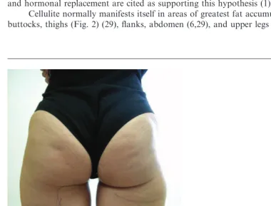
incidence of cellulite in the female sex, its appearance postpuberty, the worsening of the condition in relation to pregnancy, the menstrual cycle, and the use of contraceptives and hormonal replacement are cited as supporting this hypothesis (1).
Cellulite normally manifests itself in areas of greatest fat accumulation, such as the buttocks, thighs (Fig. 2) (29), flanks, abdomen (6,29), and upper legs (26,29,32).
Table 1
Various Terms Describing Cellulite
& Nodular liposclerosis & Cellulitic dermohypodermosis
& Sclerotic-fibrous-edematous panniculopathy & Panniculosis of the dermis
& Cellulitic hypodermosis & Gynoid lipodystrophy & Hydrolipodystrophy
& Herniation of the fat with hypodermic tension bands
Figure 2
The lesions are essentially asymptomatic. However, in an advanced degree of cellu-lite, symptoms such as a sensation of weight and pain may occur in the affected areas (10,20,29). These probably occur as a result of compression of the nervous terminals or the presence of inflammatory reactions (16,19).
The main manifestations of clinical cellulite are:
1. flaccid ‘‘mattress-like’’ skin, with multiple depressions and some elevations, caused by irregular retraction of the skin, forming a surface where protuberances and depressed areas alternate (Fig. 1) (6,16,34);
2. ‘‘orange peel’’ skin due to the tumefaction of the epidermis and dilation of follicular pores (6,16,34).
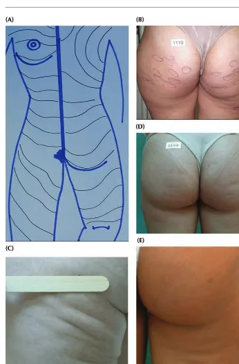
The cutaneous surface alterations that characterize cellulite are predominantly depressed, when compared to cutaneous surface of the affected area (29). These depres-sions have the same color and consistency as normal skin, and the number of ledepres-sions may vary from one to many (29). The shape of these lesions is varied (29): rounded, oval, or linear (Fig. 3). Most lesions are oval, as the longest axis of the lesions lies par-allel to the relaxed skin tension lines (Figs. 4A–E). It is interesting to note that those lesions that do not have the same disposition in relation to the relaxed skin tension lines in general originate from secondary fibrosis of the subcutaneous tissue, such as injec-tions, trauma, etc. They are usually found in the lower portion of the buttocks and the upper thigh (Fig. 4). In both the buttocks and the upper thigh, just below the gluteal fold, the longest axis is in the horizontal direction, with the lateral extremities slightly elevated. In these locations, cellulite may be more evident due to flaccidity of the epider-mis, which tends to become aggravated with age. This can be demonstrated by the diminishing or even the disappearance of the lesions when the buttocks are lifted to their original position.
CLASSIFICATION
Several authors have classified cellulite into four clinical stages or degrees (Table 2), based on the clinical alterations observed with the patient at rest and after the application of the pinch test or muscular contraction (6,29,34).
Because cellulite is diagnosed by clinical alterations, without histopathological find-ings or anatomical or pathognomonic characteristics, it can also be classified into primary and secondary cellulite. In primary cellulite, there are no aggravating factors involved. In secondary cellulite, the alterations are provoked by secondary factors such as localized fat, flaccidity, surgical or accident trauma mainly from liposuction, after injections that cause lipoatrophy, or after subcutaneous fibrosis from any inflammatory or infectious process. These circumstances may aggravate or even bring about primary cellulite and should be detected through the medical history and physical examination. Treatment, in this case, implies the correction of the primary factor.
CLINICAL APPROACH
disease or surgery, family history, presence of chronic vascular or associated hormonal diseases, the occasional or regular use of medications, and previous or current history of hormonal treatment or the use of any medicine that may contribute to the increase in the deposit of fat in the affected areas, such as corticosteroids and estrogens. Other aspects that should be researched are sedentarism, diet, psychosomatic factors, smoking, prior pregnancy, and the behavior of cellulite during pregnancy.
Although smoking and circulatory problems are frequently cited as causative agents of cellulite, in the experience of the present authors, in a sample of 1200 patients with advanced cellulite, the vast majority were neither smokers (more than 80%) nor those hav-ing varicose veins or other circulatory problems.
Figure 3
Figure 4
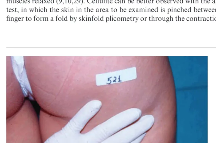
Physical Examination
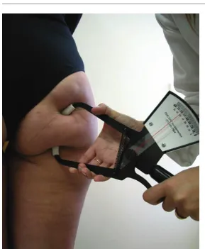
The physical examination should be performed with the patient in a standing position, with muscles relaxed (9,10,29). Cellulite can be better observed with the application of the pinch test, in which the skin in the area to be examined is pinched between the thumb and index finger to form a fold by skinfold plicometry or through the contraction of the muscles in the
Table 2
Classification of Cellulite
Classification Evaluation results
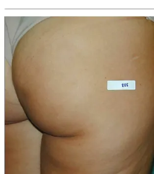
Degree or stage 0 There is no alteration to the skin surface
Degree or stage I The skin of the affected area is smooth while a subject is standing or lying down, but undulations on the skin surface can be seen on pinching the skin or during muscle contraction (Fig. 5) Degree or stage II The ‘‘orange peel’’ or ‘‘mattress’’ appearance is evident when
standing, without the use of any manipulation (skin pinching or gluteus muscle contraction) (Fig. 6)
Degree or stage III Presence of alterations described in second degree or stage II, plus presence of raised and depressed areas and nodules (Fig. 7)
Figure 5
area (Figs. 8 and 9) (9). Overhead or tangential illumination of the patient facilitates the visualization of cellulite (29). There are significant differences in the appearance of cellulite, depending on the position and the method used for its classification. For this reason, the standing position is recommended for the examination of a patient with cellulite.
Palpation should always be performed to check the elasticity of the skin (6) and sub-cutaneous tissues. However, at present there are no exact parameters for the classification of skin elasticity. Venous or lymphatic insufficiency may, in theory, aggravate cellulite and should also be checked during the physical examination (35). One should make note of the presence of varicose and telangiectatic leg veins as well as any pitting edema or induration of the skin. A Doppler or duplex ultrasound examination of the superficial venous system will also help to classify the significance of venous insufficiency. Even if venous insuffi-ciency is not found to be an etiologic factor in the pathogenesis of cellulite, its presence or absence will help direct appropriate treatment regarding graduated compression.
Figure 6
AGGRAVATING FACTORS
A number of clinical conditions or circumstances frequently accompany or aggravate cel-lulite, especially obesity, localized fatty accumulations, and skin flaccidity.
Obesity promotes a generalized increase in body weight (skeletal, muscular, intersti-tial fluid, organ hypertrophy, etc.). After a return to the original baseline weight is achieved, an increased accumulation of fat is observable (36). The clinical manifestation of localized adiposity is an increase in the ill-defined symmetrical and bilateral diffuse volume, owing to an increase in the adipose tissue (29). The localized increase in adipose tissue in the subcutaneous tissue leads to the aggravation of cellulite lesions by contribut-ing to a worsencontribut-ing of the irregular undulations of the skin. The increase in fat volume leads to an augmentation of tension forces within the fat lobules. This tension is projected to the skin surface and aggravates the depressions, causing an effect similar to that of a stuffed quilt (29). These alterations contribute to the appearance of the mechanical and circulatory alterations that occur in cellulite. Greater thickness of the subcutaneous fat in the affected areas may be seen by histopathological examination and can be measured by special instruments or by the pinch test (Fig. 9) (36).
Rosenbaum et al. described the exacerbation of cellulite with weight gain and its cor-relation with the body mass index (BMI). This study demonstrates the protrusion of adi-pose tissue into the dermis when the volume of subcutaneous fat is augmented, which explains the mattress-like appearance (31).
Flaccidity is caused by physiological ptosis of subcutaneous structures, making the skin permanently distended and loose. This condition frequently occurs in the buttocks,
Figure 7
thighs, the region above the knee, and the inner surface of the arms, regions where the skin probably has less retentive capacity and suffers the mechanical action of weight exerted by the adipose tissue and by the other subcutaneous structures (29). The weight of these struc-tures increases the effect of gravity, causing alterations to the skin surface in these areas, which is seen as laxity and looseness (29). The reduced elasticity of the skin and sudden loss of weight (29) or subcutaneous fat due to liposuction (37) are conditions that can bring about or aggravate skin flaccidity.
Although it is of great importance, the presence of flaccidity or other aggravating conditions is usually not mentioned in present day classifications of cellulite. In the absence of flaccidity, a distension test in the antigravity direction tends not to diminish the lesions. In the presence of flaccidity, however, such a test can lead to a reduction or even disappearance of cellulite lesions (Fig. 10). The pinch test causes an increase in
Figure 8
Figure 10
The patient shown in Figure 9 showing improvement to the skin surface when stretching the skin in the direction opposite to forces of gravity.
Figure 9
tension inside the lobes, and the cellulite becomes apparent as the lobes bulge and aggra-vate the traction of the septa in the pinched area (Fig. 11). Moreover, flaccidity has an effect similar to that of pinching by compressing the lobes and, thus, augmenting the ten-sion within them. This situation is responsible for the emergence or worsening of cellulite lesions, especially after the fourth or fifth decade of life when the elastic properties of the skin diminish (38). This, together with the weight of the subcutaneous fat, determines the worsening of distension of the skin.
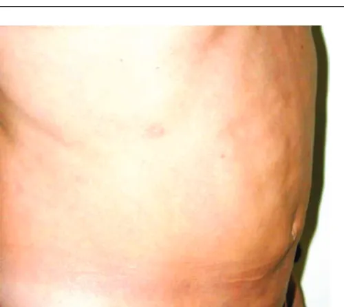
Other notable conditions that cause secondary cellulite or that aggravate cellulite are subcutaneous fibrosis caused by previous surgery, mainly liposuction, and the subcuta-neous fibrosis and lipoatrophy originating from the trauma caused by injections in the affected areas. Alterations to the cutaneous surface resulting from liposuction usually appear late, from three months to one year after surgery. They may be slight, moderate, or severe, and always emerge in previously treated areas, such as the lateral and posterior thighs, buttocks, abdomen (Fig. 12), flanks, and the region above the knees. Like cellulite, the cutaneous sequelae from liposuction are predominantly depressed subcutaneous tissue, but raised and depressed areas may intercalate and vary in number and shape as a reflec-tion of the number and variety of liposculpture cannula inserreflec-tions, as well as the size and type of cannulas. Generally, they form larger depressions with bizarre shapes and do not necessarily follow the direction of the relaxed skin tension lines. Instead, they follow the direction of cannula insertion (Fig. 12).
The cutaneous surface alterations caused by previous injections (such as insulin injections in diabetics) occur in places where the injections are normally applied, that is, in the upper, outer quarter of the buttocks. They also vary in number and shape, and do not follow the force lines of the skin.
The presence of atrophic scars in the areas frequently affected by cellulite can also simulate or aggravate cellulite.
Many factors can cause cellulite, and other factors can make it worse. The classifica-tion in Table 2 is useful for generic diagnostic purposes, but is not appropriate for an accu-rate measure of the results of treatments, other than surgical treatment. To evaluate the results of other treatments, such as topical or systemic treatments, alternative objective and subjective measures are needed; these are presented in the appendix to this chapter in the form of a protocol used in our clinics.
COMPLEMENTARY EXAMINATIONS
The BMI is widely used and cited by some authors as a simple, low-cost examination considered fundamental for the evaluation of the clinical cellulite (6,39). This is a quan-titative method that uses measures of weight and height to assess the degree of obesity (39). By using this index, it is not possible to distinguish the percentage of body fat in the muscular mass. BMI is an uncertain diagnostic index of obesity (40). Studies reveal that the estimated standard error of the percentage of body fat of BMI is approximately 5% to 6% (39).
Two-dimensional ultrasound is a noninvasive method of evaluating variations (41,42) and alterations of the subcutaneous fatty tissue, and with the assistance of Doppler, it evaluates the local circulation (6). This examination has been used in some studies for the evaluation of cellulite, and has demonstrated a diffuse pattern of extrusion of underlying adipose tissue into the reticular dermis in affected individuals, but not in unaffected individuals (2,31).
Computed tomography (43) and magnetic resonance imaging (44,45) are exami-nations used for measuring the thickness of adipose tissue, which do not allow evaluation of the dermis or microcirculation (6). In one study, the magnetic resonance imaging quan-tified deeper indentations of adipose tissue into the dermis and evidenced for the first time a great increase in the thickness of the inner fat layer in women with cellulite (46).
Although invasive, histological examination may be useful as a method for evaluat-ing cellulite (3,6,13). The stains used in this examination include hematoxylin–eosin for routine histological examination; Alcian blue for polysaccharides; periodic acid–Schiff for basement membranes; Weigert–Van Gieson (fuchsin–resorcin and acid fuchsin) for highlighting elastic, collagen, and flat muscle fibers; and Masson trichromic, which demonstrates contrast between collagen and muscle fibers (6). With this examination, it
Figure 11
is also possible to observe the diffuse extrusion pattern of underlying adipose tissue distending the reticular dermis in people with cellulite (31). The macroscopic aspect of sub-cutaneous fat from corpses is shown in Figure 13.
&
DIFFERENTIAL DIAGNOSIS
Of particular importance in the differential diagnosis of cellulite are the localized deposits of fat (13), flaccidity, surgical sequelae (from liposuction) (Fig. 12) or other trauma (47),
Figure 12
‘‘Cellulite-like’’ liposuction sequelae on the abdomen, one year after the surgery.
Figure 13
Figure 15
Lipomatosis from cellulite.
Figure 14
&
REFERENCES
1. Draelos ZD. Cellulite. Etiology and purported treatment. Dermatol Surg 1997; 23:1177–1181. 2. Lucassen GW, Van-Der-Sluys WLN, et al. The effectiveness of massage treatment on cellulite
as monitored by ultrasound imaging. Skin Res Technol 1997; 3:154–160.
3. Segers AM, Abulafia J, Kriner J, Cortondo O. Celulitis. Estudo histopatolo´gico e histoquı´mico de 100 casos. Med Cutan Ibero Lat Am 1984; 12:167–172.
4. Scherwitz C, Braun-Falco O. So-called cellulite. J Dermatol Surg Oncol 1978; 4(3):230–234. 5. Ronald M, Di Salvo. Controlling the appearance of cellulite. Cosmet Toilet 1995; 110:
50–58.
6. Rossi ABR, Vergnanini AL. Cellulite: a review. J Eur Acad Dermatol Vener 2000; 14:251–262. 7. Hexsel DM, Gobbato D, Mazzuco R, Hexsel CL. Lipodistrofia gino´ide. In: Kede MPV,
Saba-tovich, eds. Dermatologia Este´tica. 1st ed. Sa˜o Paulo: Atheneu, 2003:350–359.
8. Murphy GF. Histopathology of the skin. In: Elder DE, Elenitsas R, Jaworsky C, Johnson BL Jr, eds. Lever’s Histopathology of the Skin. Philadelphia: Lippincott-Raven, 1997:5–50. 9. Hexsel D, Mazzuco R. Subcision: uma alternativa ciru´rgica para a lipodistrofia gino´ide
(‘‘celulite’’) e outras alterac¸o˜es do relevo corporal. Ann Bras Dermatol 1997; 72(1):27–32. 10. Hexsel DM, De Oliveira NIM. Tratamento da celulite pela subcisa˜o. In: Horibe EK, ed.
Este´-tica Clı´nica e Ciru´rgica. Rio de Janeiro: Revinter, 2000:261–264.
11. Nurnberger F, Mu¨ller G. So-called cellulite: an invented disease. J Dermatol Surg Oncol 1978; 4:221–229.
12. Burton JL, Cunliffe WJ. Subcutaneous fat. In: Champion RH, Burton JL, Ebling FJG, eds. Textbook of Dermatology. 6th ed. Oxford: Blackwell Science, 1992:2140.
13. Braun-Falco O, Buddecke E, et al. Zellulitis. Round-Table Gesprach. Med Klin 1971; 66: 827–832.
14. Salache SJ, Bernstein G, Senkarik M. Superficial musculoaponeurotic system. In: Salache SJ, Bernstein G, Senkarik M, eds. Surgical Anatomy of the Skin. Norwalk: Appleto e Lange, 1988:89–97.
15. Franchi J, Pellicur F, Andre P, Schnebert S. The adipocyte in the history of slimming agents. Pathol Biol 2003; 51(5):244–247.
16. Bacci PA, Leibaschoff G. La Cellulite. Gasgo´n: Medical Books, 2000; 19:196. 17. Laguese P. Sciatique et infiltration cellulalgique. These Me´d Lyon, 1929.
18. Curri SB. Las paniculopatias de estasis venosa: diagno´stico clı´nico e instrumental. Barcelona: Hausmann, 1991.
19. Curri SB. Aspects morphohistochimiques e bioquimiques du tissue adipeaux dans la dermo hypodermose cellulitique. J Med Esth 1976; 5:183.
20. Medeiros LB. Lipodistrofia gino´ide. Abordagem terapeˆutica. In: Kede MP, Sabatovich, eds. Dermatologia Este´tica. 1st ed. Rio de Janeiro: Atheneu, 2003:337–342.
21. Di Salvo RM. Controlling the appearance of cellulite: surveying the cellulite reduction effective-ness of xantines, silanes, Coa, 1-carnitina and herbal extracts. Cosmet Toilet 1995; 110:50–59. 22. Binazzi M, Grilli-Cicioloni E. A propo´sito della cosidetta cellulite e della
dermato-paniculopa-tia edemato fibrosclero´tica. Ann It Derm Clin Sper 1977; 31:121–125.
24. Ciporkin H, Paschoal LHC. Clı´nica da L.D.G. In: Atualizac¸a˜o Terapeˆutica e Fisiopatogeˆnica da Lipodistrofia Gino´ide (LDG) ‘‘celulite’’. Sa˜o Paulo: Santos, 1992:141–154.
25. Francischelli RT, Francischelli MN. Hidrolipodistrifia. Avaliac¸a˜o epidemiolo´gica e uma pro-posta de classificac¸a˜o. SBME 2001; 12:27–36.
26. Pierard GE, Nizet JL, Pierard-Franchimont C. Cellulite: from standing fat herniation to hypo-dermal stretch marks. Am J Dermatopathol 2000; 22(1):34–37.
27. Hay RJ, Adriaans BM. Bacterial infections. In: Champion RH, Burton JL, Ebling FJG, eds. Rook/Wilkinson/Ebling, Textbook of Dermatology. Vol 2. 6th ed. Oxford: Blackwell Science, 1998:1112–1116.
28. Sanches CF. Celulitis. 3rd ed. Buenos Aires: Celsius, 1992:3–225.
29. Hexsel DM. Body repair. In: Parish LC et al., eds. Women’s Dermatology. Nova Iorque: Parthenon Publishing, 2001:586–595.
30. Garder AS. New insight on the etiology and treatment of cellulite according to Chinese medi-cine: more than skin deep. Am J Acupunct 1995; 23(4):339–346.
31. Rosenbaum M, Prieto V, Hellmer J, et al. An exploratory investigation of the morphology and biochemistry of cellulite. Plast Reconstr Surg 1998; 07(101):1934–1939.
32. Hexsel DM, Mazzuco R. Subcision: a treatment for cellulite. Int J Dermatol 2000; 39:539–544. 33. Gruber DM, Huber JC. Gender-specific medicine: the new profile of gynecology. Gynecol
Endocrinol 1999; 13(1):1–16.
34. Paschoal LHC. Tratamento da ‘‘celulite’’-lipodistrofia gino´ide (LDG). In: Horibe EK, ed. Este´-tica Clı´nica e Ciru´rgica. Rio de Janeiro: Revinter, 2000:257–260.
35. Bertin C, Zunino H, Pittet JC, et al. A double-blind evaluation of the activity of an anti-cellulite product containing retinol, caffeine, and ruscogenine by a combination of several non-invasive methods. J Cosmet Sci 2001; 52:199–210.
36. Coleman WP. Liposuction. In: Wheeland RG, ed. Cutaneous Surgery. Philadelphia: WB Saun-ders Company, 1994:549–567.
37. Matarasso A, Matarasso SL. When does your liposuction patient require an abdominoplasty?
Dermatol Surg 1997; 23:1151–1160.
38. Benaiges A, Marcet P, Armengol R, Betes C, Girone´s E. Study of the refirming effect of a plant complex. Int J Cosmet Sci 1998; 20(4):223–233.
39. Fernades JF. Avaliac¸a˜o antropome´trica. In: Fernades JF, ed. A Pra´tica da Avaliac¸a˜o Fı´sica. Sa˜o Paulo: Shape, 2003:99–100.
40. Wellens RI, Roche AF, Khamis HJ, Jackson AS, Pollock ML, Siervogel RM. Relationships between the body mass index and body composition. Obes Res 1996; 4(1):35–44.
41. Radie R, Nikolic V, Karner I, Kurbel S, Selthofer R. Ultrasound measurement in defining the regional distribution of subcutaneous fat tissue. Coll Antropol 2002; 26:59–68.
42. Perin F, Pittet JC, Schnebert S, Perrier P, Tranquart F, Beau P. Ultrasonic assessment of varia-tions in thickness of subcutaneous fat during the normal menstrual cycle. Eur J Ultrasound 2000; 11(1):7–14.
43. Ferland M, Depres JP. Assessment of adipose tissue by computed axial tomography in obese women: association with body density and anthropometric measurements. Br J Nutr 1989; 61(2):139–148.
45. Thomas EL, Saeed N, Hajnal JV, et al. Magnetic resonance imaging of total body fat. J Appl Physiol 1998; 85:1778–1785.
46. Querleux M, Cornillon C, Jolivet O, Bittoun J. Anatomy and physiology of subcutaneous adi-pose tissue by in vivo magnetic resonance imaging and spectroscopy: relationships with sex and presence of cellulite. Skin Res Technol 2002; 8(2):118–124.
47. Gruber PC, Fuller LC. Lipoatrophy semicircularis induced by trauma. Clin Exp Dermatol 2001; 26(3):269–271.
&
APPENDIX
Cellulite family history:&Yes &No
Age of onset: __________________________________________________________________
1. Predominant lesions and shapes (over 75%):
&depressions &round
&elevations &linear
&mixed &orange peel appearance
2. Number of lesions:
&less than 5
&over 5 and less than 10
&over 10 and less than 20
&over 20
3. Relief in relation to normal skin: a. Depressed:
&superficial (up to 1 mm underneath the cutaneous surface)
&medium (1 to 3 mm underneath the cutaneous surface)
&profound (over 3 mm underneath the cutaneous surface)
b. Elevated:
&discrete elevation (up to 1 mm over the cutaneous surface)
&moderate elevation (1 to 3 mm over the cutaneous surface)
4. Associated factors:
a. Localized fat:&Yes &No
Localization: ______________________________________________________________
Thickness by skinfold plicometry: ____________________________________________
b. Flaccidity:&Yes &No
&unapparent (only evidenced by the distension test)
&apparent (noticeable without the distension test)
&slight (does not determine relief alterations)
&moderate (determines relief alterations classified as cellulite degree II)
&severe (determines relief alterations classified as cellulite degree III)
5. Other lesions:
a. Surgical sequelae:&Absent&Present
Localization: ______________________________________________________________
b. Scars:&Absent&Present
Localization: ______________________________________________________________