Post–Cardiac Arrest Syndrome
Epidemiology, Pathophysiology, Treatment, and Prognostication
A Consensus Statement From the International Liaison Committee on
Resuscitation (American Heart Association, Australian and New Zealand
Council on Resuscitation, European Resuscitation Council, Heart and
Stroke Foundation of Canada, InterAmerican Heart Foundation,
Resuscitation Council of Asia, and the Resuscitation Council of Southern
Africa); the American Heart Association Emergency Cardiovascular Care
Committee; the Council on Cardiovascular Surgery and Anesthesia; the
Council on Cardiopulmonary, Perioperative, and Critical Care; the
Council on Clinical Cardiology; and the Stroke Council
Endorsed by the American College of Emergency Physicians, Society for Academic Emergency
Medicine, Society of Critical Care Medicine, and Neurocritical Care Society
Robert W. Neumar, MD, PhD, Co-Chair; Jerry P. Nolan, FRCA, FCEM, Co-Chair;
Christophe Adrie, MD, PhD; Mayuki Aibiki, MD, PhD; Robert A. Berg, MD, FAHA;
Bernd W. Böttiger, MD, DEAA; Clifton Callaway, MD, PhD; Robert S.B. Clark, MD;
Romergryko G. Geocadin, MD; Edward C. Jauch, MD, MS; Karl B. Kern, MD;
Ivan Laurent, MD; W.T. Longstreth, Jr, MD, MPH; Raina M. Merchant, MD;
Peter Morley, MBBS, FRACP, FANZCA, FJFICM; Laurie J. Morrison, MD, MSc;
Vinay Nadkarni, MD, FAHA; Mary Ann Peberdy, MD, FAHA; Emanuel P. Rivers, MD, MPH;
Antonio Rodriguez-Nunez, MD, PhD; Frank W. Sellke, MD; Christian Spaulding, MD;
Kjetil Sunde, MD, PhD; Terry Vanden Hoek, MD
The American Heart Association makes every effort to avoid any actual or potential conflicts of interest that may arise as a result of an outside relationship or a personal, professional, or business interest of a member of the writing panel. Specifically, all members of the writing group are required to complete and submit a Disclosure Questionnaire showing all such relationships that might be perceived as real or potential conflicts of interest.
This statement was approved by the American Heart Association Science Advisory and Coordinating Committee on August 31, 2008.
When this document is cited, the American Heart Association requests that the following citation format be used: Neumar RW, Nolan JP, Adrie C, Aibiki M, Berg RA, Böttiger BW, Callaway C, Clark RSB, Geocadin RG, Jauch EC, Kern KB, Laurent I, Longstreth WT Jr, Merchant RM, Morley P, Morrison LJ, Nadkarni V, Peberdy MA, Rivers EP, Rodriguez-Nunez A, Sellke FW, Spaulding C, Sunde K, Vanden Hoek T. Post– cardiac arrest syndrome: epidemiology, pathophysiology, treatment, and prognostication: a consensus statement from the International Liaison Committee on Resuscitation (American Heart Association, Australian and New Zealand Council on Resuscitation, European Resuscitation Council, Heart and Stroke Foundation of Canada, InterAmerican Heart Foundation, Resuscitation Council of Asia, and the Resuscitation Council of Southern Africa); the American Heart Association Emergency Cardiovascular Care Committee; the Council on Cardiovascular Surgery and Anesthesia; the Council on Cardiopulmonary, Perioperative, and Critical Care; the Council on Clinical Cardiology; and the Stroke Council.
Circulation. 2008;118:2452–2483.
This article has been copublished inResuscitation.
Copies: This document is available on the World Wide Web site of the American Heart Association (my.americanheart.org). A single reprint is available by calling 800-242-8721 (US only) or by writing the American Heart Association, Public Information, 7272 Greenville Ave, Dallas, TX 75231-4596. Ask for reprint No. 71-0455. A copy of the statement is also available at http://www.americanheart.org/presenter.jhtml?identifier⫽3003999 by selecting either the “topic list” link or the “chronological list” link. To purchase additional reprints, call 843-216-2533 or e-mail [email protected].
Expert peer review of AHA Scientific Statements is conducted at the AHA National Center. For more on AHA statements and guidelines development, visit http://www.americanheart.org/presenter.jhtml?identifier⫽3023366.
Permissions: Multiple copies, modification, alteration, enhancement, and/or distribution of this document are not permitted without the express permission of the American Heart Association. Instructions for obtaining permission are located at http://www.americanheart.org/presenter.jhtml? identifier⫽4431. A link to the “Permission Request Form” appears on the right side of the page.
(Circulation. 2008;118:2452-2483.) © 2008 American Heart Association, Inc.
Circulationis available at http://circ.ahajournals.org DOI: 10.1161/CIRCULATIONAHA.108.190652
2452
by guest on August 16, 2017
http://circ.ahajournals.org/
I. Consensus Process
T
he contributors to this statement were selected to ensure expertise in all the disciplines relevant to post– cardiac arrest care. In an attempt to make this document universally applicable and generalizable, the authorship comprised clini-cians and scientists who represent many specialties in many regions of the world. Several major professional groups whose practice is relevant to post– cardiac arrest care were asked and agreed to provide representative contributors. Planning and invitations took place initially by e-mail, followed a series of telephone conferences and face-to-face meetings of the cochairs and writing group members. Inter-national writing teams were formed to generate the content of each section, which corresponded to the major subheadings of the final document. Two team leaders from different coun-tries led each writing team. Individual contributors were assigned by the writing group cochairs to work on 1 or more writing teams, which generally reflected their areas of expertise. Relevant articles were identified with PubMed, EMBASE, and an American Heart Association EndNote master resuscitation reference library, supplemented by hand searches of key papers. Drafts of each section were written and agreed on by the writing team authors and then sent to the cochairs for editing and amalgamation into a single docu-ment. The first draft of the complete document was circulated among writing team leaders for initial comment and editing. A revised version of the document was circulated among all contributors, and consensus was achieved before submission of the final version for independent peer review and approval for publication.II. Background
This scientific statement outlines current understanding and identifies knowledge gaps in the pathophysiology, treatment, and prognosis of patients who regain spontaneous circulation after cardiac arrest. The purpose is to provide a resource for optimization of post– cardiac arrest care and to pinpoint the need for research focused on gaps in knowledge that would potentially improve outcomes of patients resuscitated from cardiac arrest.
Resumption of spontaneous circulation (ROSC) after pro-longed, complete, whole-body ischemia is an unnatural pathophysiological state created by successful cardiopulmo-nary resuscitation (CPR). In the early 1970s, Dr Vladimir Negovsky recognized that the pathology caused by complete whole-body ischemia and reperfusion was unique in that it had a clearly definable cause, time course, and constellation of pathological processes.1–3 Negovsky named this state “postresuscitation disease.” Although appropriate at the time, the term “resuscitation” is now used more broadly to include treatment of various shock states in which circulation has not ceased. Moreover, the term “postresuscitation” implies that the act of resuscitation has ended. Negovsky himself stated that a second, more complex phase of resuscitation begins when patients regain spontaneous circulation after cardiac arrest.1 For these reasons, we propose a new term: “post– cardiac arrest syndrome.”
The first large multicenter report on patients treated for cardiac arrest was published in 1953.4The in-hospital mor-tality rate for the 672 adults and children whose “heart beat was restarted” was 50%. More than a half-century later, the location, cause, and treatment of cardiac arrest have changed dramatically, but the overall prognosis after ROSC has not improved. The largest modern report of cardiac arrest epide-miology was published by the National Registry of Cardio-pulmonary Resuscitation (NRCPR) in 2006.5 Among the 19 819 adults and 524 children who regained any spontane-ous circulation, in-hospital mortality rates were 67% and 55%, respectively. In a recent study of 24 132 patients in the United Kingdom who were admitted to critical care units after cardiac arrest, the in-hospital mortality rate was 71%.6 In 1966, the National Academy of Sciences–National Research Council Ad Hoc Committee on Cardiopulmonary Resuscitation published the original consensus statement on CPR.7 This document described the original ABCDs of resuscitation, in which A represents airway; B, breathing; C, circulation; and D, definitive therapy. Definitive therapy includes not only the management of pathologies that cause cardiac arrest but also those that result from cardiac arrest. Post– cardiac arrest syndrome is a unique and complex combination of pathophysiological processes, which include (1) post– cardiac arrest brain injury, (2) post– cardiac arrest myocardial dysfunction, and (3) systemic ischemia/reperfu-sion response. This state is often complicated by a fourth component: the unresolved pathological process that caused the cardiac arrest. A growing body of knowledge suggests that the individual components of post– cardiac arrest syn-drome are potentially treatable. The first intervention proved to be clinically effective is therapeutic hypothermia.8,9These studies provide the essential proof of concept that interven-tions initiated after ROSC can improve outcome.
Several barriers impair implementation and optimization of post– cardiac arrest care. Post– cardiac arrest patients are treated by multiple teams of providers both outside and inside the hospital. Evidence exists of considerable variation in post– cardiac arrest treatment and patient outcome between institutions.10,11 Therefore, a well-thought-out multidisci-plinary approach for comprehensive care must be established and executed consistently. Such protocols have already been shown to improve outcomes at individual institutions com-pared with historical controls.12–14Another potential barrier is the limited accuracy of early prognostication. Optimized post– cardiac arrest care is resource intensive and should not be continued when the effort is clearly futile; however, the reliability of early prognostication (⬍72 hours after arrest) remains limited, and the impact of emerging therapies (eg, hypothermia) on accuracy of prognostication has yet to be elucidated. Reliable approaches must be developed to avoid premature prognostication of futility without creating unrea-sonable hope for recovery or consuming healthcare resources inappropriately.
The majority of research on cardiac arrest over the past half-century has focused on improving the rate of ROSC, and significant progress has been made; however, many interven-tions improve ROSC without improving long-term survival. The translation of optimized basic life support and advanced
by guest on August 16, 2017
http://circ.ahajournals.org/
life support interventions into the best possible outcomes is contingent on optimal post– cardiac arrest care. This requires effective implementation of what is already known and enhanced research to identify therapeutic strategies that will give patients who are resuscitated from cardiac arrest the best chance for survival with good neurological function.
III. Epidemiology of Post–Cardiac
Arrest Syndrome
The tradition in cardiac arrest epidemiology, based largely on the Utstein consensus guidelines, has been to report percent-ages of patients who survive to sequential end points such as ROSC, hospital admission, hospital discharge, and various points thereafter.15,16Once ROSC is achieved, however, the patient is technically alive. A more useful approach to the study of post– cardiac arrest syndrome is to report deaths during various phases of post– cardiac arrest care. In fact, this approach reveals that rates of early mortality in patients achieving ROSC after cardiac arrest vary dramatically be-tween studies, countries, regions, and hospitals.10,11The cause of these differences is multifactorial but includes variability in patient populations, reporting methods, and, potentially, post– cardiac arrest care.10,11
Epidemiological data on patients who regain spontaneous circulation after out-of-hospital cardiac arrest suggest re-gional and institutional variation in in-hospital mortality rates. During the advanced life support phase of the Ontario Prehospital Advanced Life Support Trial (OPALS), 766 patients achieved ROSC after out-of-hospital cardiac arrest.17 In-hospital mortality rates were 72% for patients with ROSC and 65% for patients admitted to the hospital. Data from the Canadian Critical Care Research Network indicate a 65% in-hospital mortality rate for 1483 patients admitted to the intensive care unit (ICU) after out-of-hospital arrest.18In the United Kingdom, 71.4% of 8987 patients admitted to the ICU after out-of-hospital cardiac arrest died before being dis-charged from the hospital.6 In-hospital mortality rates for patients with out-of-hospital cardiac arrest who were taken to 4 different hospitals in Norway averaged 63% (range 54% to 70%) for patients with ROSC, 57% (range 56% to 70%) for patients who arrived in the emergency department with a pulse, and 50% (range 41% to 62%) for patients admitted to the hospital.10In Sweden, the 1-month mortality rate for 3853 patients admitted with a pulse to 21 hospitals after out-of-hospital cardiac arrest ranged from 58% to 86%.11In Japan, 1 study reported that patients with ROSC after witnessed out-of-hospital cardiac arrest of presumed cardiac origin had an in-hospital mortality rate of 90%.19Among 170 children with ROSC after out-of-hospital cardiac arrest, the in-hospital mortality rate was 70% for those with any ROSC, 69% for those with ROSC⬎20 minutes, and 66% for those admitted to the hospital.20In a comprehensive review of nontraumatic out-of-hospital cardiac arrest in children, the overall rate of ROSC was 22.8%, and the rate of survival to discharge was 6.7%, which resulted in a calculated post-ROSC mortality rate of 70%.21
The largest published in-hospital cardiac arrest database (the NRCPR) includes data from⬎36 000 cardiac arrests.5
Recalculation of the results of this report reveals that the in-hospital mortality rate was 67% for the 19 819 adults with any documented ROSC, 62% for the 17 183 adults with ROSC ⬎20 minutes, 55% for the 524 children with any documented ROSC, and 49% for the 460 children with ROSC
⬎20 minutes. It seems intuitive to expect that advances in critical care over the past 5 decades would result in improve-ments in rates of hospital discharge after initial ROSC; however, epidemiological data to date fail to support this view.
Some variability between individual reports may be attrib-uted to differences in the numerator and denominator used to calculate mortality. For example, depending on whether ROSC is defined as a brief (approximately ⬎30 seconds) return of pulses or spontaneous circulation sustained for⬎20 minutes, the denominator used to calculate postresuscitation mortality rates will differ greatly.15 Other denominators include sustained ROSC to the emergency department or hospital/ICU admission. The lack of consistently defined denominators precludes comparison of mortality among a majority of the studies. Future studies should use consistent terminology to assess the extent to which post– cardiac arrest care is a contributing factor.
The choice of denominator has some relationship to the site of post– cardiac arrest care. Patients with fleeting ROSC are affected by interventions that are administered within seconds or minutes, usually at the site of initial collapse. Patients with ROSC that is sustained for⬎20 minutes receive care during transport or in the emergency department before hospital admission. Perhaps it is more appropriate to look at mortality rates for out-of-hospital (or immediate post-ROSC), emer-gency department, and ICU phases separately. A more physiological approach would be to define the phases of post– cardiac arrest care by time rather than location. The immediate postarrest phase could be defined as the first 20 minutes after ROSC. The early postarrest phase could be defined as the period between 20 minutes and 6 to 12 hours after ROSC, when early interventions might be most effec-tive. An intermediate phase might be between 6 to 12 hours and 72 hours, when injury pathways are still active and aggressive treatment is typically instituted. Finally, a period beyond 3 days could be considered the recovery phase, when prognostication becomes more reliable and ultimate out-comes are more predictable (Figure). For both epidemiolog-ical and interventional studies, the choice of denominator should reflect the phases of post– cardiac arrest care that are being studied.
Beyond reporting post– cardiac arrest mortality rates, epi-demiological data should define the neurological and func-tional outcomes of survivors. The updated Utstein reporting guidelines list cerebral performance category (CPC) as a core data element.15 For example, examination of the latest NRCPR database report reveals that 68% of 6485 adults and 58% of 236 children who survived to hospital discharge had a good outcome, defined as CPC 1 (good cerebral perfor-mance) or CPC 2 (moderate cerebral disability). In one study, 81% of 229 out-of-hospital cardiac arrest survivors were categorized as CPC 1 to 2, although this varied between 70% and 90% in the 4 hospital regions.10In another study, 75% of
by guest on August 16, 2017
http://circ.ahajournals.org/
51 children who survived out-of-hospital cardiac arrest had either pediatric CPC 1 to 2 or returned to their baseline neurological state.20 The CPC is an important and useful outcome tool, but it lacks the sensitivity to detect clinically significant differences in neurological outcome. The report of the recent Utstein consensus symposium on post– cardiac arrest care research anticipates more refined assessment tools, including tools that evaluate quality of life.16
Two other factors related to survival after initial ROSC are limitations set on subsequent resuscitation efforts and the timing of withdrawal of therapy. The perception of a likely adverse outcome (correct or not) may well create a self-fulfilling prophecy. The timing of withdrawal of therapy is poorly documented in the resuscitation literature. Data from the NRCPR on in-hospital cardiac arrest indicate that “do not attempt resuscitation” (DNAR) orders were given for 63% of patients after the index event, and in 43% of these, life support was withdrawn.22 In the same report, the median survival time of patients who died after ROSC was 1.5 days, long before futility could be accurately prognosticated in most cases. Among 24 132 comatose survivors of either in- or out-of-hospital cardiac arrest who were admitted to critical care units in the United Kingdom, treatment was withdrawn in 28.2% at a median of 2.4 days (interquartile range 1.5 to 4.1 days).6The reported incidence of inpatients with clinical brain death and sustained ROSC after cardiac arrest ranges
from 8% to 16%.22,23Although this is clearly a poor outcome, these patients can and should be considered for organ donation. A number of studies have reported no difference in transplant outcomes whether the organs were obtained from appropriately selected post– cardiac arrest patients or from other brain-dead donors.23–25Non– heart-beating organ dona-tion has also been described after failed resuscitadona-tion attempts after in- and out-of-hospital cardiac arrest,26,27but these have generally been cases in which sustained ROSC was never achieved. The proportion of cardiac arrest patients dying in the critical care unit and who might be suitable non– heart-beating donors has not been documented.
Despite variability in reporting techniques, surprisingly little evidence exists to suggest that the in-hospital mortality rate of patients who achieve ROSC after cardiac arrest has changed significantly in the past half-century. To minimize artifactual variability, epidemiological and interventional post– cardiac arrest studies should incorporate well-defined standardized methods to calculate and report mortality rates at various stages of post– cardiac arrest care, as well as long-term neurological outcome.16Overriding these issues is a growing body of evidence that post– cardiac arrest care impacts mortality rate and functional outcome.
IV. Pathophysiology of Post–Cardiac
Arrest Syndrome
The high mortality rate of patients who initially achieve ROSC after cardiac arrest can be attributed to a unique pathophysiological process that involves multiple organs. Although prolonged whole-body ischemia initially causes global tissue and organ injury, additional damage occurs during and after reperfusion.28,29 The unique features of post– cardiac arrest pathophysiology are often superimposed on the disease or injury that caused the cardiac arrest, as well as underlying comorbidities. Therapies that focus on individ-ual organs may compromise other injured organ systems. The 4 key components of post– cardiac arrest syndrome are (1) post– cardiac arrest brain injury, (2) post– cardiac arrest myo-cardial dysfunction, (3) systemic ischemia/reperfusion re-sponse, and (4) persistent precipitating pathology (Table 1). The severity of these disorders after ROSC is not uniform and will vary in individual patients based on the severity of the ischemic insult, the cause of cardiac arrest, and the patient’s prearrest state of health. If ROSC is achieved rapidly after onset of cardiac arrest, the post– cardiac arrest syndrome will not occur.
Post–Cardiac Arrest Brain Injury
Post– cardiac arrest brain injury is a common cause of morbidity and mortality. In 1 study of patients who survived to ICU admission but subsequently died in the hospital, brain injury was the cause of death in 68% after out-of-hospital cardiac arrest and in 23% after in-hospital cardiac arrest.30 The unique vulnerability of the brain is attributed to its limited tolerance of ischemia and its unique response to reperfusion. The mechanisms of brain injury triggered by cardiac arrest and resuscitation are complex and include
Immediate
Early
Intermediate
Recovery
ROSC
20 min
6-12 hours
72 hours
Disposition
Lim
it ongoi
n
g inj
u
ry
O
rga
n s
uppor
t
Rehabilitation
Goals
Phase
Rehabi
li
tation
Prevent Recurrence
Prognostication
Figure.Phases of post– cardiac arrest syndrome.
by guest on August 16, 2017
http://circ.ahajournals.org/
excitotoxicity, disrupted calcium homeostasis, free radical formation, pathological protease cascades, and activation of cell-death signaling pathways.31–33 Many of these pathways are executed over a period of hours to days after ROSC. Histologically, selectively vulnerable neuron subpopulations in the hippocampus, cortex, cerebellum, corpus striatum, and thalamus degenerate over a period of hours to days.34 –38Both neuronal necrosis and apoptosis have been reported after cardiac arrest. The relative contribution of each cell-death pathway remains controversial, however, and is dependent in part on patient age and the neuronal subpopulation under examination.39 – 41The relatively protracted duration of injury cascades and histological change suggests a broad therapeutic window for neuroprotective strategies after cardiac arrest.
Prolonged cardiac arrest can also be followed by fixed or dynamic failure of cerebral microcirculatory reperfusion de-spite adequate cerebral perfusion pressure (CPP).42,43 This impaired reflow can cause persistent ischemia and small infarctions in some brain regions. The cerebral microvascular occlusion that causes the no-reflow phenomenon has been attributed to intravascular thrombosis during cardiac arrest and has been shown to be responsive to thrombolytic therapy in preclinical studies.44 The relative contribution of fixed no-reflow is controversial, however, and appears to be of limited significance in preclinical models when the duration of untreated cardiac arrest is⬍15 minutes.44,45Serial mea-surements of regional cerebral blood flow (CBF) by stable xenon/computed tomography (CT) after 10.0 to 12.5 minutes
Table 1. Post–Cardiac Arrest Syndrome: Pathophysiology, Clinical Manifestations, and Potential Treatments
Syndrome Pathophysiology Clinical Manifestation Potential Treatments
Post– cardiac arrest brain injury
● Impaired cerebrovascular autoregulation
● Cerebral edema (limited)
● Postischemic neurodegeneration
● Coma
● Seizures
● Myoclonus
● Cognitive dysfunction
● Persistent vegetative state
● Secondary Parkinsonism
● Cortical stroke
● Spinal stroke
● Brain death
● Therapeutic hypothermia177 ● Early hemodynamic
optimization
● Airway protection and mechanical ventilation
● Seizure control
● Controlled reoxygenation (SaO294% to 96%) ● Supportive care Post–cardiac arrest myocardial
dysfunction
● Global hypokinesis (myocardial stunning)
● ACS
● Reduced cardiac output
● Hypotension
● Dysrhythmias
● Cardiovascular collapse
● Early revascularization of AMI171, 373
● Early hemodynamic optimization
● Intravenous fluid97 ● Inotropes97 ● IABP13,160 ● LVAD161 ● ECMO361
Systemic ischemia/reperfusion response
● Systemic inflammatory response syndrome
● Impaired vasoregulation
● Increased coagulation
● Adrenal suppression
● Impaired tissue oxygen delivery and utilization
● Impaired resistance to infection
● Ongoing tissue hypoxia/ischemia
● Hypotension
● Cardiovascular collapse
● Pyrexia (fever)
● Hyperglycemia
● Multiorgan failure
● Infection
● Early hemodynamic optimization
● Intravenous fluid
● Vasopressors
● High-volume hemofiltration374 ● Temperature control
● Glucose control223,224 ● Antibiotics for documented
infection
Persistent precipitating pathology
● Cardiovascular disease (AMI/ACS,
cardiomyopathy)
● Pulmonary disease (COPD, asthma)
● CNS disease (CVA)
● Thromboembolic disease (PE)
● Toxicological (overdose, poisoning)
● Infection (sepsis, pneumonia)
● Hypovolemia (hemorrhage, dehydration)
● Specific to cause but complicated by concomitant PCAS
● Disease-specific interventions guided by patient condition and concomitant PCAS
AMI indicates acute myocardial infarction; ACS, acute coronary syndrome; IABP, intra-aortic balloon pump; LVAD, left ventricular assist device; EMCO, extracorporeal membrane oxygenation; COPD, chronic obstructive pulmonary disease; CNS, central nervous system; CVA, cerebrovascular accident; PE, pulmonary embolism; and PCAS, post– cardiac arrest syndrome.
by guest on August 16, 2017
http://circ.ahajournals.org/
of untreated cardiac arrest in dogs demonstrated dynamic and migratory hypoperfusion rather than fixed no-reflow.43,46In the recent Thrombolysis in Cardiac Arrest (TROICA) trial, tenecteplase given to patients with out-of-hospital cardiac arrest of presumed cardiac origin did not increase 30-day survival compared with placebo (B.J.B., personal communi-cation, February 26, 2008).
Despite cerebral microcirculatory failure, macroscopic reperfusion is often hyperemic in the first few minutes after cardiac arrest because of elevated CPP and impaired cerebro-vascular autoregulation.47,48These high initial perfusion pres-sures can theoretically minimize impaired reflow.49 Yet, hyperemic reperfusion can potentially exacerbate brain edema and reperfusion injury. In 1 human study, hyperten-sion (mean arterial pressure [MAP]⬎100 mm Hg) in the first 5 minutes after ROSC was not associated with improved neurological outcome, but MAP during the first 2 hours after ROSC was positively correlated with neurological outcome.50 Although resumption of oxygen and metabolic substrate delivery at the microcirculatory level is essential, a growing body of evidence suggests that too much oxygen during the initial stages of reperfusion can exacerbate neuronal injury through production of free radicals and mitochondrial injury (see section on oxygenation).51,52
Beyond the initial reperfusion phase, several factors can potentially compromise cerebral oxygen delivery and possi-bly secondary injury in the hours to days after cardiac arrest. These include hypotension, hypoxemia, impaired cerebrovas-cular autoregulation, and brain edema; however, human data are limited to small case series. Autoregulation of CBF is impaired for some time after cardiac arrest. During the subacute period, cerebral perfusion varies with CPP instead of being linked to neuronal activity.47,48In humans, in the first 24 to 48 hours after resuscitation from cardiac arrest, in-creased cerebral vascular resistance, dein-creased CBF, de-creased cerebral metabolic rate of oxygen consumption (CMRO2), and decreased glucose consumption are pres-ent.53–56Although the results of animal studies are contradic-tory in terms of the coupling of CBF and CMRO2during this period,57,58human data indicate that global CBF is adequate to meet oxidative metabolic demands.53,55 Improvement of global CBF during secondary delayed hypoperfusion using the calcium channel blocker nimodipine had no impact on neurological outcome in humans.56These results do not rule out the potential presence of regional microcirculatory reper-fusion deficits that have been observed in animal studies despite adequate CPP.43,46Overall, it is likely that the CPP necessary to maintain optimal cerebral perfusion will vary among individual post– cardiac arrest patients at various time points after ROSC.
Limited evidence is available that brain edema or elevated intracranial pressure (ICP) directly exacerbates post– cardiac arrest brain injury. Although transient brain edema is ob-served early after ROSC, most commonly after asphyxial cardiac arrest, it is rarely associated with clinically relevant increases in ICP.59 – 62 In contrast, delayed brain edema, occurring days to weeks after cardiac arrest, has been attrib-uted to delayed hyperemia; this is more likely the conse-quence rather than the cause of severe ischemic
neurodegen-eration.60 – 62No published prospective trials have examined the value of monitoring and managing ICP in post– cardiac arrest patients.
Other factors that can impact brain injury after cardiac arrest are pyrexia, hyperglycemia, and seizures. In a small case series, patients with temperatures⬎39°C in the first 72 hours after out-of-hospital cardiac arrest had a significantly increased risk of brain death.63 When serial temperatures were monitored in 151 patients for 48 hours after out-of-hospital cardiac arrest, the risk of unfavorable outcome increased (odds ratio 2.3, 95% confidence interval [CI] 1.2 to 4.1) for every degree Celsius that the peak temperature exceeded 37°C.64 A subsequent multicenter retrospective study of patients admitted after out-of-hospital cardiac arrest reported that a maximal recorded temperature⬎37.8°C was associated with increased in-hospital mortality (odds ratio 2.7, 95% CI 1.2 to 6.3).10Recent data demonstrating neuro-protection with therapeutic hypothermia further support the role of body temperature in the evolution of post– cardiac arrest brain injury.
Hyperglycemia is common in post– cardiac arrest patients and is associated with poor neurological outcome after out-of-hospital cardiac arrest.10,65–70Animal studies suggest that elevated postischemic blood glucose concentrations ex-acerbate ischemic brain injury,71,72 and this effect can be mitigated by intravenous insulin therapy.73,74Seizures in the post– cardiac arrest period are associated with worse progno-sis and are likely to be caused by, as well as exacerbate, post– cardiac arrest brain injury.75
Clinical manifestations of post– cardiac arrest brain injury include coma, seizures, myoclonus, various degrees of neu-rocognitive dysfunction (ranging from memory deficits to persistent vegetative state), and brain death (Table 1).75– 83Of these conditions, coma and related disorders of arousal and awareness are a very common acute presentation of post– cardiac arrest brain injury. Coma precipitated by global brain ischemia is a state of unconsciousness that is unresponsive to both internal and external stimuli.84,85This state represents extensive dysfunction of brain areas re-sponsible for arousal (ascending reticular formation, pons, midbrain, diencephalon, and cortex) and awareness (bilat-eral cortical and subcortical structures).84,86 – 89 The lesser vulnerability or earlier recovery of the brain stem and diencephalon90,91may lead to either a vegetative state, with arousal and preservation of sleep-wake cycles but with persistent lack of awareness of self and environment,92or a minimally conscious state showing inconsistent but clearly discernible behavioral evidence of consciousness.93 With heightened vulnerability of cortical areas, many survivors will regain consciousness but have significant neuropsychological impairment,94 myoclonus, and sei-zures. Impairment in movement and coordination may arise from motor-related centers in the cortex, basal ganglia, and cerebellum.95These clinical conditions, which represent most of the poor functional outcome (CPC 3 and 4), continue to challenge healthcare providers and should be a major focus of research.
by guest on August 16, 2017
http://circ.ahajournals.org/
Post–Cardiac Arrest Myocardial Dysfunction Post– cardiac arrest myocardial dysfunction also contributes to the low survival rate after in- and out-of-hospital cardiac arrest.30,96,97 A significant body of preclinical and clinical evidence, however, indicates that this phenomenon is both responsive to therapy and reversible.97–102Immediately after ROSC, heart rate and blood pressure are extremely variable. It is important to recognize that normal or elevated heart rate and blood pressure immediately after ROSC can be caused by a transient increase in local and circulating catecholamine concentrations.103,104 When post– cardiac arrest myocardial dysfunction occurs, it can be detected within minutes of ROSC by appropriate monitoring. In swine studies, the ejection fraction decreases from 55% to 20%, and left ventricular end-diastolic pressure increases from 8 to 10 mm Hg to 20 to 22 mm Hg as early as 30 minutes after ROSC.101,102During the period with significant dysfunction, coronary blood flow is not reduced, which indicates a true stunning phenomenon rather than permanent injury or infarc-tion. In 1 series of 148 patients who underwent coronary angiography after cardiac arrest, 49% of subjects had myo-cardial dysfunction manifested by tachycardia and elevated left ventricular end-diastolic pressure, followed ⬇6 hours later by hypotension (MAP ⬍75 mm Hg) and low cardiac output (cardiac index⬍2.2 L · min⫺1· m⫺2).97
This global dysfunction is transient, and full recovery can occur. In a swine model with no antecedent coronary or other left ventricular dysfunction features, the time to recovery appears to be between 24 and 48 hours.102Several case series have described transient myocardial dysfunction after human cardiac arrest. Cardiac index values reached their nadir at 8 hours after resuscitation, improved substantially by 24 hours, and almost uniformly returned to normal by 72 hours in patients who survived out-of-hospital cardiac arrest.97More sustained depression of ejection fraction among in- and out-of-hospital post– cardiac arrest patients has been reported with continued recovery over weeks to months.99The respon-siveness of post– cardiac arrest global myocardial dysfunction to inotropic drugs is well documented in animal studies.98,101 In swine, dobutamine infusions of 5 to 10g · kg⫺1· min⫺1 dramatically improve systolic (left ventricular ejection frac-tion) and diastolic (isovolumic relaxation of left ventricle) dysfunction after cardiac arrest.101
Systemic Ischemia/Reperfusion Response
Cardiac arrest represents the most severe shock state, during which delivery of oxygen and metabolic substrates is abruptly halted and metabolites are no longer removed. CPR only partially reverses this process, achieving cardiac output and systemic oxygen delivery (DO2) that is much less than normal. During CPR, a compensatory increase in systemic oxygen extraction occurs, which leads to significantly decreased central (ScvO2) or mixed venous oxygen saturation.105 Inad-equate tissue oxygen delivery can persist even after ROSC because of myocardial dysfunction, pressor-dependent hemo-dynamic instability, and microcirculatory failure. Oxygen debt (the difference between predicted oxygen consumption [normally 120 to 140 mL · kg⫺1· min⫺1] and actual
consump-tion multiplied by time duraconsump-tion) quantifies the magnitude of exposure to insufficient oxygen delivery. Accumulated oxy-gen debt leads to endothelial activation and systemic inflam-mation106 and is predictive of subsequent multiple organ failure and death.107,108
The whole-body ischemia/reperfusion of cardiac arrest with associated oxygen debt causes generalized activation of immunologic and coagulation pathways, which increases the risk of multiple organ failure and infection.109 –111 This condition has many features in common with sepsis.112,113As early as 3 hours after cardiac arrest, blood concentrations of various cytokines, soluble receptors, and endotoxin increase, and the magnitude of these changes is associated with outcome.112Soluble intercellular adhesion molecule-1, solu-ble vascular cell adhesion molecule-1, and P- and E-selectins are increased during and after CPR, which suggests leukocyte activation or endothelial injury.114,115 Interestingly, hypore-sponsiveness of circulating leukocytes, as assessed ex vivo, has been studied extensively in patients with sepsis and is termed “endotoxin tolerance.” Endotoxin tolerance after car-diac arrest may protect against an overwhelming proinflam-matory process, but it may induce immunosuppression with an increased risk of nosocomial infection.112,116
Activation of blood coagulation without adequate activa-tion of endogenous fibrinolysis is an important pathophysio-logical mechanism that may contribute to microcirculatory reperfusion disorders.117,118Intravascular fibrin formation and microthromboses are distributed throughout the entire micro-circulation, which suggests a potential role for interventions that focus on hemostasis. Coagulation/anticoagulation and fibrinolysis/antifibrinolysis systems are activated in patients who undergo CPR,117particularly those who recover sponta-neous circulation.118Anticoagulant factors such as antithrom-bin, protein S, and protein C are decreased and are associated with a very transient increase in endogenous activated protein C soon after the cardiac arrest-resuscitation event.118 Early endothelial stimulation and thrombin generation may be responsible for the tremendous increase in protein C activa-tion, followed rapidly by a phase of endothelial dysfunction in which the endothelium may be unable to generate an adequate amount of activated protein C.
The stress of total-body ischemia/reperfusion affects adre-nal function. Although an increased plasma cortisol level occurs in many patients after out-of-hospital cardiac arrest, relative adrenal insufficiency, defined as failure to respond to corticotrophin (ie,⬍9 g/mL increase in cortisol), is com-mon.119,120Furthermore, basal cortisol levels measured from 6 to 36 hours after the onset of cardiac arrest were lower in patients who subsequently died of early refractory shock (median 27g/dL, interquartile range 15 to 47g/dL) than in patients who died later of neurological causes (median 52
g/dL, interquartile range 28 to 72g/dL).119
Clinical manifestations of systemic ischemic-reperfusion response include intravascular volume depletion, impaired vasoregulation, impaired oxygen delivery and utilization, and increased susceptibility to infection. In most cases, these pathologies are both responsive to therapy and reversible. Data from clinical research on sepsis suggest that outcomes
by guest on August 16, 2017
http://circ.ahajournals.org/
are optimized when interventions are both goal-directed and initiated as early as possible.
Persistent Precipitating Pathology
The pathophysiology of post– cardiac arrest syndrome is commonly complicated by persisting acute pathology that caused or contributed to the cardiac arrest itself. Diagnosis and management of persistent precipitating pathologies such as acute coronary syndrome (ACS), pulmonary diseases, hemorrhage, sepsis, and various toxidromes can complicate and be complicated by the simultaneous pathophysiology of the post– cardiac arrest syndrome.
A high probability exists of identifying an ACS in the patient who is resuscitated from cardiac arrest. In out-of-hospital cardiac arrest studies, acute myocardial infarction has been documented in⬇50% of adult patients.13,121,122An acute coronary occlusion was found in 40 (48%) of 84 consecutive patients who had no obvious noncardiac cause but had undergone coronary angiography after resuscitation from out-of-hospital cardiac arrest.123 Nine of the patients with acute coronary occlusion did not have chest pain or ST-segment elevation. Elevations in troponin T measured during treatment of cardiac arrest suggest that an ACS precedes out-of-hospital cardiac arrest in 40% of patients.124 Injury to the heart during initial resuscitation reduces the specificity of cardiac biomarkers for identifying ACS after ROSC. At 12 hours after ROSC from out-of-hospital cardiac arrest, troponin T has been reported to be 96% sensitive and 80% specific for diagnosis of acute myocardial infarction, whereas creatine kinase-MB is 96% sensitive and 73% specific.125 In the NRCPR registry, only 11% of adult in-hospital arrests were attributed to myocardial infarction or acute ischemia.5 The proportion of in-hospital patients who achieve ROSC and are diagnosed with ACS has not been reported in this population.
Another thromboembolic disease to consider after cardiac arrest is pulmonary embolism. Pulmonary emboli have been reported in 2% to 10% of sudden deaths.5,126 –129No reliable data are available to estimate the likelihood of pulmonary embolism among patients who achieve ROSC after either in-or out-of-hospital cardiac arrest.
Hemorrhagic cardiac arrest has been studied extensively in the trauma setting. The precipitating causes (multiple trauma with and without head injury) and methods of resuscitation (blood volume replacement and surgery) differ sufficiently from other situations causing cardiac arrest that hemorrhagic cardiac arrest should be considered a separate clinical syndrome.
Primary pulmonary disease such as chronic obstructive pulmonary disease, asthma, or pneumonia can lead to respi-ratory failure and cardiac arrest. When cardiac arrest is caused by respiratory failure, pulmonary physiology may be worse after restoration of circulation. Redistribution of blood into pulmonary vasculature can lead to frank pulmonary edema or at least increased alveolar-arterial oxygen gradients after cardiac arrest.130Preclinical studies suggest that brain injury after asphyxiation-induced cardiac arrest is more se-vere than after sudden circulatory arrest.131 For example,
acute brain edema is more common after cardiac arrest caused by asphyxia.60It is possible that perfusion with hypoxemic blood during asphyxia preceding complete circulatory col-lapse is harmful.
Sepsis is a cause of cardiac arrest, acute respiratory distress syndrome, and multiple organ failure. Thus, a predisposition for exacerbation of post– cardiac arrest syndrome exists when cardiac arrest occurs in the setting of sepsis. Multiple organ failure is a more common cause of death in the ICU after initial resuscitation from in-hospital cardiac arrest than after out-of-hospital cardiac arrest. This may reflect the greater contribution of infections to cardiac arrest in the hospital.30
Other precipitating causes of cardiac arrest may require specific treatment during the post– cardiac arrest period. For example, drug overdose and intoxication may be treated with specific antidotes, and environmental causes such as hypo-thermia may require active temperature control. Specific treatment of these underlying disturbances must then be coordinated with specific support for post– cardiac arrest neurological and cardiovascular dysfunction.
V. Therapeutic Strategies
Care of the post– cardiac arrest patient is time-sensitive, occurs both in and out of the hospital, and is provided sequentially by multiple diverse teams of healthcare provid-ers. Given the complex nature of post– cardiac arrest care, it is optimal to have a multidisciplinary team develop and execute a comprehensive clinical pathway tailored to avail-able resources. Treatment plans for post– cardiac arrest care must accommodate a spectrum of patients, ranging from the awake, hemodynamically stable survivor to the unstable comatose patient with persistent precipitating pathology. In all cases, treatment must focus on reversing the pathophysi-ological manifestations of the post– cardiac arrest syndrome with proper prioritization and timely execution. Such a plan enables physicians, nurses, and other healthcare professionals to optimize post– cardiac arrest care and prevents premature withdrawal of care before long-term prognosis can be estab-lished. This approach improved outcomes at individual insti-tutions compared with historical controls.12,13,132
General Measures
The general management of post– cardiac arrest patients should follow the standards of care for most critically ill patients in the ICU setting. This statement focuses on the components of care that specifically impact the post– cardiac arrest syndrome. The time-sensitive nature of therapeutic strategies will be highlighted, as well as the differential impact of therapeutic strategies on individual components of the syndrome.
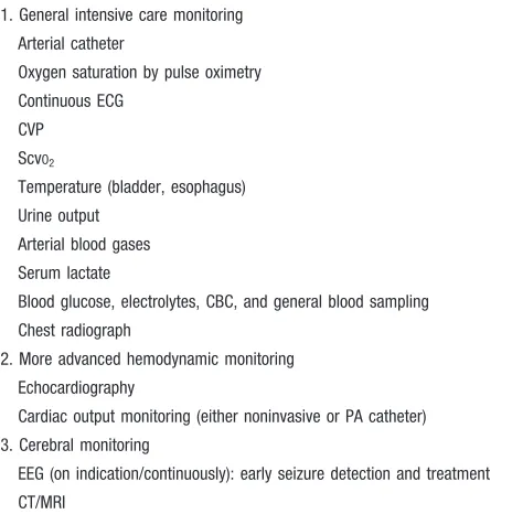
Monitoring
Post– cardiac arrest patients generally require intensive care monitoring. This can be divided into 3 categories (Table 2): general intensive care monitoring, more advanced hemodynamic monitoring, and cerebral monitoring.
by guest on August 16, 2017
http://circ.ahajournals.org/
eral intensive care monitoring (Table 2) is the minimum requirement; additional monitoring should be added depend-ing on the status of the patient and local resources and experience. The impact of specific monitoring techniques on post– cardiac arrest outcome, however, has not been validated prospectively.
Early Hemodynamic Optimization
Early hemodynamic optimization or early goal-directed ther-apy is an algorithmic approach to restoring and maintaining the balance between systemic oxygen delivery and demands. The key to the success of this approach is initiation of monitoring and therapy as early as possible and achievement of goals within hours of presentation. This approach focuses on optimization of preload, arterial oxygen content, afterload, contractility, and systemic oxygen utilization. Early goal-directed therapy has been studied in randomized prospective clinical trials of postoperative patients and patients with severe sepsis.133–135The goals in these studies have included a central venous pressure of 8 to 12 mm Hg, MAP of 65 to 90 mm Hg, ScvO2⬎70%, hematocrit⬎30% or hemoglobin ⬎8 g/dL, lactateⱕ2 mmol/L, urine outputⱖ0.5 mL · kg⫺1· h⫺1, and oxygen delivery index⬎600 mL · min⫺1· m⫺2. The primary therapeutic tools are intravenous fluids, inotropes, vasopressors, and blood transfusion. The benefits of early goal-directed therapy include modulation of inflammation, reduction of organ dysfunction, and reduction of healthcare resource consumption.133–135 In severe sepsis, early goal-directed therapy has also been shown to reduce mortality.133 The systemic ischemia/reperfusion response and myocar-dial dysfunction of post– cardiac arrest syndrome have many
characteristics in common with sepsis.112 Therefore, it has been hypothesized that early hemodynamic optimization might improve the outcome of post– cardiac arrest patients. The benefit of this approach has not been studied in random-ized prospective clinical trials, however. Moreover, the opti-mal goals and the strategies to achieve those goals could be different in post– cardiac arrest syndrome, given the concom-itant presence of post– cardiac arrest brain injury, myocardial dysfunction, and persistent precipitating pathologies.
The optimal MAP for post– cardiac arrest patients has not been defined by prospective clinical trials. The simultaneous need to perfuse the postischemic brain adequately without putting unnecessary strain on the postischemic heart is unique to the post– cardiac arrest syndrome. The loss of cerebrovas-cular pressure autoregulation makes cerebral perfusion de-pendent on CPP (CPP⫽MAP⫺ICP). Because sustained ele-vation of ICP during the early post– cardiac arrest phase is uncommon, cerebral perfusion is predominantly dependent on MAP. If fixed or dynamic cerebral microvascular dysfunc-tion is present, an elevated MAP could theoretically increase cerebral oxygen delivery. In 1 human study, hypertension (MAP⬎100 mm Hg) during the first 5 minutes after ROSC was not associated with improved neurological outcome50; however, MAP during the first 2 hours after ROSC was positively correlated with neurological outcome. Good out-comes have been achieved in published studies in which the MAP target was as low as 65 to 75 mm Hg13or as high as 90 to 100 mm Hg9,12for patients admitted after out-of-hospital cardiac arrest. The optimal MAP in the post– cardiac arrest period might be dependent on the duration of cardiac arrest, with higher pressures needed to overcome the potential no-reflow phenomenon observed with ⬎15 minutes of un-treated cardiac arrest.42,43,136 At the opposite end of the spectrum, a patient with an evolving acute myocardial infarc-tion or severe myocardial dysfuncinfarc-tion might benefit from the lowest target MAP that will ensure adequate cerebral oxygen delivery.
The optimal central venous pressure goal for post– cardiac arrest patients has not been defined by prospective clinical trials, but a range of 8 to 12 mm Hg has been used in most published studies. An important consideration is the potential for persistent precipitating pathology that could cause ele-vated central venous pressure independent of volume status, such as cardiac tamponade, right-sided acute myocardial infarction, pulmonary embolism, and tension pneumothorax or any disease that impairs myocardial compliance. A risk also exists of precipitating pulmonary edema in the presence of post– cardiac arrest myocardial dysfunction. The post– cardiac arrest ischemia/reperfusion response causes intravas-cular volume depletion relatively soon after the heart is restarted, and volume expansion is usually required. No evidence is available to indicate an advantage for any specific type of fluid (crystalloid or colloid) in the post– cardiac arrest phase. Some animal data are available indicating that hyper-tonic saline may improve myocardial and cerebral blood flow when given during CPR,137,138but no clinical data indicate an advantage for hypertonic saline in the post– cardiac arrest phase.
Table 2. Post–Cardiac Arrest Syndrome: Monitoring Options
1. General intensive care monitoring
Arterial catheter
Oxygen saturation by pulse oximetry
Continuous ECG
CVP
ScvO2
Temperature (bladder, esophagus)
Urine output
Arterial blood gases
Serum lactate
Blood glucose, electrolytes, CBC, and general blood sampling
Chest radiograph
2. More advanced hemodynamic monitoring
Echocardiography
Cardiac output monitoring (either noninvasive or PA catheter)
3. Cerebral monitoring
EEG (on indication/continuously): early seizure detection and treatment
CT/MRI
CVP indicates central venous pressure; ScvO2, central venous oxygen saturation;
CBC, complete blood count; PA, pulmonary artery; EEG, electroencephalogram; and CT/MRI, computed tomography/magnetic resonance imaging.
by guest on August 16, 2017
http://circ.ahajournals.org/
The balance between systemic oxygen delivery and consumption can be monitored indirectly with mixed venous oxygen saturation (SvO2) or ScvO2. The optimal ScvO2 goal for post– cardiac arrest patients has not been defined by prospective clinical trials, and the value of continuous ScvO2monitoring remains to be demonstrated. One important caveat is that a subset of post– cardiac arrest patients have elevated central or mixed venous oxygen saturations despite inadequate tissue oxygen delivery, a phenomenon that is more common in patients given high doses of epinephrine during CPR.139 This phenomenon, termed “venous hyperoxia,” can be attributed to impaired tissue oxygen utilization caused by microcirculatory fail-ure or mitochondrial failfail-ure.
Additional surrogates for oxygen delivery include urine output and lactate clearance. Two of the randomized prospec-tive trials of early goal-directed therapy described above used a urine output target ofⱖ0.5 mL · kg⫺1· h⫺1.133,135A higher urine output goal of ⬎1 mL · kg⫺1 · h⫺1 is reasonable in postarrest patients treated with therapeutic hypothermia, given the higher urine production during hypothermia13; however, urine output could be misleading in the presence of acute or chronic renal insufficiency. Lactate concentrations are elevated early after ROSC because of the total-body ischemia of cardiac arrest. This limits the usefulness of a single measurement during early hemodynamic optimization. Lactate clearance has been associated with outcome in patients with ROSC after out-of-hospital cardiac arrest140,141; however, lactate clearance can be impaired by convulsive seizures, excessive motor activity, hepatic insufficiency, and hypothermia.
The optimal goal for hemoglobin concentration in the post– cardiac arrest phase has not been defined. The original study of early goal-directed therapy in sepsis used a transfu-sion threshold hematocrit of 30%, but relatively few patients received a transfusion, and the use of this transfusion thresh-old, even for septic shock, is controversial.133 Subgroup analysis of patients with a closed head injury enrolled in the Transfusion Requirements in Critical Care trial showed no difference in mortality rates when hemoglobin concentration was maintained at 10 to 12 g/dL compared with 7 to 9 g/dL.142A post– cardiac arrest care protocol published by a group from Norway included a hemoglobin target of 9 to 10 g/dL.13
In summary, the value of hemodynamic optimization or early goal-directed therapy in post– cardiac arrest care has yet to be demonstrated in randomized prospective clinical trials, and little evidence is available about the optimal goals in post– cardiac arrest syndrome. On the basis of the limited available evidence, reasonable goals for post– cardiac arrest syndrome include an MAP of 65 to 100 mm Hg (taking into consideration the patient’s normal blood pressure, cause of arrest, and severity of any myocardial dysfunction), central venous pressure of 8 to 12 mm Hg, ScvO2⬎70%, urine output ⬎1 mL · kg⫺1 · h⫺1, and a normal or decreasing serum or blood lactate level. Goals for hemoglobin concentration during post– cardiac arrest care remain to be defined.
Oxygenation
Existing guidelines emphasize the use of a fraction of inspired oxygen (FIO2) of 1.0 during CPR, and clinicians will frequently maintain ventilation with 100% oxygen for vari-able periods after ROSC. Although it is important to ensure that patients are not hypoxemic, a growing body of preclinical evidence suggests that hyperoxia during the early stages of reperfusion harms postischemic neurons by causing excessive oxidative stress.51,52,143,144 Most relevant to post– cardiac arrest care, ventilation with 100% oxygen for the first hour after ROSC resulted in worse neurological outcome than immediate adjustment of the FIO2 to produce an arterial oxygen saturation of 94% to 96%.145
On the basis of preclinical evidence alone, unnecessary arterial hyperoxia should be avoided, especially during the initial post– cardiac arrest period. This can be achieved by adjusting the FIO2to produce an arterial oxygen saturation of 94% to 96%. However, controlled reoxygenation has yet to be studied in randomized prospective clinical trials.
Ventilation
Although cerebral autoregulation is either absent or dysfunc-tional in most patients in the acute phase after cardiac arrest,47 cerebrovascular reactivity to changes in arterial carbon diox-ide tension appears to be preserved.53,55,146,147 Cerebrovascu-lar resistance may be elevated for at least 24 hours in comatose survivors of cardiac arrest.55 No data exist to support the targeting of a specific PaCO2 after resuscitation from cardiac arrest; however, extrapolation of data from studies of other cohorts suggests ventilation to normocarbia is appropriate. Studies in brain-injured patients have shown that the cerebral vasoconstriction caused by hyperventilation may produce potentially harmful cerebral ischemia.148 –150 Hyper-ventilation also increases intrathoracic pressure, which will decrease cardiac output both during and after CPR.151,152 Hypoventilation may also be harmful, because hypoxia and hypercarbia could increase ICP or compound metabolic acidosis, which is common shortly after ROSC.
High tidal volumes cause barotrauma, volutrauma,153and biotrauma154in patients with acute lung injury. The Surviving Sepsis Campaign recommends the use of a tidal volume of 6 mL/kg (predicted) body weight and a plateau pressure ofⱕ30 cm H2O during mechanical ventilation of patients with sepsis-induced acute lung injury or acute respiratory distress syn-drome.155However, no data are available to support the use of a specific tidal volume during post– cardiac arrest care, and the use of this protective lung strategy will often result in hypercapnia, which may be harmful in the post– cardiac arrest patient. In these patients, it may be necessary to use tidal volumes⬎6 mL/kg to prevent hypercapnia. When therapeu-tic hypothermia is being induced, additional blood gases may be helpful to adjust tidal volumes, because cooling will decrease metabolism and the tidal volumes required. Blood gas values can either be corrected for temperature or left uncorrected. No evidence exists to suggest that one strategy is significantly better than the other.
In summary, the preponderance of evidence indicates that hyperventilation should be avoided in the post– cardiac arrest
by guest on August 16, 2017
http://circ.ahajournals.org/
period. Ventilation should be adjusted to achieve normocar-bia and should be monitored by regular measurement of arterial blood gas values.
Circulatory Support
Hemodynamic instability is common after cardiac arrest and manifests as dysrhythmias, hypotension, and low cardiac index.97 Underlying mechanisms include intravascular vol-ume depletion, impaired vasoregulation, and myocardial dysfunction.
Dysrhythmias can be treated by maintenance of normal electrolyte concentrations and use of standard drug and electrical therapies. No evidence exists to support the pro-phylactic use of antiarrhythmic drugs after cardiac arrest. Dysrhythmias are commonly caused by focal cardiac ische-mia, and early reperfusion treatment is probably the best antiarrhythmic therapy. Ultimately, survivors of cardiac ar-rest attributed to a primary dysrhythmia should be evaluated for placement of a pacemaker or an implantable cardioverter-defibrillator.
The first-line intervention for hypotension is to optimize right-heart filling pressures by use of intravenous fluids. In 1 study, 3.5 to 6.5 L of intravenous crystalloid was required in the first 24 hours after ROSC after out-of-hospital cardiac arrest to maintain right atrial pressures in the range of 8 to 13 mm Hg.97In a separate study, out-of-hospital post– cardiac arrest patients had a positive fluid balance of 3.5⫾1.6 L in the first 24 hours, with a central venous pressure goal of 8 to 12 mm Hg.13
Inotropes and vasopressors should be considered if hemo-dynamic goals are not achieved despite optimized preload. Myocardial dysfunction after ROSC is well described in both animal101,102,156,157 and human97,99,112 studies. Post– cardiac arrest global myocardial dysfunction is generally reversible and responsive to inotropes, but the severity and duration of the myocardial dysfunction may impact survival.97 Early echocardiography will enable the extent of myocardial dys-function to be quantified and may guide therapy. Impaired vasoregulation is also common in post– cardiac arrest pa-tients; this may require treatment with vasopressors and is also reversible. Persistence of reversible vasopressor depen-dency has been reported for up to 72 hours after out-of-hospital cardiac arrest despite preload optimization and re-versal of global myocardial dysfunction.97No individual drug or combination of drugs has been demonstrated to be superior in the treatment of post– cardiac arrest cardiovascular dys-function. Despite improving hemodynamic values, the effect on survival of inotropes and vasopressors in the post– cardiac arrest phase has not been studied in humans. Furthermore, inotropes have the potential to exacerbate or induce focal ischemia in the setting of ACS and coronary artery disease (CAD). The choice of inotrope or vasopressor can be guided by blood pressure, heart rate, echocardiographic estimates of myocardial dysfunction, and surrogate measures of tissue oxygen delivery such as ScvO2, lactate clearance, and urine output. If a pulmonary artery catheter or some form of noninvasive cardiac output monitor is being used, therapy can be further guided by cardiac index and systemic vascular
resistance. No evidence exists that the use of a pulmonary artery catheter or noninvasive cardiac output monitoring improves outcome after cardiac arrest.
If volume expansion and treatment with vasoactive and inotropic drugs do not restore adequate organ perfusion, mechanical circulatory assistance should be considered.158,159 This treatment can support circulation in the period of transient severe myocardial dysfunction that often occurs for 24 to 48 hours after ROSC.97The intra-aortic balloon pump is the most readily available device to augment myocardial perfusion; it is generally easy to insert with or without radiological imaging, and its use after cardiac arrest has been documented recently in some studies.13,160If additional car-diac support is needed, more invasive treatments such as percutaneous cardiopulmonary bypass, extracorporeal mem-brane oxygenation (ECMO), or transthoracic ventricular as-sist devices can be considered.161,162In a recent systematic review of published case series in which percutaneous car-diopulmonary bypass was initiated during cardiac arrest and then gradually weaned after ROSC (n⫽675), an overall in-hospital mortality rate of 55% was reported.162The clinical value of initiating these interventions after ROSC for cardio-vascular support has not been determined.
Management of ACS
CAD is present in the majority of out-of-hospital cardiac arrest patients,163–165 and acute myocardial infarction is the most common cause of sudden cardiac death.165One autopsy study reported coronary artery thrombi in 74 of 100 subjects who died of ischemic heart disease within 6 hours of symptom onset and plaque fissuring in 21 of 26 subjects in the absence of thrombus.166A more recent review reported acute changes in coronary plaque morphology in 40% to 86% of cardiac arrest survivors and in 15% to 64% of autopsy studies.167
The feasibility and success of early coronary angiography and subsequent percutaneous coronary intervention (PCI) after out-of-hospital cardiac arrest are well described in a number of relatively small case series and studies with historical controls.13,14,123,160,168 –172A subset of these studies focuses on early primary PCI in post– cardiac arrest patients with ST-elevation myocardial infarction.14,168 –171 Although inclusion criteria and the outcomes reported were variable, average intervals from symptom onset or CPR to balloon inflation ranged from 2 to 5 hours, angiographic success rates ranged from 78% to 95%, and overall in-hospital mortality ranged from 25% to 56%. In several of these studies, PCI was combined with therapeutic hypothermia. One retrospective study reported 25% in-hospital mortality among 40 consec-utive comatose post– cardiac arrest patients with ST-elevation myocardial infarction who received early coronary angiogra-phy/PCI and mild therapeutic hypothermia compared with a 66% in-hospital mortality rate for matched historical control subjects who underwent PCI without therapeutic hypother-mia.14 In this study, 21 (78%) of 27 hypothermia-treated 6-month survivors had a good neurological outcome (CPC of 1 or 2) compared with only 6 (50%) of 12 non– hypothermia-treated 6-month survivors.
by guest on August 16, 2017
http://circ.ahajournals.org/
Studies with broader inclusion criteria (not limited to ST-elevation myocardial infarction) have also shown prom-ising results. In 1 such study, 77% of all survivors of out-of-hospital cardiac arrest with presumed cardiac origin underwent immediate coronary angiography, which revealed CAD in 97%; of these,⬎80% had total occlusion of a major coronary artery.13 Nearly half of these patients underwent reperfusion interventions, with the majority by PCI and a minority by coronary artery bypass graft. Among patients admitted after ROSC, the overall in-hospital mortality rate decreased from 72% before the introduction of a comprehen-sive post– cardiac arrest care plan (which included this intensive coronary reperfusion strategy and therapeutic hypo-thermia) to 44% (P⬍0.001), and ⬎90% of survivors were neurologically normal.13
Chest pain and ST elevation may be poor predictors of acute coronary occlusion in post– cardiac arrest patients.123 Given that acute coronary occlusion is the most common cause of out-of-hospital cardiac arrest, prospective studies are needed to determine whether immediate coronary angiogra-phy should be performed in all patients after ROSC. It is feasible to initiate cooling before coronary angiography, and patients can be transported to the angiography laboratory while cooling continues.13,14,160
If no facilities are available for immediate PCI, in-hospital thrombolysis is recommended for patients with ST elevation who have not received prehospital thrombolysis.173,174 Al-though the efficacy and risk of thrombolytic therapy have been well characterized in post– cardiac arrest patients,174 –176 the potential interaction of mild therapeutic hypothermia and thrombolytic therapy has not been studied formally. Theoret-ical considerations include a possible impact on the efficacy of thrombolysis and the risk of hemorrhage. Coronary artery bypass graft is indicated in the post– cardiac arrest phase for patients with left main coronary artery stenosis or 3-vessel CAD. In addition to acute reperfusion, management of ACS and CAD should follow standard guidelines.
In summary, patients resuscitated from cardiac arrest who have electrocardiographic criteria for ST-elevation myocar-dial infarction should undergo immediate coronary angiogra-phy, with subsequent PCI if indicated. Furthermore, given the high incidence of ACS in patients with out-of-hospital car-diac arrest and limitations of electrocardiography-based di-agnosis, it is appropriate to consider immediate coronary angiography in all post– cardiac arrest patients in whom ACS is suspected. If PCI is not available, thrombolytic therapy is an appropriate alternative for post– cardiac arrest manage-ment of ST-elevation myocardial infarction. Standard guide-lines for management of ACS and CAD should be followed.
Other Persistent Precipitating Pathologies
Other causes of out-of-hospital cardiac arrest include pulmo-nary embolism, sepsis, hypoxemia, hypovolemia, hypokale-mia, hyperkalehypokale-mia, metabolic disorders, accidental hypother-mia, tension pneumothorax, cardiac tamponade, toxins, intoxication, and cerebrovascular catastrophes. The incidence of these causes is potentially higher for in-hospital cardiac
arrest.5 These potential causes of cardiac arrest that persist after ROSC should be diagnosed promptly and treated.
Therapeutic Hypothermia
Therapeutic hypothermia should be part of a standardized treatment strategy for comatose survivors of cardiac ar-rest.13,177,178Two randomized clinical trials and a meta-anal-ysis showed improved outcome in adults who remained comatose after initial resuscitation from out-of-hospital ven-tricular fibrillation (VF) cardiac arrest and who were cooled within minutes to hours after ROSC.8,9,179 Patients in these studies were cooled to 33°C or the range of 32°C to 34°C for 12 to 24 hours. The Hypothermia After Cardiac Arrest (HACA) study included a small subset of patients with in-hospital cardiac arrest.8Four studies with historical control groups reported benefit after therapeutic hypothermia in comatose survivors of out-of-hospital non-VF arrest180and all rhythm arrests.12,13,132 Other observational studies provide evidence of a possible benefit after cardiac arrest from other initial rhythms and in other settings.181,182Mild hypothermia is the only therapy applied in the post– cardiac arrest setting that has been shown to increase survival rates. The patients who may benefit from this treatment have not been fully elucidated, and the ideal induction technique (alone or in combination), target temperature, duration, and rewarming rate have yet to be established.
Animal studies demonstrate a benefit of very early cooling either during CPR or within 15 minutes of ROSC when cooling is maintained for only a short duration (1 to 2 hours).183,184When prolonged cooling is used (⬎24 hours), however, less is known about the therapeutic window. Equiv-alent neuroprotection was produced in a rat model of cardiac arrest when a 24-hour period of cooling was either initiated at the time of ROSC or delayed by 1 hour.185 In a gerbil forebrain ischemia model, sustained neuroprotection was achieved when hypothermia was initiated at 1, 6, or 12 hours after reperfusion and maintained for 48 hours186; however, neuroprotection did decrease when the start of therapy was delayed. The median time to achieve target temperature in the HACA trial was 8 hours (interquartile range 6 to 26 hours),8 whereas in a study by Bernard et al,9average core tempera-ture was reported to be 33.5°C within 2 hours of ROSC. Clearly, additional clinical studies are needed to optimize this therapeutic strategy.
The practical approach of therapeutic hypothermia can be divided into 3 phases: induction, maintenance, and rewarm-ing. Induction can be instituted easily and inexpensively with intravenous ice-cold fluids (saline 0.9% or Ringer’s lactate, 30 mL/kg)187–191or traditional ice packs placed on the groin and armpits and around the neck and head. In most cases, it is easy to cool patients initially after ROSC, because their temperature normally decreases within the first hour.10,64 Initial cooling is facilitated by concomitant neuromuscular blockade with sedation to prevent shivering. Patients can be transferred to the angiography laboratory with ongoing cool-ing by use of these easily applied methods.13,14 Surface or internal cooling devices (as described below) can also be used
by guest on August 16, 2017
http://circ.ahajournals.org/
either alone or in combination with the above measures to facilitate induction.182,192
In the maintenance phase, effective temperature monitor-ing is needed to avoid significant temperature fluctuations. This is best achieved with external or internal cooling devices that include continuous temperature feedback to achieve a target temperature. External devices include cooling blankets or pads with water-filled circulating systems or more ad-vanced systems in which cold air is circulated through a tent. Intravascular cooling catheters are internal cooling devices that are usually inserted into a femoral or subclavian vein. Less sophisticated methods, such as cold, wet blankets placed on the torso and around the extremities or ice packs combined with ice-cold fluids, can also be effective, but these methods may be more time consuming for nursing staff, result in greater temperature fluctuations, and do not enable controlled rewarming.193Ice-cold fluids alone cannot be used to main-tain hypothermia.194
The rewarming phase can be regulated with the external or internal devices used for cooling or by other heating systems. The optimal rate of rewarming is not known, but current consensus is to rewarm at approximately 0.25°C to 0.5°C per hour.181 Particular care should be taken during the cooling and rewarming phases, because metabolic rate, plasma elec-trolyte concentrations, and hemodynamic conditions may change rapidly.
Therapeutic hypothermia is associated with several com-plications.195 Shivering is common, particularly during the induction phase.196 Mild hypothermia increases systemic vascular resistance, which reduces cardiac output. A variety of arrhythmias may be induced by hypothermia, but brady-cardia is the most common.182Hypothermia induces a diure-sis, and coexisting hypovolemia will compound hemodynam-ic instability. Diuresis may produce electrolyte