The journal homepage www.jpacr.ub.ac.id p-ISSN : 2302 – 4690 | e-ISSN : 2541 – 0733
Antimicrobial, antioxidant, and cytotoxic activities of endhopitic
fungi
Chaetomium sp.
isolated from
Phyllanthus niruri
Linn: in
vitro and in silico studies
Rollando1,*, Dion Notario1, Eva Monica1, Martanty Aditya1, Rehmadanta Sitepu1
1
Program of Pharmacy, Faculty of Science and Technology, Ma Chung University, Malang 65151, Indonesia
*
Corresponding email: [email protected]
Received 17 January 2017 ; Revised 7 March 2017 ; Accepted 8 March 2017
ABSTRACT
Endophytic fungi Chaetomium sp isolated from Phyllanthus niruri Linn. Mycelium powder was extracted by using ethyl acetate. Extract was fractionated using n-hexane, dichloromethane and ethanol 96%. The antimicrobial test was carried out using disc diffusion and microdilution methods. The antioxidant activity of the fraction was determined using hydrogen peroxide free radical scavenging and reducing power capacity activities. The cytotoxicity assay of the fraction against T47D breast cancer cell was carried out using dimethylthiazol-2-yl-2,5-diphenyltetrazolium bromide method (MTT). The in silico prediction of chemical substances which are reported exist in Chaetomium sp.
performed using AutoDockVina embedded in PyRx version 8.0. Dichloromethane fraction was found as the most active sample against Escherichia coli (IC50 20.76 g/mL), Staphylococcus aureus (IC50 70.15 g/mL), Salmonella typhi (49.13 g/mL) and was
found as the most high phenolic content with value 47.44 mg GAE/g fraction, whereas the best antioxidant activity was performed by ethanol 96% fraction (85%). Cytotoxicity assay against T47D cell line showed dichloromethane fraction have highest activity with IC50
10.76 g/mL. The docking studies showed that compounds bearing xanthone structure were potential for maltose binding periplasmic and human aromatase associating with their potencies as antibacteria and anticancer. Endophytic fungi Chaetomium sp. was isolated from Phyllanthus niruri using n-hexane, dichloromethane and ethanol fractions was studied its various biological activities as antimicrobial, antioxidant and cytotoxic agent against breast cancer cell.
Key word: Endophytic fungi, Chaetomium sp., Phyllanthus niruri Linn, Biological activities, Molecular docking
INTRODUCTION
In the same family of Phyllanthaceae, Phyllanthus niruri also has a potential to be a new source for producing fungal endophyte. Originally, this herb contains chemical substances including lignin, glycoside, alkaloid, ellagitannin, terpenoid, phenylpropanoid, flavonoid and polyphenol [6]. Instead of this herb has been studied its pharmacological activity in the treatment of bladder infection, as immunomodulator, anti-inflammatory and antiviral agents [7, 8], to date, there is no report about this species to be used as an endophyte source. Hence, it is interesting to study more about extraction and biological testing of fungal endophyte from Phyllanthus niruri.
Resistance in some antimicrobial therapeutics needs a high concern in discovering and developing new antibacterial which are more susceptible to the mutant and selective to eradicate the bacteria cells with no effects to the host [6]. Subsequently, antioxidant currently becomes a hot topic related with oxidative stress associating with cells damage, indicating some diseases such as cancer and coronary heart disorder [9]. Furthermore, the searching of cancer drug that is less toxic has been a lengthy challenge with the results of no anticancer agent is perfectly killing the cancer cells growth without affecting the normal cell [10]. In this present study, we explore the opportunities of Phyllanthus niruri as the new source for mining fungal endophyte in organic fractions such as n-hexane, dichloromethane and ethanol to be further studied in vitro antibacterial, antioxidant and anticancer due to its cytotoxicity effects towards breast cancer cells type T47D.
In rational drug design, the proteins which are responsible towards the pathogenesis can be used as the target in combating the microorganism as well as the malign cells such as tumor. In this study, we have not identified the compounds contained in fungal endophyte of Phyllanthus niruri and we also have not tested the fractions against E. coli and T47D cells at protein levels due to our limitations. However, we tried to gain an insight mechanism of compounds in the most active fraction by using compounds reported in other Chaetomium sp. as the model. In particular, an in silico prediction of a possible mechanism as antibacterial and its cytotoxic activity against the breast cancer cell using molecular docking was also discussed.
EXPERIMENT
Chemicals and instrumentation
Phyllanthus niruri leaves were collected from Materia Medica, Malang, Indonesia with specimen code was MN 02371. Potato dextrose broth (PDB) was prepared from potatoes collected from Batu, Malang. Dextrose, muller hinton, potato dextrose agar (PDA), nutrient agar (NA) and nutria broth (NB) were purchased from Merck. The microbes used were Escherichia coli (E. coli) K-12, Staphylococcus aureus (S. aureus) NCTC 8325 and Salmonella typhi (S. typhi) Ty2 purchased from Microbiology Laboratory, Brawijaya University, Indonesia. All chemicals used were analytical grade i.e., ethyl acetate, n-hexane, methanol, chloroform, hydrogen peroxide, phosphate buffer pH 7.0, potassium iron (III) cyanide, iron (III) chloride, Thin Layer Chromatography Silica F254 plate, and silica 60 for column chromatography were purchased from Merck. T47D cell was courtesy from Parasitology Laboratory of Medical Faculty, Gadjah Mada University, Dulbecco’s Modified Eagle Media (DMEM), Fetal Bovine Serum (FBS) 10% (v/v), penicillin-streptomycin, fungizone 0,5% v/v and trypsin-EDTA 0,25% were purchased from Gibco, while Tissue Culture Dish was from Iwaki.
Tissue culture of endophytic fungi
The colony of endophytic fungi was taken from PDA using ose to have 5 plugs with 0.5 cm in diameter. A volume of 200 mL of the colony was inoculated into a sterilized conical flask and then incubated at room temperature for 14 days. The mycelium was then collected from the filtrate by filtration, followed by drying them at 50oC. Thereafter, the dried mycelium was then pulverized.
Extraction
The mycelium powder was macerated using ethyl acetate (1:3) for 48 hours with a frequent agitation. The liquid extract was then filtrated and the residue was macerated using new ethyl acetate for 24 hours with the same frequent agitation. The filtrate was then collected and concentrated under reduced pressure.
Fractionation
The concentrated extract (200 mg) was then fractionated using 200 mL of each n-hexane (100%), dichloromethane (100%) and ethanol 96% through chromatography column with an isocratic technique. Each fraction was collected and concentrated under reduced pressure followed by drying them at 50oC.
Antimicrobial test
The antimicrobial test was carried out using disc diffusion (Kirby-Bauer Test) [11] method whereas the minimum inhibition concentration (MIC) was performed using micro-dilution test [12].
Screening of active fraction
The series of concentration for each fraction was prepared to make 100, 50, 25, 12.5 and 6.25 g/mL of working solution. Ten L of each was dropped into paper disc which have amounts of the isolate for each paper disc i.e. 1000, 500, 250, 125 and 62.5 g/mL and left it to dry leading to the paper disc attachment on the media. Streptomycin 10 mg/mL (10 L) and a sterile anhydrous ethanol were used as the positive control and the negative control, respectively. The bacterial culture was then incubated at 37oC for 18-24 h followed by viewing the inhibition zone around the paper disc. The larger diameter of the inhibition zone was selected as the active fraction.
Determination of Minimum Inhibition Concentration (MIC)
Determination of a total phenolic content
The total phenolic content was determined using Folin-Ciocalteu reagent with gallic acid as the reference [14]. Each of fractions was dissolved into methanol which had 2 mg/mL of concentration followed by adding 500 L of Folin-Ciocalteu (50%) and 2 mL of Na2CO3 20%. This mixture was top up with 5 mL of distilled water and then stored at room temperature for 20 min by maintained its absorbance at 765 nm. The total phenolic content was observed by applying the absorbance value to the linear regression equation generated from the gallic acid standard curve (30; 45; 60; 75; and 90 μg/mL).
Determination of antioxidant activity
Two methods were used in determining the antioxidant activity of the fraction, i. e., hydrogen peroxide free radical scavenging [3] and reducing power capacity [15].
Hydrogen peroxide free radical scavenging
A solution of hyrogen peroxide (40 mmol/L) was prepared using phosphate buffer pH 7.5. The fraction was dissolved into distilled water to have 2 mg/mL of concentration and then added by H2O2 solution. After 10 minutes of reaction, the sample was maintained its absorbance at 230 nm. The capacity of the fraction to scavenge the H2O2 radical was calculated based on the formula as followed:
Scavenging capacity (%) =
Where Ai = absorbance of the control, At = absorbance of the sample.
Reducing power capacity
Each of fractions was dissolved into 1 mL of distilled water and then mixed up with 2.5 mL of phosphate buffer pH 6.6 and 2.5 mL of potassium iron (III) cyanide 1 % w/v. The mixture was then centrifuged at 3000 rpm for 10 min. The upper phase was taken 2.5 mL and then added with another 2.5 mL of distilled water, follwed by adding 0.5 mL of FeCl3 0.1% w/v. The absorbance was then measured at 700 nm using UV spectrophotometer.
Cytotoxicity assay
The cytotoxicity assay of the fraction against T47D breast cancer cell was carried out using (3-(4,5-Dimethylthiazol-2-yl)-2,5-Diphenyltetrazolium Bromide (MTT) method [16]. The T47D cell was cultured on the RPMI media, whereas the normal cell (Vero) was cultured on the M199 media. Each of them contained FBS 10%, penicillin-streptomycin 1% and fungizone 0.5%. A series of working solution was prepared with concentrations as followed: 7.81, 15.62, 31.25, 62.5, 125, 250 and 500 µg/ml. Cisplatin (15 µM) was used as the positive control.The cell viability was determined at 595 nm and the absorbance was converted into the percentage viability using plate reader. The IC50 was calculated by plotting the % viability against the concentration of the fraction and the selectivity index (SI) was defined according to the ratio between the IC50 of Vero cell and the IC50 of T47D cell.
Molecular Docking
prepared using AutoDockTools 1.5.6 (www.autodock.scripps.edu). The proteins were added with polar hydrogen and given by kollman charge whereas the ligands were given by gasteiger charges. The grid was centered on each protein binding site and the docking was
then performed using AutoDock Vina embedded in PyRx version 8.0
(www.autodock.scripps.edu). The docking results were observed as free energy of binding (Gbind) and the selected docking pose was visualized using Discovery Studio 3.5 (www.accelyrs.com).
RESULT AND DISCUSSION Fungal endophyte culture
Fungal endophyte of Chaetomium sp. (Fig. 1a) which was isolated from Phyllanthus niruri leaves, characterized as white fungal colony (Fig. 1b). The fungal morphology was identified by Mycology Laboratory, Agriculture Faculty of Gadjah Mada University, Indonesia. Genus Chaetomium sp. had characteristics as followed: a helix solid mycelium, composed of dark hyphae pigment and had uniform sclerotia. These characters were in agreement with fungal endophyte from genus Chaetomium sp., which was reported by Sharma [17] .
(a) (b)
Figure 1. (a) Fungal culture of Chaetomium sp. in 14th day of incubation and (b) the morphology of Chaetomium sp. with 7 days in age
Screening of active fraction
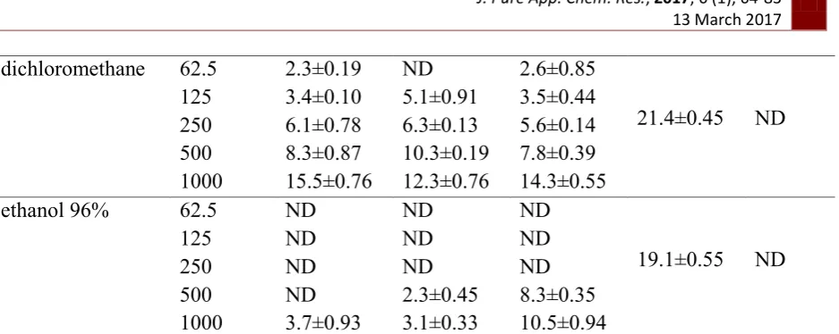
The preliminary determination of antibacterial activity of the fractions was necessary to select which fraction would be potential for further testing. Table 1 presented the inhibition zone of three fractions (n-hexane, dichloromethane and ethanol 96%) towards S. aureus, E. coli dan S. typhi.
Table 1. The results of the disc diffusion assay of the fractions against the bacteria
Fraction Loading (µg)
Zone of inhibition (mm)(mean ± SD) n = 5 experiments
S. aureus E. coli S. typhi
(+) control
(-) control
n-hexane 62.5 ND ND ND
26.3±0.45 ND
125 ND ND ND
250 ND ND ND
500 10.5±0.16 ND 5.2±0.42
dichloromethane 62.5 2.3±0.19 ND 2.6±0.85
21.4±0.45 ND
125 3.4±0.10 5.1±0.91 3.5±0.44
250 6.1±0.78 6.3±0.13 5.6±0.14
500 8.3±0.87 10.3±0.19 7.8±0.39
1000 15.5±0.76 12.3±0.76 14.3±0.55
ethanol 96% 62.5 ND ND ND
19.1±0.55 ND
125 ND ND ND
250 ND ND ND
500 ND 2.3±0.45 8.3±0.35
1000 3.7±0.93 3.1±0.33 10.5±0.94
ND: not detected, SD: Standard deviation, S. aureus: Staphylococcus aureus, E. coli: Escherichia coli, S. typhi: Salmonella typhi
As results, dichloromethane fraction demonstrated a higher inhibition towards the bacterial growth than other two fractions. In the lowest concentration (62.5 g/mL), dichloromethane fraction inhibited S. aureus and S. typhi, respectively; while neither n-hexane nor ethanol 96% fractions performed the inhibitions. The inhibition of all bacteria by dichloromethane fraction was observed at concentration 125 g/mL, thereafter, the inhibition was proportionally increased along with the higher concentration of the fraction. Towards this fraction, all bacteria are similarly susceptible as shown by its close values of inhibition zone starting from 125 to 1000 g/mL. On the other hand, n-hexane and ethanol 96% fractions were more likely susceptible towards E. coli and S. typhi, respectively, as shown by its higher inhibitions at 1000 g/mL. Although they can strongly inhibit the bacteria at the highest concentration only, however, we decided to include them for further IC50 calculation due to its potential selective antimicrobial activity to more specific bacteria as discussed.
Minimum Inhibition Concentration (MIC) and IC50 calculation
The potent fraction was justified from its capability to inhibit the bacteria at the minimum concentration. When the fraction was able to inhibit at least 50% of bacterial growth, only the IC50 value would be defined. Table 2 presented the MIC and IC50 value of each fraction against three bacteria. According to the susceptibility, the dichloromethane was the best fraction due to its capability to inhibit all bacteria at lower concentration than two other fractions.
Table 2. The IC50 and MIC value of three fractions against S. aureus, E. coli and S. typhi
Bacteria
Samples(mean ± SD) n = 5 experiments
n-hexane dichloromethane ethanol 95% IC50
(µg/mL)
MIC (µg/mL)
IC50 (µg/mL)
MIC (µg/mL)
IC50 (µg/mL)
MIC (µg/mL)
E. coli 124.88±0.34 >500 20.76±0.56 125 101.54±0.87 >500 S. aureus 233.86±0.98 >500 70.15±0.87 250 118.09±0.88 >500 S. typhi 101.98±0.23 >500 49.13±0.55 125 88.12±0.46 >500
SD: Standard deviation, E. coli: Escherichia coli, S. aureus: Staphylococcus aureus, S. typhi: Salmonella typhi, IC50 =Inhibition concentration 50%, MIC = Minimum inhibition concentration.
dichloromethane fraction as it showed the lowest IC50 value (20.76 g/mL) among three bacteria. Either n-hexane or ethanol 96% was less sensitive than dichloromethane, as shown by its IC50 value with more than 100 g/mL. However, as indicated in the preliminary antibacterial assay, ethanol 96% fraction selectively inhibited S. typhi more than other bacteria due to its IC50 value i.e., 88.12 g/mL, which was potential for a narrow spectrum antibacterial. The least susceptible fraction was detected on n-hexane that showed more than 100 g/mL of IC50 to all bacteria.
The total phenolic content
The total phenolic content is associated with the reducing capacity of the compounds under redox reaction. Table 3 presented the total phenolic content of the three fractions which was found in a quite broad range. Due to its polar character, phenolic compounds were more soluble in a polar solvent such as ethanol 96% than in a semipolar (dichloromethane) and nonpolar (n-hexane) solvents. However, in the dichloromethane, the total phenolic content was quite high that might be contributing in the bacterial growth inhibition as known that phenol could denature the protein structure leading to bacterial eradication [18].
Table 3.The total phenolic content of fractions
Fraction Total phenolic content (mg GAE/g fraction) (mean ± SD) n = 5 experiments
n-hexane 10.11 ± 0.43
dichloromethane 47.44 ± 0.11
ethanol 96% 63.45 ± 0.87
SD: Standard deviation, mg GAE/g fraction: Miligram gallic acid eqivalent per gram fraction
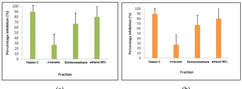
Antioxidant activity
Hydrogen peroxide is rapidly decomposed into oxygen and water, which may form hydroxyl radical that can modulate the lipid peroxidation process leading to the cell damage. From this assay, all fractions possessed the free radical scavenging activity at 34-85% (Fig. 2a).
(a) (b)
Figure 2. The antioxidant activity of three fractions using methods of (a) H2O2 free radical scavenging and (b) reducing power capacity, n = 5 experiments
order among the three fractions. The ethanol 96% fraction was able to reduce the reduction of Fe (III) to Fe (II) in such a way those compounds donated electron to stabilize the radicals up to 80% in the ethanol, which was almost compare to that vitamin C (90%) as the positive control. These two antioxidant activities corresponded to the total phenolic content, in which ethanol as a polar solvent able to fractionate phenolic based compounds. This might possess the antioxidant activity associating with lipid peroxidation inhibition.
Cytotoxicity assay against T47D breast cancer cells
T47D is a breast cancer cell lines that is primarily used in breast cancer research [19]. The inhibition of this cell line up to 50% was associated with the potential anticancer from the fractions (Table 4). All fractions were observed IC50 less than 100 g/mL indicating its potential as anticancer with dichloromethane fraction re-showing the lowest IC50 value than two others. Fortunately, this fraction showed the highest selectivity index (SI) indicating its selectivity was more to cancer than the normal cell. There have been an agreement between antibacteria and cytotoxicity assay, hence, this fraction was promiscuous for chemotherapeutics against bacteria and cancer cell.
Table 4. The results of cytotoxicity assay against T47D and Vero cell line
Fraction
Samples (mean ± SD) n = 5 experiments
IC50 T47D(µg/mL) IC50 Vero(µg/mL) SI n-hexane 122.64±0.76 253.88±1.65 2.07
dichloromethane 10.76±0.23 221.77±0.96 20.61
ethanol 96% 57.16±2.34 323.77±1.65 5.66
SD: Standard deviation, IC50 =Inhibition concentration 50%, SI : Selectivity index
Figure 3. The effect of dichloromethane fraction towards T47D (a-c) and Vero cells (d-f). The viewing was performed under inverted microscope with 100x of magnitudes. (a) negative control (b) dichloromethane fraction 5 g/mL, (c) dichloromethane10 g/mL, (d) negative control, (e) dichloromethane fraction 250 g/mL and (f) dichloromethane fraction 500 g/mL. The arrow indicated the living cell whereas the dashed arrow indicated the morphological alteration
In silico prediction of compounds in Chaetomium sp. using molecular docking
In silico activity against protein targets in E. coli
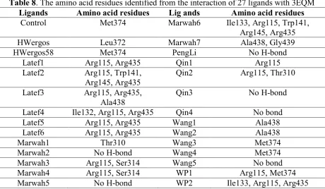
As results, active ligands showed free energy of binding at range of -12.1 to -3.9 kcal/mol while inactive ligands were found at -0.7 to 7.9 kcal/mol. The positive value of inactive ligands indicated no-affinity between ligand and the protein target in E. coli. According to the mean of the Gbind, the lowest free energy of binding went to the interaction between maltose binding periplasmic (MBP) protein and the ligand, indicated its strongest binding interaction among 10 protein targets. Maltose binding periplasmic was a huge bacterial periplasmic protein (370 amino acid residues) involved in active transport and chemotaxis towards maltose. This enzyme worked under substantial conformational changes to let maltose binds to the ATP –binding cassette (ABC)-7 transporter, initiating downstream signaling for either transport or chemotaxis [38]. Therefore, the MBP could be the selected protein target for further study. Table 6 presented the amino acid residues involved in the binding of 27 ligands.
Table 6.The amino acid residues identified from the interaction of 27 ligands with 1VY
Ligands Amino acid residues Ligands Amino acid residues
Control Lys15, Arg66, Tyr155, Asp65, Glu153, Asp14, Glu111
Marwah6 Asn12
HWergos No H-bond Marwah7 Asn12
HWergos58 No H-bond PengLi Asp65
Latef1 Arg345, Glu44, Qin1 No H-bond
Latef2 Trp62, Arg66 Qin2 No H-bond
Latef3 Trp62, Arg66 Qin3 Lys42
Latef4 Asn12 Qin4 Lys42, Tyr210, Ser211
Latef5 Asn12 Wang1 Glu153, Lys42
Latef6 Arg345, Glu153 Wang2 Glu153
Marwah1 Lys42 Wang3 Tyr155, Arg66, Arg345
Marwah2 Asn12, Lys15 Wang4 Arg66, Glu111
Marwah3 Asn12 Wang5 No H-bond
Marwah4 Lys42, Leu43, Arg66, Ser211 WP1 Asn12
Marwah5 No H-bond WP2 Asn12
1JVY: Maltose binding periplasmic protein, Lys: Lysine, Arg: Arginine, Tyr: Tyrosine, Asp: Aspartica acid, Glu: Glutamic acid, Trp: Typtophan, Asn: Aspagine, Leu: Leucine, Ser: Serine, No H-bond: No hydrogen bond
From these 27 ligands, 20 of them were characterized hydrogen bond interaction with MBP, whereas the remained six ligands were absent. The absent hydrogen bond interaction might be due to the nonpolar characters of the ligands bearing steroid, pentadienylfuranol, azaphilone and chromone (Supplementary data table 1), therefore, the Gbind of those compounds were significantly contributed by hydrophobic and other van der waals interactions. It is well studied that the strength of van der waals interaction is weaker than hydrogen bond interaction due to their distance bonds, i.e. > 2.2 Å and 1.5 – 2.2Å, respectively [39].
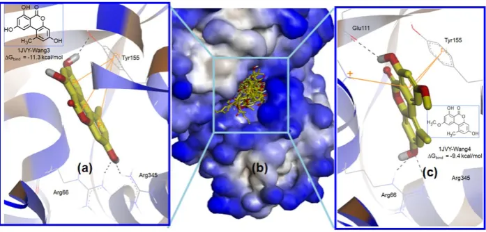
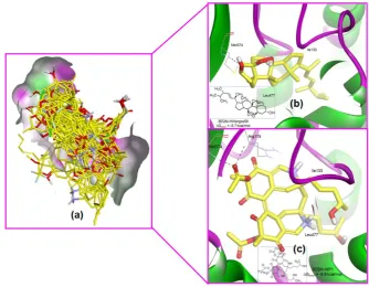
with same residues of the positive control associating with its more potential ligands to be optimized as MBP inhibitors. Figure 4 illustrated the superposition of 27 ligands at the binding site of MBP and the most active ligand-MBP molecular interaction.
Figure 4. The pose of (a) Wang3, (b) the superposition of 27 ligands in Chaetomium sp., and the pose of (c) Wang4 at the binding site of 1JVY. The individual poses of Wang3 and Wang4 was visualized as stick forms with carbon in yellow and standard colors of O and H whereas the protein was performed in ribbon, whereas the superposition was as surface. The protein was colored representing blue as hydrophobic region, brown as hydrophilic region and white as neutral region. The picture was visualized using Discovery Studio 3.5 (www.accelrys.com)
As visualized in Fig. 4a and 4c, Wang3 and Wang4 showed the similar binding mode towards MBP. On one hand, Wang3 showed hydrogen bond interaction with Arg66, Tyr155 and Arg345 and its pose was strongly supported by - interaction between three aromatic rings of the ligand with the phenol moiety of Tyr155. On the other hand, Wang4 possessed hydrogen bond interaction with Arg66 and Glu111. The conformation of Wang4 was stabilized by the presence of - as well as -cation interaction between one phenyl ring of the ligand with Tyr155 and Lys15, respectively. Either Wang3 or Wang4 had xanthone like structure, which could be a good lead compounds for further MBP inhibitor associating with its potency as antibacterial against E. coli.
In silico activity against protein targets in T47D cell
Likewise, the range of Gbind for predicting active ligands against 10 protein targets in T47D was at-11.3 to -4.2 kcal/mol, whereas predicted inactive ligands were at -2.8 to 8.8 kcal/mol. Overall proteins, human placental aromatase CYP450 (PDB 3EQ) was identified having the lowest value in the mean of Gbind among all 27 ligands. This indicated its potential as the protein target in blocking T47D cell growth. Human placental aromatase is an integral membrane enzyme, which catalyzes the demethylation and aromatization of androgen, which is a precursor to synthesize human estrogen [50]. Estrogen is the primary messenger in control of breast cancer, thereby inhibition of human aromatase is one of multiple ways to degenerate the breast cancer cell replication. Table 8 presented the amino acid residues involved in the binding of 27 ligands-human aromatase.
Table 8. The amino acid residues identified from the interaction of 27 ligands with 3EQM
Ligands Amino acid residues Lig ands Amino acid residues
Control Met374 Marwah6 Ile133, Arg115, Trp141,
Arg145, Arg435
HWergos Leu372 Marwah7 Ala438, Gly439
HWergos58 Met374 PengLi No H-bond
Latef1 Arg115, Arg435 Qin1 Arg115
Latef2 Arg115, Trp141,
Arg145, Arg435
Qin2 Arg115, Thr310
Latef3 Arg115, Arg435,
Ala438
Qin3 No H-bond
Latef4 Ile132, Arg115, Arg435 Qin4 No bond
Latef5 Arg115, Arg435 Wang1 Ala438
Latef6 Arg115, Arg435 Wang2 Ala438
Marwah1 Thr310 Wang3 Met374
Marwah2 No H-bond Wang4 Met374
Marwah3 Arg115, Ser314 Wang5 No bond
Marwah4 Arg115, Ser314 WP1 Arg115, Met374
Marwah5 No H-bond WP2 Ile133, Arg115, Arg435
3EQM: Human placental aromatase, Met: Methionine, Leu: Leucine, Arg: Arginine: Trp: Tryptophan, Ala: Alanine, Ile: Isoleucine, Thr: Threonine, Ser: Serine, Gly: Glycine, Met: Methionine, No H-bond: No hydrogen bond
PengLi, Qin3, Qin4 and Wang5) had no hydrogen bond interaction with the aromatase, however they still occupied the binding site via hydrophobic interaction. Figure 5 illustrated the superposition of 27 ligands at the binding site of the human aromatase and the most active ligand-aromatase molecular interaction.
Figure 5. The superposition of (a) 27 ligands in Chaetomium sp., the pose of (b) HWergos58, and (c) HP1 at the binding site of 3EQM. The individual poses of HWergos58 and HP1 was visualized as stick forms with carbon in yellow and standard colors of O and H whereas the protein was performed as ribbon, whereas the superposition was as surface. The protein was colored representing green as hydrogen bond acceptor (HB, magenta as hydrogen bond donor (HBD) and white as neutral region. The picture was visualized using Discovery Studio 3.5 (www.accelrys.com)
CONCLUSION
isolating compounds from the corresponding endophyte and test them against the selected protein targets. The docking studies showed that compounds bearing xanthone structure were potential for maltose binding periplasmic and human aromatase associated with their potencies as antibacteria and anticancer, respectively. Beside, compounds bearing steroid is also potential for human aromatase inhibitor due to it is disclose scaffold with the control ligand of human aromatase. We are ongoing to isolate the compounds from the endophyte thus hoping that this preliminary docking simulation could make the isolation of active compounds being more guided.
REFERENCES
[1] Strobel, G.A., Microb Infect, 2003, 5(6), 535-544.
[2] Tanaka, M., Sukiman, H., Takebayashi, M., Saito, K., Suto, M., Prana, T. K., Prana, M. S., Tomita, F., Microbes Environ, 1999, 14(4), 237-241.
[3] Ruch, R.J., S. Cheng, and J. E. Klaunig, Carcinogenesis, 1989, 10(6), 1003-1008. [4] Kandavel, D. and S. Sekar, Int J Pharm Pharm Sci, 2015, 7(5), 253-257.
[5] Sowparthani, K. and G. Kathiravan, J Pharm Biomed Sci., 2011, 10(10), 1-10.
[6] Paithankar V. V., Raut K. S ., Charde R. M., Vyas J. V., Res. Pharm., 2015, 1(4), 1-9. [7] Colpo, E., Vilanova, C. D. D. A., Pereira., R. P., Reetz, L. G. B., Oliveira, L., Farias,
I. L. G., Boligon, A. A., Athayde, M. L., Rocha, J. B. T., Asian Pac J Trop Med.,
2014, 7(2), 113-118.
[8] Mediani, A., Abas, F., Khatib, A., Tan, C. P., Ismail, I. S., Lajis, N. H., Plant Foods Hum Nutr, 2015, 70(2), 184-192.
[9] Sies, H., Exp Physiol, 1997, 82(2), 291-295.
[10] Mans, D. R., A. B. Da Rocha, and G. Schwartsmann, Oncologist, 2000, 5(3),185-198. [11] Hudzicki, J., Am Soc Microbiol, 2009, 1-23.
[12] Schwarz, S., Silley, P., Simjee, S., Woodford, N., Duijkeren, E. v., Johnson, A. P., Gaastra, W., J Antimicrob Chemother, 2010, 65(4), 601-604.
[13] Bressan, W. and M. T. Borges, BioControl, 2004, 49(3), 315-322. [14] Singleton, V. L. and J. A. Rossi, Am J Enol Vitic, 1965, 16(3), 144-158. [15] Oyaizu, M., Jpn J Nutr, 1986, 44, 307-315.
[16] Hayon, T., Dvilansky, A., Shpilberg, O., and Nathan, I., Leuk Lymphoma, 2003, 44(11), 1957-1962.
[17] Sharma, O.P., Textbook of Fungi. 1989, Tata McGraw-Hill.
[18] Ares, M., Bacterial RNA isolation, Cold Spring Harb Protoc, 2012, 2012(9), 1024-1027.
[19] Karey, K.P. and D.A. Sirbasku, Cancer Res, 1988, 48(14), 4083-4092.
[20] Baars, L., Protein targeting, translocation and insertion in Escherichia coli: Proteomic analysis of substrate-pathway relationships, 2007, Stockholms Universitet, Sweden. [21] Abdel-Lateff, A., Tetrahedron Lett, 2008, 49(45), 6398-6400.
[22] Marwah, R. G., Fatope, M. O., Deadman, M. L., Al-Maqbali, Y. M., Husband, J., Tetrahedron, 2007, 63(34), 8174-8180.
[23] Li, P., Yang, G., Qiu, Y., Lin, L., Dong, F., Phytochem Lett, 2015, 13, 334-342. [24] Wang, M. H., Li, L., Jiang, T., Wang, X. -W., Sun, B.-D., Song, B., Zhang, Q.-B., Jia,
H. –M., Ding, G., Zou, Z.-M., Chin Chem Lett, 2015, 26(12), 1507-1510.
[25] Wang, Y., Xu, L., Ren, W., Zhao, D., Zhu, Y., Wu, X., Phytomedicine, 2012, 19(3), 364-368.
[27] Qin, J. -C., Zhang, Y. -M., Gao, J. -M., Bai, M. -S., Yang, S. -X., Bioorg Med Chem Lett, 2009, 19(6), 1572-1574.
[28] Kemp, L. E., C. S. Bond, and W. N. Hunter, Acta Crystallogr Sect D-Biol, 2003, 59(3), 607-610.
[29] Mascarenhas, N. M. and J. Kästner, Proteins: Struct Funct Bioinf, 2013, 81(2), 185-198.
[30] McKnight, G. L., Mudri, S. L., Mathewes, S. L., Traxinger, R. R., Marshall, S., Sheppard, P. O. and O'Hara, P. J., J Biol Chem, 1992, 267(35), 25208-25212.
[31] Dover, S. and Halpern, Y. S., J Bacteriol, 1972, 109(2), 835-843.
[32] Brvar, M., Perdih, A., Renko, M., Anderluh, G., Turk, D., Solmajer, T., J Med Chem,
2012, 55(14), 6413-6426.
[33] Seefeld, M.A., Miller, W. H., Newlander, K. A., Burgess, W. J., Payne, D. J., Rittenhouse, S. F., Moore, T. D., DeWolf Jr., W. E., Keller, P. M., Qiu, X., Janson, C. A., Vaidya, K., Fosberry, A. P., Smyth, M. G., Jaworski, D. D., Slater-Radosti, C., Huffman, W. F., Bioorg Med Chem Lett, 2001, 11(17), 2241-2244.
[34] Nakama, T., O. Nureki, and S. Yokoyama, J Biol Chem, 2001, 276(50), 47387-47393. [35] Hayashi, H., Mizuguchi, H., Miyahara, I., Nakajima, Y., Hirotsu, K., and
Kagamiyama, H., J Biol Chem, 2003, 278(11), 9481-9488.
[36] Gil-Ortiz, F., Ramón-Maiques, S., Fita, I., Rubio, V., J Mol Biol, 2003, 331(1), 231-244.
[37] Schmitt, E., Mechulam, Y., Fromant, M., Plateau, P., Blanquet, S., EMBO J, 1997, 16(15), 4760-4769.
[38] Davidson, A. L., H. A. Shuman, and H. Nikaido, Proc Natl Acad Sc, 1992, 89(6), 2360-2364.
[39] Patrick, G. L., An introduction to medicinal chemistry. 2013, Oxford university press. [40] Ghosh, D., Griswold, J., Erman, M., Pangborn, W., Nature, 2009, 457(7226),
219-223.
[41] Thoma, R., Schulz-Gasch, T., D'Arcy, B., Benz, J., Aebi, J., Dehmlow, H., Hennig, M., Stihle, M., & Ruf, A., Nature, 2004, 432(7013), 118-122.
[42] e l . e . m d . . l e . b lle . ode . . obub A., and Rauh, D., J Med Chem, 2009, 52(13), 3915-3926.
[43] Qiu, C., Tarrant, M. K., Choi, S. H., Sathyamurthy, A., Bose, R., Banjade, S., Pal, A., Bornmann, W. G., Lemmon, M. A., Cole, P. A., Leahy, D. J., Structure, 2008, 16(3), 460-467.
[44] Ravelli, R. B. G., Gigant, B., Curmi, P. A., Jourdain, I., Lachkar, S., Sobel, A., Knossow, M., Nature, 2004, 428(6979), 198-202.
[45] Warnmark, A., Treuter, E., Gustafsson, J., Hubbard, R. E., Brzozowski, A. M., Pike, A. C. W., J Biol Chem, 2002, 277(24), 21862-21868.
[46] Gampe, R.T., Montana, V. G., Lambert, M. H., Miller, A. B., Bledsoe, R. K., Milburn, M. V., Kliewer, S. A., Willson, T. M., Xu, H. E., Mol Cell, 2000, 5(3), 545-555.
[47] Shiau, A.K., Barstad, D., Loria, P. M., Cheng, L., Kushner, P. J., Agard, D. A., Greene, G. L., Cell, 1998, 95(7), 927-37.
[48] Namboodiri, H.V., Bukhtiyarova, M., Ramcharan, J., Biochemistry, 2010, 49(17), 3611-3618.
[49] Mezzetti, A., Schrag, J. D., Cheong, C. S., Kazlauskas, R. J., Chem Biol, 2005, 12(4), 427-437.
SUPPLEMENTARY DATA
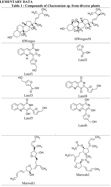
Table 1 : Compounds of Chaetomium sp. from diverse plants