How to cite:
Vierlia WV, Wulandari LR, Sujuti H, Effendi M (2018) Rho- Kinase Inhibitor Y-27632 Decreases the Thickness of Trabecular Meshwork in Juvenile Rats Model Injected with Sodium Hyaluronate. J. Trop. Life. Science 8 (2): 187 – 191. *Corresponding author:
Wino Vrieda Vierlia
Department of Ophthalmology, Faculty of Medicine Universitas Brawijaya
Jalan Veteran, Malang, Indonesia 65145 E-mail: [email protected]
VOL. 8, NO. 2, pp. 187 – 191, April 2018 Submitted February 2018; Revised March 2018; Accepted April 2018
Rho- Kinase Inhibitor Y-27632 Decreases the Thickness of Trabecular Meshwork in Juvenile Rats
Model Injected with Sodium Hyaluronate
Wino Vrieda Vierlia *, Lely Retno Wulandari, Hidayat Sujuti, Ma’sum Effendi Department of Ophthalmology, Faculty of Medicine, Universitas Brawijaya, Malang, Indonesia
ABSTRACT
Most glaucoma drugs lower the intraocular pressure (IOP) by decreasing the aqueous humor production and in-creasing the outflow through uveoscleral pathway. None of these drugs work mainly on inin-creasing outflow through the trabecular pathway. Consequently, the experiment to develop glaucoma drugs directly target at the trabecular outflow pathway is highly required. The purpose of this study is to reveal the effect of rho-kinase inhibitor Y-27632 on the thickness of trabecular meshwork in juvenile rats model injected with sodium hyaluronate. This study was an experimental study with posttest only control group design. Twenty-four rats were included in this study. Each eye of the rat would be considered as one sample. Samples were divided into 6 groups, negative control group, positive control I group with intracameral sodium hyaluronate injection, positive control II group with topical Y-27632 10 mM, and three experimental groups with intracameral injection of sodium hyaluronate and Y-Y-27632 10-1 mM, 1 mM, and 10 mM respectively. After the procedures all rats were sacrificed and enucleated. Trabecular mesh-work tissue was stained with Hematoxilene-Eosin and evaluated under 400× microscopic magnification. Quantitative measurements were taken using computerized image analysis with dot slide program. There were significant statistic differences among the positive control I group and the experimental groups (p-value < 0.05) as well as the positive control II group and the experimental groups (p-value < 0.05). The highest mean of decreasing trabecular meshwork thickness was noted in the group given by sodium hyaluronate and Y-27632 10 mM with value of 118.42 µm. There was decreasing thickness of trabecular meshwork due to the effect of rho- kinase inhibitor Y-27632 in juvenile rats injected with sodium hyaluronate.
Keywords: Y-27632, trabecular meshwork, sodium hyaluronate INTRODUCTION
Primary open angle glaucoma (POAG) is one of the leading cause of visual impairment worldwide. In most cases the aqueous humor secretion remains normal in glaucoma and the main risk factors related to the disease are the elevation of Intraocular Pressure (IOP) and high levels of aqueous humor outflow resistance. Despite the precise underlying mechanism regarding the increasing outflow resistance in POAG remains unclear, most con-sensus stated that the basic resistance located in trabec-ular meshwork (TM) outflow pathway. Presently, most glaucoma medications decrease the IOP by targeting the aqueous humor formation and uveoscleral pathway re-
sistance. Even so, none of the first line medication ther-apy option obtainable for patients with trabecular mesh-work outflow pathway resistance. There are three im-portant structures of the trabecular meshwork related to aqueous humor outflow regulation, extracellular matrix in the juxtacanalicular region, inner wall of Schlemm’s canal and its lumen [1, 2, 3].
Rho-kinase specific inhibitor has been shown to induce inhibition of actomyosin activity and modulate the tra-becular meshwork and Schlemm’s canal morphology. Y -27632 is noted as one of those protein inhibitors. Honjo (2001) concluded that Y-27632 had reduced the intraoc-ular pressure significantly in rabbits. Cellintraoc-ular changes of trabecular meshwork and ciliary muscle relaxation were considered to be the underlying mechanism [4, 5, 6].
Y-27632 has been used widely in experimental re-search both in vivo and in vitro in adults. This fact en-courages our study to evaluate the morphology of tra-becular meshwork in juvenile age. Benozzi (2002) stated that sodium hyaluronate injection had increased the density of extracellular matrix and decreased the diame-ter of flow channels [7].
The purpose of this study is to evaluate the effect of rho-kinase inhibitor (Y-27632) on trabecular meshwork thickness in juvenile rat model with sodium hyaluronate injection.
MATERIALS AND METHOD
Animals sampling
This study was conducted in Physiology Laboratory of Faculty of Medicine Universitas Brawijaya Malang from July 2012 to October 2012. Twenty-four juvenile Wistar rats aged 4 – 6 weeks with 100 ± 50 gr in weight were included in this study.
Preparation of the eyes
Samples were divided into six groups, first group as negative control without any treatment, second group as positive control I group received only intracameral sodium hyaluronate injection, and third to fifth groups given intracameral sodium hyaluronate injection and topical rho-kinase inhibitor (Y-27632) with concentra-tion of 0.1 mM, 1 mM, and 10 mM respectively. The sixth group considered as positive control II group re-ceived only topical rho-kinase inhibitor (Y-27632) with 10 mM concentration. The second to fifth group were anesthetized with ketamine hydrochloride (50 mg/kg) (Ketamine-hameln 50 mg/mL, Hameln pharmaceuti-cals, Germany) administered intraperitoneally. Fifteen µL of sodium hyaluronate (BBTVisc 1.5%, Bohus Bio-Tech) was injected into one eye of anesthe-tized rats us-ing a syrus-inge and a 30-gauge needle. The eyes were fo-cused under a surgical microscope (DECA-21, Inami & Co.Ltd, Model-0940SD, made in Japan) with coaxial light. The needle moved through the corneoscleral lim-bus to the anterior chamber with the bevel down. When the tip of the bevel reached the anterior chamber, the
liquid progressively increased the chamber’s depth, sep-arating the needle from the iris and avoiding needle– lens contact. Applications were made slowly but using a force sufficient to just empty the syringe content. Antibiotic eye ointment was applied after injection.
Application of 1 drop of 0.5% proparacaine hydro-chloride were performed 24 hours later on each eye. The second group was then received no other treatment. The third to fifth group were given topical rho-kinase inhib-itor (Y-27632) with concentration of 0.1 mM, 1 mM, dan 10 mM respectively. Each treatment was administered to the central cornea as four 3-ml drops at intervals of 30 seconds, with blinking prevented between and after the last drops. The sixth group was only treated with Y-27632 10 mM topically. All animals were sacri-ficed 3 hours after treatment.
Eyes were enucleated immediately after sacrificing, then subdivided at the equator into an anterior and pos-terior segment. The eyes were fixed in 4% parafor-mal-dehyde (PFA). The anterior segments were subdi-vided into quadrants after careful removal of the lens. From all quadrants 1 – 2 mm wide specimens including trabecu-lar meshwork and Schlemm’s canal were pre-pared and the specimens were embedded in paraffin.
Histological analysis
The specimens were stained with Hematoxilene-eo-sin and histologically analyzed uHematoxilene-eo-sing light microscopic with 400× magnification. Photographs of the micros-copic sections were taken and analyzed using automated image and dot slide program. In the meridional sections, measurements were taken of the thickness of the trabec-ular meshwork. Measurements were taken four times and the average value was stated in µm.
Statistics
Data were analyzed using independent t-tests, one-way ANOVA, and multiple comparison test with Least Significance Difference (LSD). P-value < 0.05 was con-sidered to be statistically significant.
RESULTS AND DISCUSSION
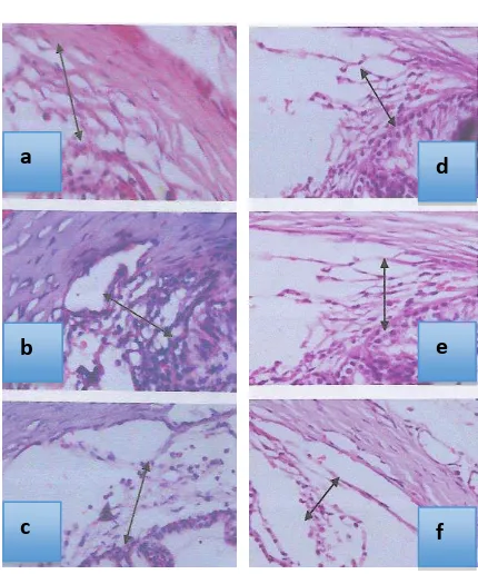
Trabecular meshwork appeared as a fenestrated tis-sue containing multilayer lamella and endothelial cells. The appearance of trabecular meshwork in control groups and the decreasing thickness of trabecular mesh-work along with the increasing concentration of rho-ki-nase inhibitor (Y-27632) were seen in Figure 1.
Figure 1. Trabecular meshwork thickness (arrows) in HE stain-ing under 400× microscopic magnification (Note: A. Negative control group. B. Positive control I group. C. Positive control II group. D. Y-27632 10-1 mM group. E. Y-27632 1mM group. F. Y-27632 10mM group) Table 1. The average of trabecular meshwork thickness in all groups were stated in Table 1.
One-way ANOVA test showed the differences of trabecular meshwork thickness among positive I control group and treatment groups (p < 0.05). The results were then analyzed using LSD as multiple comparisons test and the significant differences among all groups were noted (p < 0.05) (Table 2).
One-way ANOVA test showed the differences of trabec-ular meshwork thickness among positive control II group and treatment groups (p < 0.05). The results were then analyzed using LSD as multiple comparisons test and the significant differences among all groups were noted (p < 0.05) (Table 3).
Table 2. Results of LSD test between positive I control group and treatment groups
Variables Comparison groups p-value
Trabecular Table 3. Results of LSD test between positive control ii group
and treatment groups
Variables Comparison groups p-value
Trabecular In this study, measurements were performed using a computerized dot-slide program and done by one ob-server repeated 4 times and then taken on average in every sample. Instrumentation in a reliable and objective measurement would provide a good internal validity. Randomization was also performed on sample selection so that all rats in this study were drawn at random ac-cording to the inclusion criteria. There were no samples that died or were excluded from this study. The exist-ence of mortality or drop out sample will reduce the in-ternal validity of the study. The use of control groups in experimental research will support the external validity. There were two positive control groups and one negative control group used in this study. Given all the reasoning above it might be assumed that the validity of the re-search is quite good.
Trabecular meshwork tissue is recognized to have the highest responsibility in regulating aqueous humor outflow as conventional pathway. Trabecular meshwork consists of three zones, the uveal meshwork, corneoscle-ral meshwork, and juxtacanalicular connective tissue. Trabecular meshwork tissue is considered to obtain reg-ulatory mechanism as it also has smooth-muscle-like properties. The actomyosin system has contributed to the regulation of contraction as well as relaxation of the tissue [8].
Rho-kinase is identified to be involved in various physiological roles in correlation with cytoskeletal sys-tem such as the cell morphology, motility, and smooth muscle contraction. This protein is also expressed in the
a d
b e
trabecular meshwork tissue. Y-27632 is currently identi-fied as specific inhibitor of the rho-protein kinase [9].
The concentration of Y-27632 used in this study based on the previous experiment. Honjo (2001) con-ducted the study with Y-27632 chosen concentration of 10 and 100 mM to provide the desired concentration with assumption that only 1% of the drug penetrated intracamerally in approximately 125 µL rabbit anterior chamber volume. As much as 15 µL anterior chamber volume in rats were included in this study therefore we used 1/
10 concentration of the previous study
concentra-tion [6].
The increasing intraocular pressure effect caused by sodium hyaluronate injection were noted after 24 hours and lasted for 5 days in the study conducted by Benozzi (2002). We also used the same 24 hours after injection to perform topical application of Y-27632. Honjo (2001) also stated that Y-27632 decreased the intraocular pres-sure after 30 minutes and reached the peak after 1 to 3 hours of application. In this study the evaluation was conducted after 3 hours of Y-27632 administration in order to gain optimal effect [6, 7].
The lowest average thickness of the trabecular meshwork was noted in the group receiving Y-27632 10 mM. Zhang (2008) stated that trabecular meshwork has components that resemble smooth muscle cells and other proteins related to cytoskeletal processes. Giving agents that cause an increase in actin depolymerization and decreased the interactions among cells in the trabec-ular meshwork may induce relaxation of the trabectrabec-ular meshwork so that the tissue is reduced in thickness. Rao (2001) stated that the Y-27632 increased permeability and changes in cell adhesion as well as relaxation of the trabecular meshwork. Germano (2015) identified that the precise mechanism of Rho-kinase inhibitor in in-creasing the conventional outflow of aqueous humor is not utterly understood. It was assumed that the cytoskel-eton system is disturbed by the inhibition of rho-kinase activity. The main effect of this process would be the reducing of trabecular meshwork contractility that lead to the improvement of aqueous humor outflow [8, 10, 11].
There were significant differences among the groups treated with various concentrations of Y-27632 and the control groups. The data showed that the con-centration range of Y-27632 have given enough morpho-logical changes with significant difference. The most substantial changes found in the group receiving Y-27632 with the highest concentration of 10 mM.
We may conclude from this study that there was a tendency of a greater morphological changes of trabec-ular meshwork along with the increasing concentration of Y-27632 given. However, we might not be able to de-termine the amount of optimal concentration of Y-27632 to provide the most effective morphological changes of trabecular meshwork. It might be affected by the changes that were increased linearly in a row with the increasing concentration given. So further research is still needed with higher concentration and various range in order to evaluate the effectiveness of optimal concen-tration of Y-27632 to morphological changes in the tra-becular meshwork. The mechanism of tratra-becular mesh-work morphological changes due to administration of Y-27632 are not directly evaluated in this study. This is because the research has not included the evaluation of molecular changes yet. However, some related re-searches might have explained the possible underlying mechanisms. It is expected that this study is able to be a basic research for further evaluation of molecular changes in the trabecular meshwork due to Y-27632.
CONCLUSION
It can be concluded from this study that the admin-istration of rho-kinase inhibitor (Y-27632) on juvenile rats model injected with sodium hyaluronate had re-sulted in the decreasing thickness of trabecular mesh-work. We hoped that this agent might be an alternative medical therapy in lowering intraocular pressure related to the aqueous humor facilitation through the trabecular meshwork modulation. Further research is needed to evaluate the effectiveness of rho-kinase inhibitor (Y-27632) on the role of trabecular meshwork modulation in molecular basis.
ACKNOWLEDGMENT
The authors would like to thank Prof. Dr. dr. Loeki Enggar Fitri, M.Kes., Sp.ParK. from the Faculty of Med-icine Universitas Brawijaya sincerely for her constructive suggestion and correction of this manuscript.
REFERENCES
1. Kim JW (2017) Comparative study of the effects of trabecu-lar meshwork outflow drugs on the permeability and nitric oxide production in trabecular meshwork cells. Korean Jour-nal of Ophthalmology 31 (5): 452 – 459. doi: 10.3341/kjo. 2017.0020.
tigative Ophthalmology and Visual Science 57 (14): 6197 – 6209. doi:10.1167/iovs.16-20189.
3. Yu WY, Sheridan C, Grierson I et al. (2011) Progenitors for the corneal endothelium and trabecular meshwork: A pottial source for personalized stem cell therapy in corneal en-dothelial diseases and glaucoma. Journal of Biomedicine and Biotechnology 2011 (2011). doi:10.1155/412743.
4. Tian B, Gabelt BT, Geiger B, Kaufman PL (2009) The role of the actomyosin system in regulating trabecular fluid out-flow. Experimental Eye Research 88 (4): 713 – 717. doi: 10.1016/j.exer.2008.08.008.
5. Ishizaki T, Uehata M, Tamechika I et al. (2000) Pharmaco-logical properties of Y-27632, a specific inhibitor of rho-as-sociated kinases. Molecular Pharmacology 57 (5): 976 – 983. 6. Honjo M (2001) Effects of Rho-Associated Protein Kinase Inhibitor Y-27632 on Intraocular Pressure and Outflow Fa-cility. Investigative Ophthalmology and Visual Science 42 (1): 137 – 144.
7. Benozzi J, Nahum LP, Campanelli JL, Rosenstein RE. (2002) Effect of Hyaluronic Acid on Intraocular Pressure in Rats.
Investigative Ophthalmology and Visual Science 43 (7): 2196 – 2200.
8. Germano RAS, Finzi S, Challa P, Juniro RS (2015) Rho ki-nase inhibitors for glaucoma treatment-review. Arquivos Brasileiros de Oftalmologia 78 (6): 388 – 391. doi: 10.5935/0004-2749.20150103.
9. Moon JS, Kim HK, Shin SY (2017) Effects of the rho-kinase inhibitor Y-27632 on extraocular muscle surgery in rabbits. Journal of Ophthalmology 2017 (2017). doi:10.1155/8653130.
10. Zhang M, Maddala R, Rao PV (2008) Novel molecular in-sights into RhoA GTPase-induced resistance to aqueous hu-mor outflow through the trabecular meshwork. American Journal of Physiology Cell Physiology 295 (5): 1057 – 1070. doi: 10.1152/ajpcell.00481.2007.
11. Rao PV, Deng PF, Kumar J, Epstein DL (2001) Modulation of aqueous humor outflow facility by the rho-kinase-specific inhibitor Y-27632. Investigative Ophthalmology and Visual Science 42 (5): 1029 – 1037.