DISERTASI
PENGARUH ASAP ROKOK TERHADAP GSH,
MMP-8 DAN MMP-9 PADA PATOGENESIS
EMFISEMA
JONI ANWAR
PROGRAM PASCASARJANA
UNIVERSITAS AIRLANGGA
SURABAYA
DISERTASI
PENGARUH ASAP ROKOK TERHADAP GSH, MMP-8 DAN MMP-9 PADA PATOGENESIS EMFISEMA
JONI ANWAR
PROGRAM PASCASARJANA
PENGARUH ASAP ROKOK TERHADAP GSH, MMP-8 DAN MMM-9 PADA PATOGENESIS EMFISEMA
DISERTASI
Untuk memperoleh Gelar Doktor
Dalam Program Studi Ilmu Kedokteran pada program pasca Sarjana Universitas
Airlangga dan Dipertahankan Dihadapan Panitia Ujian Doktor Terbuka
Pada hari : Kamis
Tanggal : 29 September 2011
Pukul : 1000 - 1200
Oleh : JONI ANWAR NIM : 090315289.D
PROGRAM PASCASARJANA
LEMBAR PENGESAHAN
DESERTASI INI TELAH DISETUJUI PADA 5 OKTOBER 2011
Oleh:
Promotor
NIP: 19470810.197412.1.002
Prof. Dr. H. Muhammad Amin, dr., SpP(K)
Ko – promotor I Ko – promotor II
Prof. Soetjipto, dr., MS., Ph.D Prof.
Telah diuji pada Ujian Tertutup
Tanggal 24 Agustus 2011
Panitia penguji Disertasi
Ketua : Prof. Dr. Yoes Prijatna D, dr., MSc., SpPar
Anggota :
1. Prof. Dr. H. Muhammad Amin, dr., SpP (K)
2. Prof. Soetjipto,dr.,MS.,Ph.D
3. Prof. Retno Handajani, dr, M.S.,Ph
4. Prof. Purnomo Suryohudoyo, dr., SpBK
5. Prof. H Kuntoro, dr., MPH., Dr,PH
6. Prof. Dr. Ida Bagus Ngurah Rai, dr., Sp.P
7. Dr. Sunaryo, dr., MS.,MSc
Ditetapkan denganSurat keputusan Rektor
Universitas Airlangga
Nomor : 1998/H3/KR/2001
RINGKASAN
PENGARUH ASAP ROKOK TERHADAP GSH, MMP-8 DAN MMP-9 PADA PATOGENESIS EMFISEMA
Emfisema ada lah kelainan paru yang di tandai ol eh adanya pe lebaran abnormal dan permanen ruang udara distal dari bronkiolus terminalis disertai destruksi dinding alveoli dan t anpa f ibrosis yang n yata. E mfisema da n bronkitis kr onik tidak dimasukkan kedalam P POK ka rena em fisema a dalah kelainan anatomis s edangkan bronkitis kr onik a dalah diagnosis kl inis. S elain i tu ke duanya t idak m enggambarkan kelainan faal paru pada saluran napas (Senior dan Shapiro, 1998; PDPI, 2011).
Menurut pr ediksi W HO (World Health Organization) tahun 2020, pr evalensi
PPOK aka n meningkat dari peringkat ke-12 menjadi peringkat ke-5 untuk penyakit yang p aling s ering di temukan sedangkan mortalitas j uga m eningkat d ari pe nyebab kematian ke-6 menjadi penyebab kematian ke-3 di dunia (Barnes dkk, 2003; Barnes, 2004).
Tidak terdapat data yang akurat di Indonesia tentang prevalensi PPOK ini, tetapi pada Survei Kesehatan Rumah Tangga (SKRT) DEPKES RI tahun 1986 didapatkan penyakit a sma, br onkitis kr onik da n e mfisema menduduki pe ringkat ke -10 s ebagai penyebab kematian. S urvei K esehatan R umah T angga t ahun 1992 m enunjukkan angka ke matian ka rena pe nyakit a sma b ronkial, br onkitis kr onik da n e mfisema menduduki peringkat ke-7 dari 10 pe nyebab kematian di Indonesia dan pada SKRT 1995 pe nyakit s istem pe rnapasan m erupakan pe nyebab k ematian k e-2 da ri 10 penyakit penyebab kematian (Litbangkes, 1999).
Paru merupakan organ yang paling sering terpapar dengan oksidan atau radikal bebas terutama asap rokok yang dapat menyebabkan kerusakan pada protein, lipid dan DNA. Asap r okok t erdiri da ri f ase gas d an fase s olid. F ase gas m engandung 1015
oksidan ( terutama alkil da n pe roksil) dan 500 – 1000 ppm ni trit oksida ( NO) pada setiap hisapan sedangkan fase solid atau fase tar mengandung 1018 radikal bebas per
gram yang t erdiri da ri r adikal hi droksil da n hidrogen pe roksida (H2O2), metal
Anion superoksida akan dinetralisir oleh superoksid dismutase (SOD) menjadi hidrogen p eroksida ( H2O2). H idrogen p eroksida akan dinetralisir ol eh katalase da n
glutation pe roksidase d engan ba ntuan glutation ( GSH). Glutation m erupakan pertahanan antioksidan ut ama pa da pa ru. Penurunan ka dar G SH menyebabkan sebahagian H2O2
Untuk m elihat pe ngaruh a sap r okok t erhadap pa ru di lakukan pe nelitian observasional m enggunakan de sain pot ong l intang (cross-sectional) t erhadap 18
orang perokok dengan emfisema sebagai kelompok studi dan 19 orang perokok tanpa emfisema sebagai kelompok kontrol. Variabel yang diteliti adalah GSH, MMP-8 dan MMP-9 dari sputum yang induksi dengan salin hipertonik.
tidak dapat di netralisir da n be bas m asuk kedalam s el da n mendegradasi ikatan IkB de ngan faktor t ranskripsi Nuclear-factor kappa B (NFkB)
sehingga NFKB yang bebas akan memasuki nukleus, merangsang faktor transkripsi, meningkatkan produksi mediator pro-inflamasi dan menyebabkan terjadi rekruitmen netrofil da n m akrofag. N etrofil da n makrofag m emproduksi e nzim matriks metaloproteinase yang d apat m endegradasi matriks eks trasel. Netrofil m emproduksi enzim matriks metaloprotein (MMP) yaitu MMP-8 dan MMP-9 sedangkan makrofag memproduksi M MP-9 yang m erupakan enzim el astolitik utama yang da pat mendegradasi matriks ekstrasel.
Pada penelitian ini didapatkan penurunan tidak bermakna kadar GSH (12.37 ± 4.10) µM pada kelompok perokok dengan emfisema dibandingkan (16.28 ± 9.47) µM pada kelompok perokok tanpa emfisema (p>0.05)
Dari penelitian ini di dapatkan aktivitas M MP-8 yang lebih tinggi pada perokok dengan emfisema (1024.87 ± 488.49) ng/mL dibandingkan t anpa emfisema (263.50 ± 60.45) ng/mL dengan perbedaan yang sangat bermakna (p<0.002).
Pada pe nelitian i ni di dapatkan a ktivitas M MP-9 yang lebih tinggi p ada perokok dengan emfisema (2080.47 ± 1712.94) ng/mL dibandingkan perokok tanpa emfisema (749.92 ± 331.64) ng/mL dengan perbedaan yang bermakna (p<0.05)
SUMMARY
THE IMPACTS OF CIGARETTE SMOKE TO GSH, MMP-8 AND MMP-9 IN PATHOGENESIS OF EMPHYSEMA
Emphysema is defined as a condition of the lung characterized by abnormal and permanent enlargement of distal airspaces from the terminal bronchiole, accompanied by the destruction of their walls, and without obvious fibrosis.
The pr evalence and burden of C OPD ar e p rojected to increase i n the coming decades due t o c ontinued e xposure t o C OPD risk f actors a nd t he c hanging a ge structure of the world,
There i s no accurate data i n Indonesia about the prevalence of C OPD but according to National Health Survey by Indonesian Ministry of Health in 1986 were found that Asthma, Chronic Bronchitis and Emphysema is the number t en cause of death among 10 c ommon caused of deaths. National Health Survey i n 1992 were found that the disease of respiratory s ystem is t he second common caused of death among 10 common caused of deaths.
s population. The Global Burden of Disease Study has projected that COPD, which ranked sixth as the caused of death in 1990, will become the third leading caused of death worldwide by 2020.
The lung is the organ that frequently exposed to oxidants or free radicals which caused the degradation of protein, l ipid, deoxyribo nuc leic a cid ( DNA). Cigarette smoke is the most commonly encountered factors for pulmonary emphysema and the elimination of t his f actors is a n important s tep toward prevention and c ontrol of pulmonary emphysema. Oxidants from cigarette smoke consist of gas and solid phase. Gas phase contains 1015 oxidants (especially alkiyl and peroxyl) and 500–1000 ppm
nitrite oxide (NO). Solid phase contains 1017 free radicals that consist of hydroxyl
radical, hydrogen pe roxide (H2O2), metal-chelator and qui non-semiquinon.
Gluthation system will neutralized hidrogen peroxide (H2O2) into water (H2
Macrophages a nd ne utrophils produces matrix metalloproteinases (MMPs), especially ne utrophyl elastase ( MMP-8) and gelatinase B ( MMP-9) w hich degrade most of matrix extracellular (especially elastin) as t he caus ed of l ung emphysema. Because of direct impacts of s uperoxide a nion a nd h ydrogen pe roxide to the
formation of radicals hydroxyl and indirectly were the increase MMP-8 and MMP-9 activity, that’s importance to study the concentration of GSH, MMP-8 and MMP-9 activity in pathogenesis of emphysema
To study the impa cts of c igarette s moke in the pathogenesis of e mphysema, a cross-sectional analitycal study was conducted to cigarette smokers with emphysema as the study group and cigarette smokers without emphysema as the control group. The study variables were t he con centration of GSH, MMP-8 and MMP-9 activity. The statistical analyses comprised of homogeneity test, normality test, Mann-Whitney test a nd Independent T-test. All pa rticipants are m en, 18 c igarette s mokers with emphysema and 19 cigarette smokers without emphysema.
It was found that the decrease concentration of GSH (12.37 ± 4.10) µM in the study group was lower than the control group (16.28 ± 9.47) µM and the difference was not s ignificant (p>0.05). There w as s ignificant increase of M MP-8 activity (1024.87 ± 488.49) ng/ml in the study group in comparison (263.50 ± 60.45) ng/ml with the c ontrol group ( p<0,05). There w as s ignificant increase activity of MMP -9 (2080.47 ± 1712.94) ng/ml in comparison with the control group (749.92 ± 331.64) ng/ml.
Observing the results of this study, it could be concluded that there was not significant decrease of the concentration of GSH that could caused accumulation of H2O2, recruitments and activation of macrophages and neutrophils to produced more
ABSTRACT
THE IMPACTS OF CIGARETTE SMOKE TO GSH, MMP-8 AND MMP-9 IN PATHOGENESIS OF EMPHYSEMA
Background : Emphysema is a condition of lung characterized by abnormal and permanent enlargement of distal airspaces from terminal bronchiole, accompanied by destruction of their walls, and without obvious fibrosis. The aims of this study is to know the impa cts of c igarette s moke i n pa thogenesis of emphysema. A cross-sectional analytical study was conducted to cigarette smokers with emphysema as the study group and cigarette smokers without emphysema as the control group.
Methods : The study variables were GSH concentration, MMP-8 a nd M MP-9
activity. The s tatistical ana lyses c omprised of hom ogeneity, nor mality and independent T test. All participants are men, 18 patients are cigarette smokers with emphysema as a the study group and 19 cigarette smokers without emphysema as a the control group. The examination of GSH, MMP-8 and MMP-9 were using ELISA method.
Results : It was found that the decrease concentration of GSH (12.37 ± 4.10) µM in the study group was l ower t han the control gr oup (16.28 ± 9.47) µM and t he difference was not significant (p>0.05). There was a s ignificant increase of MMP-8 activity (1024.87 ± 488.49) ng/ml in the study group in comparison (263.50 ± 60.45) ng/ml with the control group (p<0.05). There was an increase activity of MMP-9 ( 2080.47 ± 1712.94) ng/ml in comparison with the control group (749.92 ± 331.64) ng/ml, the difference was significant (p<0.05).
Conclusion : There i s not s ignificant decreased of t he concentration of GS H and there are significant increased of MMP-8 and MMP-9 activity which degrade most of elastin in matrix extracellular as the cause lung emphysema.
DAFTAR ISI
PENETAPAN PANITIA PENGUJI ………v
UCAPAN TERIMA KASIH ………...vi
RINGKASAN ………..………..ix
DAFTAR SINGKATAN DAN LAMPIRAN ……….………xxiv
BAB 1 PENDAHULUAN ……….………1
1.1Latar belakang masalah ………...………….………..1
1.2Rumusan masalah ………...6
1.3Tujuan penelitian ………6
1.4Tujuan umum ………..…..………...6
1.5Tujuan khusus ……….6
1.6Manfaat penelitian ………..………7
BAB 2 TINJAUAN PUSTAKA ………..………...7
2,1 Organ paru……….………7
2.1.1 Anatomi Paru………,,,…………..9
2.1.2 Matriks ekstrasel (MES) ……..……….……10
2.1.2.1 Kolagen ……….10
2.1.3.2 Makrofag Alveolar ………...14
2.1.3.3 Limfosit T ………...15
2.2.1.2 Polusi Udara dan lingkungan Kerja ………..……. .…….19
2.2.1.3 Faktor Genetik . ………...19
2.2.1.4 Faktor Lain ………...……….20
2.2.2 Patogenesis PPOK………..22
2.2.2.1 Inflamasi………..23
2.2.2.3 Protease - antiprotease ………24
2.3 Emfisema……….….………24
2.3.1 Patogenesis Emfisema….………..………….……….24
2.3.1.1 Elastase - Antielastase ………...………25
2.3.1.2 Oksidan – Antioksidan .………..……….……..25
2.3.1.2.1 Oksidan atau Radikal bebas ………...………26
2.2.1.2.1.1 Produksi Radikal Bebas ………..……….26
2.3.1.2.1.1 Dampak terhadap inflamasi………..……….30
2.3.1.2.1,2 Dampak oksidan terhadap Netrofil ..………...….………31
2.3.1.2.1.3 Dampakoksidan terhadap Makrofag Alveolar.. ………...31
2.3.1.2.1.4 Dampak Negatif Oksidan ……….………..…...32
2.3.1.2.1.5 Dampak Negatif Terhadap terhadap Membran sel . ………...33
2.3.1.2.1.6 Dampak Negatif Terhadap DNA ……….34
2.3.1.2.1.7 Dampak Negatif Terhadap Protein ……….……….34
2.3.1.2.1.8 Dampak Positif Oksidan ………...……...35
2.3.1.2.2 Antioksidan ………..……..35
2.3.1.2.2.1 Mekanisme Kerja Antioksidan ………...36
2.3.1.2.2.1.1 Antioksidan Pencegah ……….……...……..37
2.3.1.2.2.1.2 Antioksidan Pemutus Rantai ……….………...37
2.3.1.2.2.1.3 Superoksid Dismutase ………...….…...39
2.3.1.2.2.1.4 Glutation ……….…...41
2.3.1.2.2.1.5 Glutation Peroksidase ……….….…….42
2.3.1.2.2.1.6 Vitamin E ………..………….…..44
2.3.1.2.2.1.7 Vitamin C ……….……….44
2.3.1.2.2.1.8 Vitamin A ………..……...45
2.3.1.2.2.1.8 Seruloplasmin……….45
2.3.1.2.2.2 Pertahanan Sel ………...45
2.3.1.2.2.2.1 Pertahanan Intrasel ………...46
2.3.1.2.2.2.2 Pertahanan Pada Membran Sel ………..……...46
2.3.1.2.2.2.3 Pertahanan Ekstrasel ………...46
2.3.1.3 Protease- Antiprotease ………...47
2.3.1.3.1 Klasifikasi Protease ………48
2.3.1.3.1.1 Matriks Metaloproteinase ………49
2.3.1.3.1.1.1 Struktur MMP ………...50
2.3.1.3.1.2 Kolagenase ……….………...51
2.3.1.3.1.2.1 Human Kolagenase ………..….………...52
2.3.1.3.1.2.2 Kolagenase Netrofil (MMP-8) ……….52
2.3.1.3.1.3 Gelatinase ………...52
2.3.1.3.1.3.1 Gelatinase A (MMP-2) ………...………...53
2.3.1.3.1.3.2 Gelatinase B (MMP-9)………...53
2.3.1.3.1.3.3 Makrofag Elastase (MMP-12) ………..………54
2.3.2 Diagnosis Emfisema ..………...55
2.3.2.1 Gambaran Klinis ………55
2.3.2.1.1 Anamnesis ……….….55
2.3.2.1.2 Pemeriksaan Fisik ………....55
2.3.2.2 Pemeriksaan Penunjang………...56
2.3.2.2.1 Pemeriksaan Rutin………57
2.3.2.2.2 Pemeriksaan Faal paru….……….58
2.3.2.2.3 Pemeriksaan Radiologi ………58
2.3.2.3.1 Pemeriksaan Faal Paru …….…….…….……….59
2.3.2.3.2 Uji-latih Kardiopulmoner ….…….…….……….59
2.3.2.3.3 Uji Provakasi Pronkus ……….…….………...60
2.3.2.3.4 Uji Kortikosteroid ……….…….………..60
2.3.2.3.5 Analisis Gas Darah……….…….……….60
2.3.2.3.6 Pemeriksaan CT-scan Toraks ….…….………60
2.3.2.3.7 Pemeriksaan Elektrokardiografi .………….………61
2.3.2.3.8 Pemeriksaan Ekhokardiografi….……….61
2.3.2.3.9 Pemeriksaan Bakteriologi ………..……….……….61
2.3.2.3.10 Pemeriksaan Kadar Alfa-1 Antitripsin .……….61
2.3.2.3.11 Induksi Sputum ………..61
BAB 3 KERANGKA KONSEPTUAL DAN HIPOTESIS 3.1. Kerangka konseptual Penelitian …...……….….……….63
3.2. Keterangan kerangka konseptual………..64
3.3. Hipotesis ………..………...65
BAB 4 METODEPENELITIAN 4.1 Jenis dan rancangan penelitian ………..…………..67
4.2 Populasi ………...67
4.2.1 Besar sampel ……….68
4.2.1 Tehnik pengambilan sampel ……….68
4.2.2 Variabel penelitian………..69
4.2.4 Kriteria Penerimaan ………...………...69
4.2.5 Kriteria Penolakan ………70
4.2.6 Kriteria Drop-Out ………...70
4.2.7 Definisi Operasional ……….71
4.3. Bahan penelitian ……….72
4.4. Instrumen penelitian ………...72
4.5. Lokasi dan waktu penelitian ………...73
4.6. Prosedur pengambilan dan pengumpulan data ………73
4.7. Tempat Penelitian ………76
4.8. Cara pengolahan data ………..76
4.9. Kerangka Alur Penelitian ..………..77
BAB 5 HASIL PENELITIAN DAN ANALISIS DATA 5.1 Distribusi menurut kelompok umur………..78
5.2 Distribusi menurut jenis pekerjaan…..………..79
5.3 Jumlah rokok yang dihisap setiap hari………..80
5.4 Distribusi lama merokok (tahun) ………..82
5.5 Jumlah rokok yang dihisap setiap hari dikalikan lama merokok……..…82
5.6 Hasil pemeriksaan laboratorium....………84
5.7 Hasil pemeriksaan densitas paru dengan CT-scan toraks……….85
BAB 6 PEMBAHASAN
6.1.1 Hasil penelitian GSH sputum....……….…………92
6.1.2 Hasil penelitian MMP-8 sputum.….……….………….95
6.1.3 Hasil penelitian MMP-9 sputum.….……….………….96
6.1.4 Hasil temuan baru ………..98
6.1.5 Keterbatasan penelitian ……….99
BAB 7 KESIMPULAN DAN SARAN 7.1 Kesimpulan ………..………...…100
7.2 Saran ……….………….100
DAFTAR TABEL
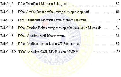
Tabel 5.1.1 Tabel Distribusi Menurut Umur………..79
Tabel 5.2 Tabel Distribusi Menurut Pekerjaan………....80
Tabel 5.3 Tabel Jumlah batang rokok yang dihisap setiap hari………...81
Tabel 5.4 Tabel Distribusi Menurut Lama Merokok (tahun)………...82
Tabel 5.5 Tabel Jumlah Rokok yang dihisap dikalikan lama Merokok………...83
Tabel 5.6 Tabel Analisis hasil laboratorium …….………..………...84
Tabel 5.7 Tabel Analisis pemeriksaan CT-Scan toraks ……….85
DAFTAR GAMBAR
Gambar 1. Kerangka Konseptual Penelitian….…..………63
Gambar 2. Bagan Rancangan Kerja………...……….76
Gambar 3. Kalibrator dan Spirometri………..………..114
Gambar 4. Pemeriksaan Spirometri………..……….115
Gambar 5. Alat Nebuliser………..………116
NAC mengandung sistein yang diperlukan untuk pembentukan GSH untuk
menetralisir H2O2 yang dapat memecah ikatan NFkB dengan IkB. Sistein juga dapat
