A Controlled Trial of Daily Left Prefrontal
Cortex TMS for Treating Depression*
Mark S. George, Ziad Nahas, Monica Molloy, Andrew M. Speer,
Nicholas C. Oliver, Xing-Bao Li, George W. Arana,
S. Craig Risch, and James C. Ballenger
Background: Transcranial magnetic stimulation (TMS) is
a new technology for noninvasively stimulating the brain. Several studies have suggested that daily stimulation of the left prefrontal cortex with TMS for 2 weeks has probable antidepressant effects. We conducted a parallel-design, double-masked, sham-controlled study to address whether 2 weeks of daily TMS over the left prefrontal cortex has antidepressant activity greater than sham.
Methods: Thirty medication-free adult outpatients with
nonpsychotic, major depressive (n5 21) or bipolar (n5
9) (depressed phase) disorder who were in a current major depression (Hamilton Rating Scale for Depression [HRSD] 21-item score of.18) were treated each weekday for 2 weeks. Subjects were randomly assigned to receive either daily active (20 subjects) or sham (10 subjects) stimulation. Additionally, the 20 active subjects were equally divided between slower (5 Hz) and faster (20 Hz) frequency treatment. Antidepressant response was defined as greater than a 50% improvement in the baseline HRSD.
Results: Active TMS resulted in significantly more
re-sponders (9/20) than did sham (0/10) (x256.42, p,.01). The number of responders did not differ significantly between the two active cells (3/10 faster and 6/10 slower). Expressed as a percent change from baseline, active TMS subjects had significantly greater improvement on the Beck Depression Inventory as well as the Hamilton Anx-iety Rating Scale than did those who received sham.
Conclusions: Daily left prefrontal TMS for 2 weeks
significantly reduced depression symptoms greater than did sham. The two forms of active TMS treatment did not differ significantly. Biol Psychiatry 2000;48:962–970
© 2000 Society of Biological Psychiatry
Key Words: Transcranial magnetic stimulation, depres-sion, prefrontal cortex, treatments, mood, emotion, clinical trials
*See accompanying Editorial, in this issue.
Introduction
T
ranscranial magnetic stimulation (TMS) is a new method for noninvasively stimulating the brain (George and Belmaker 2000; George et al 1999a). A brief but powerful electric current is passed through a small coil of wires on the scalp. This generates a powerful but local magnetic field, which passes unimpeded through the skull and induces a weaker focal electric current in the brain (Barker et al 1985; Roth et al 1991; Saypol et al 1991). The highly localized TMS magnetic field typically has a strength of about 1–1.5 T (or 30,000 times the earth’s magnetic field, or about the same intensity as the static magnetic field used in clinical magnetic reonance imaging [MRI]) (Bohning 2000). Although different coil designs allow for a focal or more diffuse stimulation, current technology is not able to stimulate deep brain structures directly. Transcranial magnetic stimulation can be per-formed in outpatient laboratory settings and, unlike elec-troconvulsive therapy (ECT), does not cause a seizure or require anesthesia. Subjects usually notice no adverse effects except for occasional mild headache and discom-fort at the site of the stimulation. Recent technologic advances led to the development of magnetic stimulators that could repeatedly stimulate faster than once per second (1 Hz). This, by convention, is called repetitive transcra-nial magnetic stimulation (rTMS). There is some evidence from work in animals (Post et al 1997) and humans (Pascual-Leone et al 1991, 1994) that stimulation at different frequencies may have divergent and even antag-onistic effects on neuronal activity (Kimbrell et al 1999; Wassermann et al 1998), with higher frequencies exciting the brain and slower frequencies inhibiting activity.Clinical depressions are very common, with one out of five Americans having an episode during their life (Kessler et al 1994). Many depressions can be treated
From the Brain Stimulation Laboratory, Department of Psychiatry (MSG, ZN, MM, NCO, X-BL, SCR, JCB) and Departments of Radiology (MSG) and Neurology (MSG), Medical University of South Carolina, and the Department of Psychi-atry, Ralph H. Johnson Veterans Affairs Hospital (MSG, ZN, GWA), Charles-ton, South Carolina; Biological Psychiatry Branch, National Institute of Mental Health, Bethesda, Maryland (AMS); and the Department of Psychiatry, Shangdong Medical University, Jinan, China (X-BL).
Address reprint requests to Mark S. George, M.D., Director, Functional Neuroim-aging Division, Psychiatry, Associate Professor of Psychiatry, Radiology and Neurology, Medical University of South Carolina, Department of Radiology, 171 Ashley Avenue, Charleston SC 29425.
Received May 5, 2000; revised August 24, 2000; accepted August 24, 2000.
effectively, but as many as 50 – 60% have incomplete recovery or a significant side effect burden with current treatments. The pathophysiology of depression is incom-pletely understood, yet certain neurotransmitters such as serotonin, norepinephrine, and dopamine are likely to be involved. Additionally, specific brain regions such as the dorsolateral prefrontal cortex (DLPFC) (George et al 1994) and deeper limbic areas have been shown to be functioning abnormally on brain imaging studies of de-pressed subjects (for reviews, see George et al 1998a; Ketter et al 1996).
The search for better treatments combined with emerg-ing knowledge of mood-regulatemerg-ing circuits prompted ex-ploratory work with TMS in depression. Initially, re-searchers used a round coil with nonfocal stimulation over the vertex, with inconclusive results (Grisaru et al 1994; Hoflich et al 1993; Kolbinger et al 1995). In 1994, George and Wassermann (1994) reasoned that focal stimulation with rTMS over the left DLPFC might have antidepressant effects. This thinking was based on abnormalities found in functional neuroimaging studies of depression, as well as from studies showing that changes in prefrontal cortex activity predict response to ECT (Nobler et al 1994). Initial open trials (Figiel et al 1998; George et al 1995) and then crossover designs (George et al 1997; Pascual-Leone et al 1996) have shown a significant antidepressant effect of left prefrontal TMS.
Building on these earlier studies, we initiated a sham-controlled parallel-design trial of left prefrontal cortex rTMS in antidepressant medication–free depressed adults. Additionally, to better understand whether the frequency of stimulation was important in this putative antidepres-sant effect we used two different stimulation frequencies with theoretically divergent brain effects.
Methods and Materials
Subjects
Thirty-two depressed adults enrolled in a 2-week double-masked sham-controlled trial. (Here we use the term mask in the same way that the older term blind was used in studies.) After full explanation of the procedures, all subjects signed a written informed consent approved by the Medical University of South Carolina Institutional Review Board and the U.S. Food and Drug Administration (FDA) Devices Section. They were randomly assigned to either sham treatment (n510) or active treatment (n520) (with two different active treatment frequencies, faster [20 Hz] or slower [5 Hz]).
Subjects were free of antidepressant medications for at least 2 weeks before study entry, although three bipolar patients re-quired ongoing mood stabilizers or benzodiazapines for anxiety. Applicants who were partially responding to their medications and who would likely significantly worsen with a medication taper were not enrolled. Other exclusionary criteria were a
substantial risk of suicide during the trial, the presence of psychosis, or a history of seizures or major head trauma.
Subjects underwent an MRI scan of the brain at the beginning and end of the study (Nahas et al, in press-a) and had three regional cerebral blood flow single photon emission computed tomography (SPECT) scans—at baseline, during the fifth rTMS session, and 3 days after completion of the trial before restarting medications (Nahas et al 1998b; Teneback et al 1999).
TMS Procedure
A trained medical doctor (AMS, ZN, or MSG) delivered TMS using a Cadwell (Kennewick, WA) Magnetic Stimulator equipped with a water-cooled figure eight–shaped coil. Subjects were seated upright in a comfortable chair and had their eyes open during TMS. Because of the potential for interaction between the subject and the nonmasked researcher administering TMS, subjects were informed at study entry that there would be limited interaction during treatment sessions.
On the initial treatment visit, we determined motor threshold (MT) at rest of the contralateral (right) abductor pollicus brevis (APB) muscle as described previously (Pridmore et al 1998). The left prefrontal cortex stimulation site was defined as the location 5 cm rostral and in a parasagittal plane from the site of maximal APB stimulation. Each morning we asked subjects about poten-tial events that could have changed the MT (medications, sleep deprivation) and quickly rechecked the MT and adjusted the daily dose accordingly. This recalculated MT never changed more than 5% from the baseline level.
Subjects were randomly assigned to receive stimulation over 20 min each weekday morning for 2 weeks at 20 Hz (2 sec on and 28 off, repeat every 30 sec) (faster), 5 Hz (8 sec on and 22 off, repeat every 30 sec) (slower), or sham. Within the sham treatment group, we again randomized the stimulation frequency (5 or 20 Hz, equally divided among the 10 subjects). During sham stimulation we raised the lateral wing of the figure eight coil 45° off the head with the edge of the medial wing of the coil still touching the scalp. The subject therefore received peripheral stimulation of skin and scalp muscles, but significant magnetic fields did not pass through the skull and affect the brain (Bohning et al 1997; Lisanby et al 1998). With the flat water-cooled coil, this results in the coil intersection being further from the scalp than with other coils. Subjects received treatment for 10 week-days (2 weeks) for 20 min/day, at 100% MT, with an equal total number of 16,000 stimulations across all cells (e.g., slower, 8 sec35 Hz540 stimuli per train32580 stimuli/min320 min51600 stimuli/day310 days for each cell).
Ratings and Response Classification
1), and at the end of the study (week 2). Trained psychiatric nurses, masked to treatment arm, performed all ratings. Hamilton Rating Scale for Depression scores were used to calculate percent improvement from the beginning to the end of treatment (2 weeks). Subjects who showed$50% improvement at 2 weeks from baseline were classified as responders.
Mask Breaking and Follow-Up
After the 2-week study, subjects were counseled to restart antidepressant medications based on a thorough past medication history. Exit interviews showed that in no case did error or behavior on the part of the person administering TMS compro-mise the mask.
Statistical Analyses
PRIMARY OUTCOME VARIABLE. Differences in the di-chotomous outcome measure of response (50% or more reduc-tion in HRSD-21 from study baseline) were assessed using x2
tests between groups (active and sham) and then between the two active subgroups (faster and slower).
SECONDARY OUTCOME VARIABLES. Other ratings (HARS, CGI, MMSE) were analyzed for differences by group (active and sham) as change from the baseline using two-group analysis of variance (ANOVA). In an exploratory attempt to determine if there were specific depression symptoms that contributed to the differences between active cells and sham, total and individual HRSD-21 and HARS items were expressed as percent change from baseline and were analyzed for differ-ences within the active group using one-way ANOVA. Within the sham group, only two items reached significance, but this is confounded by the smaller cell size and a true comparison between groups of the within-group changes over time is not justified.
Results
Subjects
Two subjects who had been randomized to receive active slower TMS did not tolerate the procedure and dropped
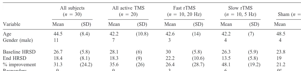
out after less than three treatments. They were excluded from final analysis. Thus, included in the final analysis were 30 subjects who completed all treatments and who met DSM-IV criteria for either major unipolar depression (21 subjects) or bipolar depression, depressive phase (nine). The baseline HRSD means and SDs by group were 23.8 (4.1), sham, and 28.1 (6.1), active (30 [5.8], faster subgroup, and 26.3 [5.9], slower subgroup) (Table 1).
Although failure to respond to other antidepressant medications was not an explicit entry criteria, this cohort was largely treatment resistant and had been ill many months before starting this trial. The average number of years since the first diagnosis of depression in this group was 18 (12.5 SD). The average length of time in the current index episode of depression was also quite high as a group (21.6 months [21.8 SD]). Seven subjects had previously been treated with ECT and had not responded (one), responded and had suffered side effects that pre-cluded future use (four), or chose to try TMS before the next ECT course (two).
Side Effects
Except for two subjects, all subjects enrolled completed the study with no unexpected side effects. Two subjects (one man, both in the active slower cell) with average MTs (60% and 70% of machine output) elected to stop the study because of the pain of stimulation. One subject tried for 2 consecutive days, whereas the other decided to stop after only 2 min of the first session. Ten subjects reported mild headaches following at least one session (beginning immediately after to 3 hours after rTMS), which were relieved by acetaminophen. The total number of headaches posttreatment was 18, out of 300 treatment sessions, for an incidence of 6%. No subjects reported problems with memory or attention. There were no seizures (a possible adverse effect of rTMS). The bipolar patients did not have a mania induced (George et al 1998b), and a woman in her
Table 1. Subject Demographics by Group with Key Outcome Variables
Variable
All subjects (n530)
All active TMS (n520)
Fast rTMS (n510, 20 Hz)
Slow rTMS
(n510, 5 Hz) Sham (n510)
Mean (SD) Mean (SD) Mean (SD) Mean (SD) Mean (SD)
Age 44.5 (8.4) 42.2 (10.8) 42.6 (14) 42.2 (7) 48.5 (8)
Gender (male) 11 7 3 4 4
Baseline HRSD 26.7 (5.8) 28.1 (6) 30 (5.8) 26.3 (5.9) 23.8 (4.1) End HRSD 18.4 (8.1) 18.3 (9) 22.2 (10.6) 13.5 (5.8) 19 (6) % improvement 31.3 (24.2) 35.6 (26) 26.4 (28.7) 48.1 (19.2) 21.2 (16)
Responders 9 9 3 6 0a
TMS, transcranial magnetic stimulation; rTMS, repetitive TMS; HRSD, Hamilton Rating Scale for Depression.
a
second trimester of pregnancy had no problems (Nahas et al 1999) (pregnancy was not an exclusion in this trial, although it has been made an exclusionary criteria for more recent trials approved by the FDA).
Clinical Response
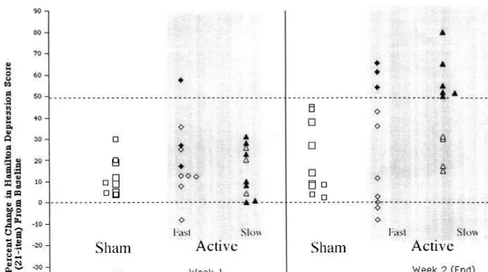
PRIMARY OUTCOME VARIABLES. Comparing Re-sponders Active rTMS resulted in significantly more responders than did sham (9/20 responders active [three faster, six slower], 0/10 sham) (x2 5 6.42, p 5 .013). Response was defined as.50% improvement in HRSD score at week 2 (end) from baseline ([(HRSD end 2 HRSD baseline)/HRSD baseline] 3 2100) (Figure 1).
Comparing Percent Change of HSRD from Entry at 2 Weeks A two-group (active and sham) ANOVA (active, 35.6% [0.26 SD], and sham, 21.2% [0.16 SD]) did not find a significant difference. However, a three-group (sham, faster, and slower) ANOVA (slower, 44.9%, and faster 26.4%) revealed a trend to a group effect (ANOVA F5 3.06, p5.06), which was driven by slower (5 Hz) rTMS as superior to sham (Fisher protected least significant difference p 5 .03) but not statistically different from 20-Hz rTMS (Fisher p 5.08).
SECONDARY OUTCOME VARIABLES. These are
ex-pressed as two-group ANOVAa of percent change from baseline.
The HARS was significantly reduced from baseline in the active TMS group relative to sham ( ANOVA critical
difference 14.5, p 5 .03). The Hamilton anxiety scores and SDs by group were 37.6 (6.3), end 27.3 (7.4), 26% (18) improvement (active entry), and 32.8 (7.8), end 29.5 (8.6), 9.6% (16) improvement (sham entry).
Beck Depression Inventory scores significantly im-proved more with active TMS than with sham (ANOVA mean difference 27.7, p 5 .05). The Beck Depression scores and SDs by group were 30.5 (11.2), end 17.1 (12), 40.2% (28.9) improvement (active entry), and 27.1 (11), end 23.2 (14.1), 12.5% (43) improvement (sham entry).
Clinical Global Improvement scores, expressed as a percentage change from baseline, did not significantly differ across the groups. The CGI scores and SDs by group were 4.5 (0.8), end 3.1 (1.2), 27% (32) improvement (active entry), and 4.2 (0.7), end 3.8 (1.5), 11% (33) improvement (sham entry).
Mini-Mental State scores improved across all cells over the 2 weeks, although significantly more in the sham arm than the active TMS (ANOVA critical difference 8.9, p5 .02). There was an unequal baseline distribution of MMSE scores across cells, with lower initial scores in the sham group. The MMSE scores (30-point scale) and SDs by group were 29.0 (1.4), end 29.3 (1.2), 1.4% (4.8) improve-ment (active entry), and 25.5 (4.8), end 28.3 (4.3), 12.5% (18) improvement (sham entry).
HRSD ITEM ANALYSIS. In an effort to understand
what aspects of depression or anxiety TMS might be influencing, and in what time domains, HRSD-21 and HARS scores were analyzed for differences by time
(baseline, week 1, and week 2) as change from baseline using ANOVA within the active group.
Table 2 presents the scores on the total and then each item of the HRSD-21 in the active TMS group. The total HRSD score was significantly reduced at week 1 and the reduction became robust at the end of TMS (week 2). Within the subitems, the depressed mood score and the insomnia score were reduced significantly at week 1, with all other items showing improvement at week 2.
Table 3 presents the scores on the total and individual items of the HARS in the active TMS group. The total HARS score reduced significantly at week 1 and the reduction became robust at the end of TMS (week 2). The insomnia score also reduced significantly at week 1, with other changes occurring at week 2.
CHARACTERISTICS OF TMS RESPONDERS. We per-formed post hoc exploratory analyses to discern if there were characteristics that distinguished TMS responders from nonresponders within the active group. Eight of the nine responders were women, whereas only four of 11 women in the total 20-subject active group were nonre-sponders, thus hinting at but not definitely revealing a gender bias, with better relative response in women. Although not statistically significant, responders also had a shorter duration of the current episode of depression (8.7 months [11.6 SD]) than nonresponders (21.6 months [17 SD]).
Finally, there were relatively more bipolar depressed patients in the responder group than in the study at large (5/9 [55%] of the responders were bipolar, and 2/11 [18%] of the nonresponders were bipolar). Bipolar subjects were roughly equally distributed across the three cells (2/10 placebo, 4/10 slower, 3/10 faster) There was no difference in age or other variables such as the severity of the initial depression or anxiety scores. Analyses of baseline SPECT (Teneback et al 1999) and MRI (Kozel et al 2000) studies found differences in responders relative to nonresponders. Specifically, TMS responders at baseline showed in-creased orbitofrontal cortex activity that improved follow-ing treatment, and TMS responders had a shorter distance into the prefrontal cortex than did nonresponders.
Discussion
In this double-masked parallel-design study, 2 weeks of TMS applied over the left prefrontal cortex was associated with a significantly greater antidepressant response (as measured by the number of subjects who responded) than sham. There were no significant differences between the two active TMS treatment arms (faster or slower TMS). Assessing antidepressant response as percent improve-ment from baseline failed to show significant group differences using the HSRD-21. However, analysis of other continuous variables in general found changes con-sistent with an antidepressant response. For example, active TMS, relative to sham, significantly reduced the Hamilton Anxiety Rating Scale score and the Beck De-pression Inventory score. An exploratory analysis of individual HRSD items found a significant improvement in sleep during the first week of treatment, with other items improving during the second week. There was no apparent subgroup of HRSD items that was driving the clinical effect; rather, many items showed significant improvement.
Therefore, this study in general adds support to the theory that repeated daily administration of left prefrontal TMS over 2 weeks has antidepressant effects greater than sham. However, this antidepressant medication–free
dou-Table 2. Scores on Each Item and Total of HRSD-21 in the Active TMS Group over Time
Time
Depressed mood 3.1060.70 2.4561.00a 2.29 61.19a Insomnia (early) 1.5260.75 0.8060.83a 0.71
60.78a Work and activities 2.7160.96 2.0060.92 2.0561.02a Retardation 0.9060.70 0.7560.72 0.4360.68a Anxiety—somatic 1.8160.75 1.7560.79 1.2960.78a Somatic symptoms
(general)
1.7160.56 1.5060.69 1.1960.87a
Hypochondriasis 1.6261.07 1.2561.12 1.0060.77a Weight loss 0.4860.81 0.2060.52 0.0060.00a Paranoid symptoms 0.5260.68 0.3060.57 0.1460.36a Total HSRD 27.4365.34 22.1566.56b 18.76
66.65b
HRSD-21, 21-item Hamilton Rating Scale for Depression; TMS, transcranial magnetic stimulation.
ap,.05, relative to baseline. bp,.01, relative to baseline.
Table 3. Scores on Each Item and Total of the HARS over Time within the Active Group
Time Intellectual 3.0560.86 2.5060.89 2.0060.97a Somatic (sensory) 2.0061.05 1.7560.72 1.3960.70a Gastrointestinal 2.6760.97 2.3060.92 1.9460.87a Symptoms, autonomic 2.1060.83 1.5060.69 1.7260.67a Symptoms, total
HARS
36.1065.70 30.4564.89b 28.26 65.59b
HARS, Hamilton Anxiety Rating Scale.
ble-masked parallel study has significant limitations that are important for proper interpretation of results.
Like many of the TMS studies to date, this trial lasted only 2 weeks. Most antidepressant medication trials re-quire 6 – 8 weeks of treatment before significant effects are seen. This trial was designed in 1996, when there were fewer published TMS studies (with two suggesting signif-icant effects after only 5 days of treatment [Figiel et al 1998; Pascual-Leone et al 1996]). At the time of study design, there was thus little justification for a prolonged duration of a sham phase or exposure of medication-free subjects to an experimental treatment. In the current study, consistent with our earlier study (George et al 1997) and others, clinically significant effects were only apparent at 2 weeks, with only one subject meeting response criteria at the end of 1 week (Figure 1). Thus, this study, like others (Berman et al 2000; Klein et al 1999; Padberg et al 1998), although finding a clinical antidepressant effect greater than sham, does not replicate a study by Pascual-Leone and colleagues (Pascual-Leone et al 1996), who found .50% reduction in HRSD scores after only 1 week of left prefrontal stimulation in 17 psychotically depressed sub-jects (see also Loo et al 1999). The literature in general has now failed to replicate the earlier studies showing acute effects at 1 week, and clearly, longer treatment studies are indicated, similar to those used in studying antidepressant medications or electroconvulsive therapy.
This study also could not assess how long the effects of TMS might persist in responders. Individuals with sub-stantial prior histories of depression require maintenance antidepressant treatment or relapse will occur (Kupfer et al 1992). We thus immediately placed patients on mainte-nance antidepressant medications following TMS. We are in the process of a follow-up study to determine long-term outcome and safety in this cohort. We are also conducting a trial of maintenance TMS intermittently over 1 year in bipolar depressed subjects who have responded to TMS in an acute-phase double-masked study.
The small sample size allowed for testing of only very large effects and is also vulnerable to effects that originate from unequal initial distribution of subjects. There were no significant differences between groups in baseline vari-ables (except the MMSE), and they were matched on age and gender. It is thus not apparent if differences in subjects between cells might have biased the results.
Another important limitation in this study, as in all previous trials with TMS in depression, is that the person administering the TMS was aware of the treatment arm of the subject. Thus, there was a potential for explicit or unconscious bias in the interaction between the subject and the person administering TMS. We explicitly told subjects beforehand that we would limit interaction and discussions during treatment sessions. Exit questioning
found no examples of failure of the mask, and we had an established routine to diminish contact between the subject and the person administering TMS, with ratings performed by trained masked raters. We are currently exploring more automated methods of TMS delivery involving sham coils where there is never any contact between the subject and anyone who knows their treatment status. We employed a sham method of angling the coil 45° off of the head, with one edge touching the scalp. With the water-cooled Cadwell coil in this study, with extra lucite casing, this places the TMS coils far from the scalp and angles the bulk of stimulation away from the brain. It is thought that this reduces the amount of magnetic stimulation, and induced electric current, to less than 25% of the active condition. For more discussion of this important area, see Loo et al (2000).
Despite these shortcomings, this study supports the theory that left prefrontal TMS has antidepressant effects and confirms other double-masked studies that have been published in the time since this study was designed and performed (Berman et al 2000; Klein et al 1999). The effect size in terms of percent change in HRSD score is larger than in the previous crossover design study by our group, where, due to concerns about safety, we stimulated with 20 Hz frequency at only 80% of MT (George et al 1995). We currently hypothesize that stimulation at higher intensity may have more robust effects (Kozel et al 2000), and formal clinical testing is needed. A recent interleaved TMS/functional MRI study by our group in 7 healthy adults has shown that left prefrontal stimulation has intensity-dependent effects both locally and in the right prefrontal cortex. Stimulation at or above MT produced significantly more activation than did stimulation below the MT (80% MT) (Nahas et al 2000). In addition, detailed measurements of MRI scans in subjects in conjunction with this clinical trial revealed that nonresponders had larger prefrontal cortex distance than responders (Kozel et al 2000). Since the TMS magnetic field declines logarith-mically with distance from the coil, small variations in the distance to cortex may play a role in TMS antidepressant response. Thus our findings in this clinical trial, combined with results from interleaved TMS/functional MRI, high-light the importance of the distance into the brain from the scalp and the intensity relative to MT. We currently are testing in a pilot clinical trial the hypothesis that higher intensities of stimulation, calibrated after measuring the distance into the prefrontal cortex for each subject, may carry greater antidepressant effect, particularly in elderly depressed subjects with prefrontal atrophy (Coffey et al 1989).
applications. However, other side effects of the treatment were minimal. Seven subjects in the world have experi-enced seizures during TMS. With more rigorous safety guidelines, there have been no seizures since 1996. With all other parameters constant, stimulation at lower fre-quencies is less likely to cause a seizure. Thus the failure to find in this study a large significant difference in the antidepressant effect of faster TMS, relative to slower, may be important for future studies and the safety of the field. These results should encourage more antidepressant studies using slower frequency TMS, which is less likely to cause seizures.
The mechanisms by which TMS over the left prefrontal cortex might improve depression are unclear. Studies showing no changes in prolactin or electroencephalo-graphic activity during or immediately after TMS indicate that TMS is not causing subclinical seizures. Prefrontal TMS, and not TMS at other sites, produces changes in peripheral thyroid hormone, hinting that the hypothalamus is secondarily affected (George et al 1996; confirmed by Zwanzger et al 1999). Functional neuroimaging studies in healthy adults during left prefrontal TMS have shown that prefrontal stimulation has remote effects at the cingulate gyrus, insula, amygdala, and other regions long hypothe-sized to be important in controlling mood (George et al 1999b). A SPECT study performed in subjects in this trial found that, after 2 weeks, TMS responders had reduced activity in the anterior cingulate cortex (Teneback et al 1999). This finding of changes in cingulate activity in TMS antidepressant responders is consistent with cingu-late changes seen following sleep deprivation (Austin et al 1992; Ebert et al 1994; Wu and Bunney 1990) and fluoxetine treatment (Mayberg et al 1997). Klaus Ebmeier and colleagues in Scotland have performed a similar SPECT study in depressed patients undergoing a TMS clinical trial and have also found that TMS reduces cingulate activity in responders (K. Ebmeier, personal communication and unpublished data). Presumably, TMS over the prefrontal cortex causes changes in resting activ-ity locally and in connected deeper regions. Further work with TMS and imaging is needed to address these theories. There are a host of unexplored variables in understand-ing TMS effects. Longer duration studies with larger samples are needed to more adequately address the issue of whether slower TMS is more effective than faster, or whether different stimulation frequencies have differential antidepressant effects depending on the baseline state of the subject (Kimbrell et al 1999). The optimal prefrontal location for stimulation is also not clear. Another parallel double-masked study has found antidepressant effects with right prefrontal stimulation (Klein et al 1999). Our recent interleaved TMS/functional MRI study revealed profound bilateral changes in blood flow following
unilat-eral stimulation, calling into question whether the effects of unilateral prefrontal stimulation are ipsilateral, con-tralateral, or bilateral (Nahas et al 2000). Even ignoring the issue of whether to stimulate the right or the left prefrontal cortex, it is not known if more precise localiza-tion of the coil, perhaps guided by an MRI, would produce more robust clinical effects. Similarly, the stimulation intensity, total number, and dosing schedule are as yet unexamined in terms of their antidepressant effects. Ex-ploring these variables in clinical trials would be lengthy and expensive. Recent advances with functional imaging and with TMS in animals offer the promise of rapid exploration of parameters that may guide clinical trials.
In conclusion, this double-masked controlled trial sug-gests that 2 weeks of daily left prefrontal TMS has antidepressant effects greater than sham, with no signifi-cant difference between faster and slower frequency stim-ulation. Further studies are needed to explore which variables are important in this effect, and to test whether TMS may have a place in clinical practice.
This study was funded by a Young Investigator Grant to Dr. George from the National Alliance for Research on Schizophrenia and Depression.
This work was performed at the Medical University of South Carolina and presented in abstract form at the 1998 annual meeting of the American Psychiatric Association (interim analysis) (Nahas et al 1998a) and the 1999 annual meeting of the Society of Biological Psychiatry (complete analysis) (Nahas et al, in press-b).
References
Austin MP, Dougall N, Ross M, Murray C, O’Carroll RE, Moffoot A, et al (1992): Single photon emission tomography with 99 mTc-exametazime in major depression and the pattern of brain activity underlying the psychotic/neurotic continuum. J Affect Disord 26:31– 43.
Barker AT, Jalinous R, Freeston IL (1985): Non-invasive mag-netic stimulation of the human motor cortex. Lancet 1:1106 – 1107.
Berman RM, Narasimhan M, Sanacora G, Miano AP, Hoffman RE, Hu XS, et al (2000): A randomized clinical trial of repetitive transcranial magnetic stimulation in the treatment of major depression. Biol Psychiatry 47:332–337.
Bohning DE (2000): Introduction and overview of TMS physics. In: George MS, Belmaker RH, editors. Transcranial
Mag-netic Stimulation in Neuropsychiatry. Washington, DC:
American Psychiatric Press, 13– 44.
Bohning DE, Pecheny AP, Epstein CM, Vincent DJ, Dannels WR, George MS (1997): Mapping transcranial magnetic stimulation (TMS) fields in vivo with MRI. Neuroreport 8:2535–2538.
Coffey CE, Fiegel GS, Djang WT (1989): Subcortical white matter hyperintensities on magnetic resonance imaging: Clin-ical and neuroanatomClin-ical correlates of depressed elderly.
J Clin Neuropsychiatry Clin Neurosci 1:135–144.
limbic flow and total sleep deprivation in major depression with melancholia. Psychiatry Res 55:101–109.
Endicott J, Spitzer RL (1978): A diagnostic interview: The schedule for affective disorders and schizophrenia. Arch Gen
Psychiatry 35:837– 844.
Figiel GS, Epstein C, McDonald WM, Amazon-Leece J, Figiel L, Saldivia A, Glover S (1998): The use of rapid rate transcranial magnetic stimulation (rTMS) in refractory de-pressed patients. J Neuropsychiatry Clin Neurosci 10:20 –25. Folstein MF, Folstein SE, McHugh PR (1975): “Mini-mental State.” A practical method for grading the cognitive state of patients for the clinician. J Psychiatr Res 12:189 –198. George MS, Belmaker RH, editors (2000): Transcranial
Mag-netic Stimulation in Neuropsychiatry. Washington, DC:
American Psychiatric Press.
George MS, Ketter TA, Post RM (1994): Prefrontal cortex dysfunction in clinical depression. Depression 2:59 –72. George MS, Lisanby SH, Sackeim HA (1999a): Transcranial
magnetic stimulation: Applications in neuropsychiatry. Arch
Gen Psychiatry 56:300 –311.
George MS, Nahas Z, Speer AM, Kimbrell TA, Wassermann EM, Teneback C, et al (1998a): Transcranial magnetic stim-ulation—a new method for investigating the neuroanatomy of depression. In: Ebert D, Ebmeier K, editors. New Models for
Depression. New York: Karger, 94 –122.
George MS, Speer AM, Molloy M, Nahas Z, Teneback CC, Risch SC, et al (1998b): Low frequency daily left prefrontal rTMS improves mood in bipolar depression: A placebo-controlled case report. Hum Psychopharmacol 13:271–275. George MS, Stallings LE, Speer AM, Spicer KM, Vincent DJ,
Bohning DE, et al (1999b): Prefrontal repetitive transcranial magnetic stimulation (rTMS) changes relative perfusion lo-cally and remotely. Hum Psychopharmacol 14:161–170. George MS, Wassermann EM (1994): Rapid-rate transcranial
magnetic stimulation (rTMS) and ECT. Convuls Ther 10: 251–253.
George MS, Wassermann EM, Williams W, Steppel J, Pascual-Leone A, Basser P, et al (1996): Changes in mood and hormone levels after rapid-rate transcranial magnetic stimu-lation of the prefrontal cortex. J Neuropsychiatry Clin
Neu-rosci 8:172–180.
George MS, Wassermann EM, Williams WA, Callahan A, Ketter TA, Basser P, et al (1995): Daily repetitive transcranial magnetic stimulation (rTMS) improves mood in depression.
Neuroreport 6:1853–1856.
George MS, Wassermann EM, Williams WE, Kimbrell TA, Little JT, Hallett M, Post RM (1997): Mood improvements following daily left prefrontal repetitive transcranial magnetic stimulation in patients with depression: A placebo-controlled crossover trial. Am J Psychiatry 154:1752–1756.
Grisaru N, Yarovslavsky U, Abarbanel J, Lamberg T, Belmaker RH (1994): Transcranial magnetic stimulation in depression and schizophrenia. Eur Neuropsychopharmacol 4:287–288. Hamilton M (1960): A rating scale for depression. J Neurol
Neurosurg Psychiatry 12:56 – 62.
Hoflich G, Kasper S, Hufnagel A, Ruhrmann S, Moller HJ (1993): Application of transcranial magnetic stimulation in treatment of drug-resistant major depression—a report of two cases. Hum Psychopharmacol 8:361–365.
Kessler RC, McGonagle KA, Zhao S, Nelson CB, Hughes M, Eshleman S, et al (1994): Lifetime and 12-month prevalence of DSM-III-R psychiatric disorders in the United States. Results from the National Comorbidity Survey. Arch Gen
Psychiatry 51:8 –19.
Ketter TA, George MS, Kimbrell TA, Benson BE, Post RM (1996): Functional brain imaging, limbic function, and affec-tive disorders. Neuroscientist 2:55– 65.
Kimbrell TA, Little JT, Dunn RT, Frye MA, Greenberg BD, Wassermann EM, et al (1999): Frequency dependence of antidepressant response to left prefrontal repetitive transcra-nial magnetic stimulation (rTMS) as a function of baseline cerebral glucose metabolism. Biol Psychiatry 46:1603–1613. Klein E, Kreinin I, Chistyakov A, Koren D, Mecz L, Marmur S, et al (1999): Therapeutic efficacy of right prefrontal slow repetitive transcranial magnetic stimulation in major depres-sion: A double-blind controlled study. Arch Gen Psychiatry 56:315–320.
Kolbinger HM, Hoflich G, Hufnagel A, Moller H-J, Kasper S (1995): Transcranial magnetic stimulation (TMS) in the treatment of major depression—a pilot study. Hum
Psycho-pharmacol 10:305–310.
Kozel FA, Nahas Z, DeBrux C, Molloy M, Lorberbaum JP, Bohning DE, et al (2000): How the distance from coil to cortex relates to age, motor threshold and possibly the antidepressant response to repetitive transcranial magnetic stimulation. J Neuropsychiatry Clin Neurosci 12:376 –384. Kupfer DJ, Frank E, Perel JM, Cornes C, Mallinger AG, Thase
ME, et al (1992): Five-year outcome for maintenance thera-pies in recurrent depression. Arch Gen Psychiatry 49:769 – 773.
Lisanby SH, Luber B, Schroeder C, Osman M, Finck D, Jalinous R, et al (1998): Intracerebral measurement of rTMS and ECS induced voltage in vivo. Biol Psychiatry 43:100S.
Loo C, Mitchell P, Sachdev P, McDarmont B, Parker G, Gandevia S (1999): Double-blind controlled investigation of transcranial magnetic stimulation for the treatment of resis-tant major depression. Am J Psychiatry 156:946 –948. Loo CK, Taylor JL, Gandevia SC, McDarmont BN, Mitchell PB,
Sachdev PS (2000): Transcranial magnetic stimulation (TMS) in controlled treatment studies: Are some “sham” forms active? Biol Psychiatry 47:325–331.
Mayberg HS, Brannan SK, Mahurin RK, Jerabek PA, Brickman JS, Tekell JL, et al (1997): Cingulate function in depression: A potential predictor of treatment response. Neuroreport 8:1057–1061.
Nahas Z, Bohning DE, Molloy M, Outz JA, Risch SC, George MS (1999): Safety and feasibility of repetitive transcranial magnetic stimulation in the treatment of anxious depression in pregnancy. J Clin Psychiatry 60:50 –52.
Nahas Z, DeBrux C, Lorberbaum JP, Speer AM, Molloy MA, Liberatos C, et al (in press-a): Safety of rTMS: MRI scans before and after 2 weeks of daily left prefrontal rTMS for the treatment of depression. J ECT.
Nahas Z, Lomarev M, Roberts DR, Shastri A, Lorberbaum JP, Vincent DJ, et al (2000): Left prefrontal transcranial magnetic stimulation produces intensity dependent bilateral effects as measured with interleaved BOLD fMRI. Hum Brain Mapp. Nahas Z, Speer AM, Molloy M, Arana GW, Risch SC, George
frequency and intensity in the antidepressant effect of daily left prefrontal rTMS. Biol Psychiatry 43:94S.
Nahas Z, Speer AM, Molloy M, Oliver NC, Arana GW, Ballenger JC, et al (in press-b): Role of stimulation frequency in the antidepressant effect of left prefrontal rTMS. Biol
Psychiatry.
Nahas Z, Stallings LE, Speer AM, Teneback CC, Vincent DJ, Bohning DE, et al (1998b): Perfusion SPECT studies of rTMS on blood flow in health and depression. Biol Psychiatry 43:19S.
Nobler MS, Sackeim HA, Prohovnik I, Moeller JR, Mukherjee S, Schnur DB, et al (1994): Regional cerebral blood flow in mood disorders, III. Treatment and clinical response. Arch
Gen Psychiatry 51:884 – 897.
Padberg F, Haag C, Zwanzger P, Thoma H, Kathmann N, Stubner S, et al (1998): Rapid and slow transcranial magnetic stimulation are equally effective in medication-resistant de-pression: A placebo-controlled study. Abstract presented at the 21st Congress of the Collegium Internationale Neuro-Psychopharmacologicum.
Pascual-Leone A, Gates JR, Dhuna A (1991): Induction of speech arrest and counting errors with rapid-rate transcranial magnetic stimulation. Neurology 41:697–702.
Pascual-Leone A, Rubio B, Pallardo F, Catala MD (1996): Beneficial effect of rapid-rate transcranial magnetic stimula-tion of the left dorsolateral prefrontal cortex in drug-resistant depression. Lancet 348:233–237.
Pascual-Leone A, Valls-Sole J, Wasserman EM, Brasil-Neto JP, Hallett M (1994): Responses to rapid-rate transcranial mag-netic stimulation of the human motor cortex. Brain 117:847– 858.
Post RM, Kimbrell TA, Frye M, George MS, McCann UD, Little JT, et al (1997): Implications of kindling and quenching for the possible frequency dependence of rTMS. CNS Spectrums
Int J Neuropsychiatr Med 2:54 – 60.
Pridmore S, Filho JAF, Nahas Z, Liberatos C, George MS (1998): Motor threshold in transcranial magnetic stimulation: A comparison of a neurophysiological and a visualization of movement method. J ECT 14:25–27.
Roth BJ, Saypol JM, Hallett M, Cohen LG. (1991): A theoretical calculation of the electric field induced in the cortex during magnetic stimulation. Electroencephalogr Clin Neurosci 81: 47–56.
Saypol JM, Roth BJ, Cohen LG, Hallett M (1991): A theoretical comparison of electric and magnetic stimulation of the brain.
Ann Biomed Eng 19:317–328.
Teneback CC, Nahas Z, Speer AM, Molloy M, Stallings LE, Spicer KM, et al (1999): Two weeks of daily left prefrontal rTMS changes prefrontal cortex and paralimbic activity in depression. J Neuropsychiatry Clin Neurosci 11:426 – 435. Wassermann EM, Wedegaertner FR, Ziemann U, George MS,
Chen R (1998): Crossed reduction of motor cortex excitabil-ity by 1 Hz transcranial magnetic stimulation. Neurosci Lett 250:141–144.
Wu JC, Bunney WE (1990): The biological basis of an antide-pressant response to sleep deprivation and relapse: Review and hypothesis. Am J Psychiatry 147:14 –21.