Mycophenolate mofetil treatment reduces cholesterol-induced
atherosclerosis in the rabbit
Freddy Romero, Bernardo Rodrı´guez-Iturbe *, He´ctor Pons, Gustavo Parra,
Yasmir Quiroz, Jaimar Rinco´n, Luisandra Gonza´lez
Department of Immunobiology,Di6ision of Organ Transplantation,Instituto de In6estigaciones Biome´dicas (FUNDACITE-Zulia),
Uni6ersidad del Zulia and the Renal Ser6ice,Hospital Uni6ersitario,Maracaibo, Venezuela
Received 20 April 1999; received in revised form 30 September 1999; accepted 3 November 1999
Abstract
Immunosuppressive therapy has been shown to either improve or, more frequently, enhance the development of atherosclerosis. We tested the effect of mycophenolate mofetil (MMF), an inhibitor of nucleotide synthesis widely used in transplant therapy, in diet-induced atherosclerosis in the rabbit. Two groups (n=10 each) of New Zealand White (NZW) rabbits were fed a 1% cholesterol diet for 12 weeks. One group received MMF (CHOL+MMF group) by gastric gavage (30 mg/kg daily) and the other group (CHOL) received the same volume of saline by the same route. There were no differences in the serum cholesterol (mean values]30 mmol/l in both groups after 2 weeks) or in the triglyceride, blood sugar, total protein, and albumin serum levels and weight gain in both groups of animals. The cholesterol-fed untreated rabbits had atherosclerotic plaques covering 43.9.19SD 16.40% of their thoracic aorta and 41.9922.59% of their abdominal aorta, while the MMF treated group had 18.597.17% and 17.799.71%, respectively (PB0.01). The cholesterol content of the aorta (mg/g) in the cholesterol-fed untreated group was 4.619SD 1.21 in the thoracic aorta and 4.5492.07 in the abdominal aorta, whereas the MMF treated group had and 2.8390.84 and 2.7791.44, respectively (PB0.01). Infiltrating macrophages (RAM 11 positive cells/100 nuclei) in the intimal layer of the aorta were 58.49SD26.16 in the CHOL group and 8.595.51 in the CHOL+MMF group: (PB0.001). CD18 positive cells/100 nuclei were 27.4917.6 in the CHOL group and 5.393.82 in the CHOL+MMF group (PB0.01), and the intima/media ratio was 0.6690.11 in the CHOL group and 0.3090.09 in the MMF treated rabbits (PB0.001). MMF also reduced proliferating smooth muscle cells (HHF35 positive) infiltrating between the macrophages. These results indicate that MMF ameliorates importantly the atherogenic potential of a high cholesterol diet and this effect is associated with a reduction in macrophage and foam cell infiltration and smooth muscle cell proliferation and infiltration. Since chronic treatment with this drug is given routinely in various clinical conditions with relatively minor side effects, consideration may be given to its use as adjuvant therapy in artheriosclerotic cardiovascular disease. © 2000 Elsevier Science Ireland Ltd. All rights reserved.
Keywords:Mycophenolate mofetil in atherosclerosis; Inflammation and atherosclerosis; Macrophages in atherosclerosis
www.elsevier.com/locate/atherosclerosis
1. Introduction
The development of atherosclerosis involves an infl-ammatory reaction with infiltration and activation of immune competent cells [1]. However, suppression of cellular immunity has resulted usually [2 – 5], though not always [6], in enhancement of atherosclerosis in animals fed with a high cholesterol diet. Mycophenolate
mofetil (MMF) is a drug whose active component, mycophenolic acid, inhibits inosine 5%-monophosphate dehydrogenase. Since this enzyme controls the synthesis of guanosine triphosphate, MMF suppresses de novo purine synthesis, thereby exerting a selective and re-versible antiproliferative activity in macrophages and lymphocytes [7]. In addition, MMF also suppresses the expression of adhesion molecules that are critical for the migration of leukocytes from circulation to the tissues [8]. We [9] and others [10] have recently shown that MMF prevents macrophage infiltration in the rem-nant kidney model, and it seemed plausible that similar * Corresponding author. Present address. Apartado Postal 1430,
Maracaibo 4001-A, Venezuela. Tel.: +58-61-519610; fax: + 58-61-524838.
E-mail address:[email protected] (B. Rodrı´guez-Iturbe).
effect could be achieved in the aorta of rabbits fed with a high cholesterol diet. If so, a reduction in atheroscle-rotic disease was a likely consequence because macrophage-derived foam cells are the most important cellular component in the atherosclerotic lesion [11 – 13]. Here we report that MMF ameliorates importantly the atherogenic potential of a high cholesterol diet suppressing macrophage infiltration and smooth muscle cell proliferation. Since chronic treatment with this drug has relatively minor side effects [14,15], these findings suggest therapeutic applications of this drug in hypercholesterolemic conditions with a high risk of potentially fatal cardiovascular complications in man.
2. Methods
2.1. Animals, diet and experimental design
Studies were done in 20 male New Zealand White (NZW) rabbits, which weighed 1.0 – 1.5 kg at the begin-ning of the experiment. After obtaibegin-ning baseline blood samples, they were fed ad libitum a 1% Cholesterol diet for 12 weeks. This diet was prepared dissolving choles-terol (Sigma, St. Louis, MO) in 100% ethanol at a temperature of 60°C, mixing this solution with standard rabbit chow (Purina) and allowing the complete evapo-ration of the ethanol.
The rabbits were divided in two groups: (1) CHOL+ MMF group (n=10) received by gastric gavage 30 mg/kg of MMF (CellCeptR, Roche Pharm. Co) in 0.4 ml of water and (2) CHOL group (n=10) which re-ceived by the same route daily 0.4 ml water. Since the MMF is insoluble in water, the drug was individually prepared as a suspension by vigorous shaking immedi-ately before administration as described in a previous paper [9]. The MMF and vehicle were given as de-scribed daily throughout the 12 experimental weeks.
Blood samples for determination of plasma choles-terol and triglycerides, serum creatinine, blood glucose levels, total protein and albumin concentration, were taken every 2 weeks. Blood chemistries were done by autoanalyzer methodology (Express Plus, CIBA, Corn-ing Diagnostic Corp.). High density lipoprotein (HDL) cholesterol was determined after precipitation of low (LDL) and very-low (VLDL) fractions with phospho-tungstic acid – MgCl2 [16]. The combined VLDL and LDL fraction was calculated as the difference between the total cholesterol and the HDL cholesterol. Rabbits were weighted every 2 weeks.
A separate set of 10 rabbits of similar weight kept for 12 weeks on a standard rabbit chow not supplemented with cholesterol were sacrificed to determine normal values.
2.2. Preparation of tissues
Animals were sacrificed at the end of the 12th week. The aorta was rapidly dissected and cut from the beginning of the aortic arch to the bifurcation of the iliac vessels. Then, thoracic and abdominal segments of the aorta were separated using as a reference the di-aphragm. Aortic rings of about 1 mm width were cut at the initiation of the aortic arch for histologic and immunohistologic analysis. Then, thoracic and abdomi-nal segments of the aorta were open longitudiabdomi-nally and photographed for evaluation of the extension of atherosclerotic plaques. The adventitia was then care-fully separated, and the aortic segments were weighted and used for determination of the total cholesterol content.
2.3. Determination of aortic cholesterol content
Lipids were isolated from the aortic segments as described by Folch et al. [17]. Briefly, tissue was ho-mogenized in a mixture of cholorophorm – methanol 2:1 (v:v) in a final vol. 20 times de mixture volume. Ho-mogenates were centrifuged at 2500 rpm for 15 min and the supernatant was washed in ionic 0.017% MgCl2 solution and then centrifuged for 20 min. Lipids were extracted from the lower layer. Cholesterol was deter-mined in the lipid extract by the method of Zlatkis et al. [18] as follows: 0.1 ml of the lipid extract was reacted with 3.0 ml acetic acid 100% and 2.0 ml ferric chloride solution. This mixture was vigorously agitated at room temperature and absorbance was measured at 560 nm (Shimatzu spectrophotometer model UV2100S, Kyoto, Japan). Calibration curves were prepared dis-solving 100 mg cholesterol (Sigma, St. Louis, MO) in 100 ml glacial acetic acid 100%.
2.4. Extension of plaque formation
Aortic ring sections were fixed in formalin as well as snap frozen in tissue freezing medium (TBS™, Triangle Biomedical Sciences, Durham, NC, USA) and kept at −70°C. Formalin-fixed tissues were stained with hema-toxylin-eosin and Periodic Acid Schiff (PAS). At least three sections from the aorta from each rabbit were analyzed and in each one of them, 15 – 20 different fields were studied using a graduated eye piece for the assessment of the intimal/medial thickness ratio. Re-sults represent the mean obtained in each animal.
All histologic and immunohistologic (see later) stud-ies were done by two investigators (GP, FR) without prior knowledge of the animal group being studied.
2.5. Immunohistologic techniques and antibodies
Immunohistology was done in 4 mm thick frozen
sections by the avidine biotine peroxidase methodology as described in previous communications [20]. The fol-lowing monoclonal antibodies (Mab) were used: Mab anti rabbit macrophages (clone RAM11, concentration 3mg/ml. Accurate Chemical and Scientific Corp.,
West-bury, NY), Mab anti CD54 (clone RR1/1, concentra-tion 25mg/ml; specificity=human and rabbit ICAM-1.
Bender Medsystems, Vienna, Austria), Mab anti rabbit CD18 (clone L13/64, concentration 5 mg/ml;
distribu-tion in rabbit macrophages, neutrophils and platelets. Research Diagnostics Inc. Flanders, NJ), Mab anti muscle cell actin (clone HHF-35, concentration 5 mg/
ml; specificity=alfa actin from skeletal, cardiac and smooth muscle cells and gamma actin from smooth muscle source. Accurate Chemical and Scientific Corp., Westbury, NY, USA) and mouse anti proliferating cell nuclear antigen (PCNA) (clone PC10, concentration 5
mg/ml. Zymed Lab. Inc., San Francisco, CA). Staining
with anti-CD54, anti-CD18 and anti-PCNA Mabs were standardized using rabbit lymph node frozen sections,
while anti-muscle cell actin Mab was standardized in frozen sections of rabbit atherosclerotic plaques (tho-racic aorta). The following negative controls were used: (a) normal rabbit aorta and (b) aorta from rabbits with atherosclerosis in which the antibodies listed above were substituted by a non-pertinent monoclonal anti-body (B1: Mab anti-human B lymphocytes, Coulter Immunology, FL) at a concentration of 5mg/ml
follow-ing the procedures described previously [20].
Positive cells and nuclei were counted and results expressed as the number of positive cells/100 nuclei. Staining for CD54 and HHF35 was graded in an arbitrary scale from 1+ to 4+, depending on the intensity and number of positive cells.
Statistical analysis was done with the help of a commercial statistical package (GraphPad Instat™). Non-parametric tests were used throughout: Wilcoxon (paired samples) and Mann – Whitney (unpaired sam-ples) were used to test differences between the baseline and the 12-week data; Kruskal – Wallis ANOVA tests were used to evaluate jointly the findings in controls and both experimental groups (MMF treated and un-treated). Serial determinations were analyzed by non-parametric Friedman’s repeated measurments test, followed by Dunn’s multiple comparisons post-tests. Correlation between cholesterol content in the aortas and extension of atheromatous plaque distribution was explored with linear regression analysis and with Spear-man’s non-parametric correlation. Results are given as mean9SD.
3. Results
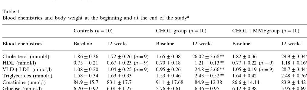
The routine blood chemistries and the body weight of the rabbits at the start and at the end of the experiment are shown in Table 1. There are no differences between
Table 1
Blood chemistries and body weight at the beginning and at the end of the studya
Controls (n=10) CHOL group (n=10) CHOL+MMFgroup (n=10)
Blood chemistries Baseline 12 weeks Baseline 12 weeks Baseline 12 weeks
Cholesterol (mmol/l) 1.8690.36 1.7290.26 (n=9) 1.6590.38 26.0293.68** 1.8290.36 29.993.34**
Triglycerides (mmol/l) 2.4390.52** 1.6490.42 2.4890.76**
Creatinine (mmol/l) 84.9915.7 83.1917.7 91.1917.68 84.9912.38 86.6914.14 83.994.42 6.2090.92 6.0191.27 5.7690.61 6.3690.95 6.1290.98
Glucose (mmol/l) 5.9590.69
56.894.34 61.892.05 59.394.00 55.292.40 52.596.12
Total protein (g/l) 56.392.90
40.094.40
aThere are no significant differences between experimental groups (CHOL, high cholesterol diet; CHOL+MMF, high cholesterol
Fig. 1. Plasma cholesterol levels. Similar hypercholesterolemia was present in the cholesterol-fed animals treated with Mycophenolate mofetil (open circles, interrupted line) and the untreated animals (closed circles, continuous line). Data are mean9SD. Values in both groups after the second week represent a significant increment (PB
0.001) above baseline determinations (B).
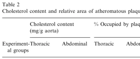
ment with MMF (Table 2). There was a significant correlation between the aortic cholesterol content and the extension of atherosclerotic plaques in the abdomi-nal aorta (r=0.494, P=0.031) and in the thoracic aorta (r=768, PB0.0001). [The later correlation is shown in Fig. 2] deleted.
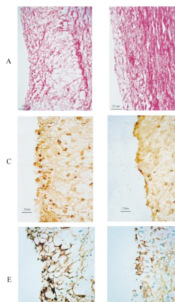
Histological evaluation showed that animals treated with MMF had about half the value of intima/media ratio than the untreated group (Fig. 3, right side). Infiltration of RAM11 positive cells and LFA1 positive cells in the intima was seven times and five times, respectively, more intense in the untreated CHO group than in the CHO+MMF group (Fig. 2, left side). Similarly, intense staining with smooth muscle cell spe-cific actin was present in the untreated rabbits and substantially reduced with MMF. Macrophage infiltra-tion in the media was also higher in the CHOL group (10.69SD6.87 RAM11 positive cells/100 nuclei) than in the CHO+MMF group (0.7690.85, PB0.01). Representative examples of histology are shown in Fig. 3(A – F).
Proliferating cells (PCNA positive cells/100 nuclei) were evaluated in six control rabbits, five cholesterol-fed rabbits untreated with MMF and in five choles-Table 2
Cholesterol content and relative area of atheromatous plaquesa
Cholesterol content % Occupied by plaques (mg/g aorta)
Abdominal
Experiment-Thoracic Thoracic Abdominal al groups
4.5492.07
4.6191.21 41.9922.59
Cholesterol 43.9916.4
Cholesterol 2.8290.84** 2.7791.47** 18.597.17** 17.799.71** +MMF
aNormal values for cholesterol content (mg/g of aortic tissue) in
rabbits with a diet not supplemented with cholesterol (n=10) are 2.1890.35 and 1.4390.15, for thoracic aorta and abdominal aorta, respectively.
**PB0.01, values are mean9SD.
Fig. 2. Effect of MMF on the intensity of macrophage infiltration, expression of adhesion molecule CD18 in the intima and intima/me-dia aortic ratio. Open bars, control rabbits in normal diet; Closed bars, high cholesterol diet; Hatched bars, high cholesterol diet and MMF treatment. Left side, macrophage infiltration indicated by the number of RAM 11 positive cells (n=9 in the untreated group,n=8 in the MMF-treated group and n=8 in the control group) and expression of CD18 adhesion molecule in the aortic intima (n=8 in all groups) are expressed as a ratio of positive cells/100 nuclei. Right side, the size and severity of the atherosclerotic lesions is reflected by the intima/media ratio, which was significantly reduced by MMF treatment. The values found in the untreated hypercholesterolemic rabbits (n=10) ranged from 0.5 to 1.0, which correspond to relatively advanced lesions; in contrast, the intimal/medial ratio in the MMF-treated rabbits (n=8), ranged from 0.1 to 0.4, which represent an early fatty streak consisting of mixed macrophages and smooth muscle cells [13]. All data shown are mean9SD. ***PB0.001 vs. MMF-treated and control groups;8PB0.001 vs control.
the untreated and the MMF-treated rabbits. Control and experimental groups gained weight similarly during the 12 weeks of the experiment. As shown in Table 1, both experimental groups increased significantly (PB
0.01) the levels of total cholesterol, VLDL+LDL, HDL, and triglycerides after 12 weeks of cholesterol supplemented diet. However, total cholesterol increased about 16 times, while HDL and tryglicerides increased 1.5 – 1.6 times. There are no significant differences be-tween the group treated with MMF and the untreated group.
Plasma cholesterol changes are shown in Fig. 1. Levels increased up to the 4th week with the high cholesterol diet and then remained at levels close to 30 mmol/l until the end of the study. There were no significant differences between the CHOL and CHOL+MMF groups.
terol-fed rabbits treated with MMF. They were scarce in the aorta of control rabbits (intima=0.790.51, media=0.791.21) but prominent in cholesterol-fed rabbits (intima=50.298.2, media=16.491.67, PB
0.01 vs. control). Treatment with MMF reduced the number of PCNA positive cells (intima 28.2910.10, media=7.093.81;PB0.05 andPB0.01, respectively, versus the corresponding findings in cholesterol-fed un-treated rabbits).
Intimal expression of CD54 adhesion molecule (graded from 0 to 4+) was higher in the untreated group (3.090.82) and in the MMF treated group (1.9391.25) but this difference did not reach the estab-lished level of significance.
4. Discussion
The cholesterol-fed rabbit is a model frequently used to study the development and modifications in the natural history of atherosclerosis. We administered a 1% cholesterol diet for 12 weeks and observed incre-ments in plasma cholesterol and, to a lesser degree, increments in plasma triglycerides, similar and to those reported in the literature with similar diets [21,22]. No other biochemical abnormality was detected in our serial studies.
Since the early events in the development of atherosclerosis include a local proliferative reaction [1] with leukocyte adhesion and infiltration of lipid-con-taining macrophages beneath the endothelium [13,23 – 25], a drug that could block these events would likely retard or diminish plaque formation. For this purpose we selected MMF because in previous experiments in rats with reduced nephron mass, MMF administration given at the dose and by the route selected here was well tolerated and resulted in a substantial reduction of the renal inflammatory infiltrate [9]. In the present studies only two rabbits presented diarrhea that sub-sided after stopping MMF for 3 days and did not reappear after restarting the drug. The rabbits taking daily MMF and the cholesterol-fed untreated rabbits, had comparable weight gain in the 12 weeks of the studies (Table 1).
Total cholesterol content of the aorta, is a good indirect measure of atherosclerotic severity in choles-terol-fed rabbits and the separation of total cholesterol in the aorta into free and esterified fractions is not superior to total cholesterol alone [26]. In fact, in the present work there was a significant correlation be-tween the aortic cholesterol content and the extension of plaque formation, particularly in the thoracic aorta. The extension of plaque formation was determined using their relative weight in magnified xerox copies of pictures of the aortas. This method combines the meth-ods used by Hata et al. [27] and Araujo et al. [28] and offers acceptable reproducibility.
The size and severity of the atherosclerotic lesions is reflected by the intima/media ratio and this ratio was significantly reduced in the MMF-treated rabbits (Fig. 2, right side and Fig. 3A and B). The values found in the cholesterol-fed untreated group ranged from 0.5 to 1.0, which, in studies of Rosenfeld and Ross [13], correspond to relatively advanced lesions that contain the beginning of a fibrous cap and a core of foam cells [29] with proliferating smooth muscle cells containing lipid laden macrophages [24]. In contrast, the intima/ media ratio in the MMF treated rabbits ranged from 0.1 to 0.4 (Fig. 2) which represent a fatty streak consist-ing of mixed macrophages and smooth muscle cells [13]. It is now recognized that the macrophage-derived foam cells represent the most important cellular compo-nent in the artheriosclerotic lesion [11] [25] [29 – 34]. Therefore, the drastic reduction in these cells in the MMF treated group is a likely explanation for the reduction in cholesterol content and plaque extension in the aorta of rabbits treated with MMF.
In man, long term administration of MMF is well tolerated in chronic conditions such as transplant im-munosuppression and refractory rheumatoid arthritis [14,15]. Our studies, showing an important reduction in atherogenesis and cellular infiltration and proliferation in cholesterol-fed rabbits, are in line with a recent preliminary report that suggested a trend towards re-duction in plaque formation by the subcutaneous ad-ministration of mycophenolic acid [35]. We suggest that MMF may be of value in clinical conditions character-ized by a very high plasma cholesterol level and a high associated risk of ischemic heart disease, such as famil-ial hypercholesterolemia. Indeed, drugs with more sig-nificant side effects and several invasive surgical procedures with a high rate of complications are presently used in the treatment of this condition.
Acknowledgements
Grant support from the Asociacio´n de Amigos del Rin˜o´n and from CONICIT.
References
[1] Ross R. Atherosclerosis — an inflammatory disease. New Engl J Med 1999;340:115 – 26.
[2] Emeson EE, Shen M-L. Accelerated atherosclerosis in hyperlipi-demic C57BL/6 mice treated with Cyclosporin A. Am J Pathol 1993;142:1906 – 15.
[3] Roselaar SE, Schoenfeld G, Daugherty A. Enhanced develop-ment of atherosclerosis in cholesterol-fed rabbits by suppression of cell-mediated immunity. J Clin Invest 1995;96:1389 – 94. [4] Bellon JM, Bujan MJ, Jurado F, Hernando A, Ga-Honduvilla
[5] Matsumoto T, Saito E, Watanabe H, Fugioka T, Yamada T, Takahashi Y, Ueno T, Tochihara T, Kanmatsuse K. Influence of FK506 on the experimental atherosclerosis in cholesterol-fed rabbits. Atherosclerosis 1998;139:95 – 106.
[6] Drew AF, Tipping PG. Cyclosporine treatment reduces early atherosclerosis in the cholesterol-fed rabbit. Atherosclerosis 1995;116:181 – 9.
[7] Allison AC, Eugui EM. Mycophenolate mofetil a rationally designed immunosuppressive drug. Clin Transplant 1993;7:96 – 112.
[8] Hauser IA, Johnson DR, Thevenod F, Goppelt-Strube M. Effect of mycophenolic acid on TNF alpha-induced expression of cell adhesion molecules in human venous endothelial cells in vitro. Br J Pharmacol 1997;122:1315 – 22.
[9] Romero F, Rodrı´guez-Iturbe B, Parra G, Gonza´lez L, Herrera-Acosta J, Tapia E. Mycophenolate mofetil prevents the progres-sive renal failure induced by 5/6 renal ablation in rats. Kidney Int 1999;55:945 – 55.
[10] Fuhijara CK, Costa-Malheiros DAM, Zatz R, Noronha IL. Mycophenolate mofetil attenuates renal injury in the rat remnant kidney. Kidney Int 1998;54:1510 – 9.
[11] Tsukada T, Rosenfeld ME, Ross R, Gown AM. Immunocyto-chemical analysis of cellular components in lesions of atheroscle-rosis in Watanabe and fat-fed rabbits using monoclonal antibodies. Arteriosclerosis 1986;6:601 – 13.
[12] Hansson GK, Seifert PS, Olsson G, Bonjers G. Immunohisto-chemical detection of macrophages and T lymphocytes in atherosclerotic lesions of cholesterol-fed rabbits. Arterioscler Thromb 1991;11:745 – 50.
[13] Rosenfeld ME, Ross R. Macrophage and smooth muscle cell proliferation in atherosclerotic lesions of WHHL and compara-bly hypercholesterolemic fat-fed rabbits. Arteriosclerosis 1990;10:680 – 7.
[14] European Mycophenolate Mofetil Cooperative Study Group. Placebo-controlled study of mycophenolate mofetil combined with cyclosporin and corticosteroids for prevention of acute rejection. Lancet 1995; 345: 1321 – 1325.
[15] Schiff MH, Gloldblum R, Rees MMC. 2-morpholinoethyl my-cophenolic acid (MEMPA) in the treatment of refractory rheumatoid arthritis (RA). Arthritis Rheum 1990;33s:155. [16] Assman G, Schriewer H, Schmitz G, Hagele E. Quantification of
high-density-lipoprotein cholesterol by precipitation with phos-photungstic acid/MgCl2. Clin Chem 1983;29:2026 – 30.
[17] Folch J, Lers M, Sloane SG. A simple method for the isolation and purification of total lipids from animal tissues. J Biol Chem 1957;266:497 – 509.
[18] Zlatkis A, Zak B, Boyle A, Mich D. A new method for the determination of cholesterol. J Lab Clin Med 1953;41:486 – 92. [19] Llera-Mora M, Rothblat GH, Glick JM, England JM.
Etoposide treatment suppresses atherosclerotic plaque develop-ment in cholesterol-fed rabbits. Arterioscler Thromb 1992;12:1363 – 70.
[20] Parra G, Moreno P, Rodrı´guez-Iturbe B. Glomerular prolifera-tive activity and T lymphocyte infiltration in acute serum sick-ness. Clin Immunol Immunopathol 1997;82:299 – 302.
[21] Morel DW, LLera-Moya M, Friday KE. Treatment of choles-terol-fed rabbits with dietary vitamins E and C inhibits lipo-protein oxidation but not development of atherosclerosis. J Nutr 1994;124:2123 – 30.
[22] Dabbagh AJ, Schwaery GT, Keaney Jr JF, Frei B. Effect of iron overload and iron deficiency on atherosclerosis in the hyperc-holesterolemic rabbit. Arterioscl Thromb Vasc Biol 1997;17:2638 – 45.
[23] Faggiotto A, Ross R. Studies of hypercholesterolemia in the nonhuman primate. II. Fatty streak conversion to fibrous plaque. Arteriosclerosis 1984;4:341 – 56.
[24] Ross R, Faggiotto A, Bowen-Pope D, Raines E. The role of endothelial injury and platelet and macrophage interactions in atherosclerosis. Circulation 1984;70:77 – 82.
[25] Rosenfeld ME, Tsukada T, Gown AM, Ross R. Fatty streak initiation in Watanabe heritable hyperlipemic and comparably hypercholesterolemic fat-fed rabbits. Arteriosclerosis 1987;7:9 – 23.
[26] Nielsen LB, Nordenstgaard BG, Stender S, Kjeldsen K. Aortic esterified cholesterol is not superior to total cholesterol as a measure of atherosclerosis severity in cholesterol-fed rabbits. Atherosclerosis 1993;99:133 – 6.
[27] Hata Y, Shigematsu H, Tsushima M, Aihara K. A xerographic method for the quantitative assessment of atherosclerotic lesions. Atherosclerosis 1978;29:251 – 8.
[28] Araujo JA, Romano EL, Brito BE, Parthe´ E, Romano M, Bracho M, Montan˜o RF, Cardier J. Iron overload augments the development of atherosclerotic lesions in rabbits. Arterioscl Thromb Vasc Biol 1955;15:1172 – 89.
[29] Tsukada T, Rosenfeld ME, Ross R, Gown AM. Immunocyto-chemical analysis of cellular components in lesions of atheroscle-rosis in Watanabe and fat-fed rabbits using monoclonal antibodies. Arteriosclerosis 1986;6:601 – 13.
[30] Gown AM, Tsukada T, Ross R. Human atherosclerosis. II. Immunocytochemical analysis of the cellular composition of human atherosclerotic lesions. Am J Pathol 1986;125:191 – 207. [31] Munro JM, Van Der Walt JD, Munro CS. An
immunohisto-chemical analysis of human aortic fatty streaks. Hum Pathol 1987;18:375 – 80.
[32] Gerrity RG. The role of the monocyte in atherogenesis. I. Transition of blood-borne monocyte into foam cell in fatty lesions. Am J Pathol 1981;103:181 – 90.
[33] Scott RF, Kim DN, Schmee J, Thomas WA. Atherosclerotic lesions in coronary arteries of hyperlipidemic swine. 2. Endothe-lial cell kinetics and leukocyte adherence associated with early lesions. Atherosclerosis 1986;62:1 – 10.
[34] Joris I, Zand T, Nunnari JJ, Krolikowski FJ, Majno G. Studies on the pathogenesis of atherosclerosis. I. Adhesion and emigra-tion of mononuclear cells in aorta of hypercholesterolemic rats. Am J Pathol 1983;113:341 – 58.
[35] Schreiber TC, Greenstein SM, Kim DY, Calderon TM, Sun S, Schechner RS, Tellis VA, Berman JW. Effect of Mycophenolate mofetil on atherosclerosis in a rabbit model: inital histologic and immunohistochemical analysis. Transplant Proc 1998;30:961 – 2.