1D AND 2D NMR STUDIES OF
1D AND 2D NMR STUDIES OF
2–(2–(BENZYLOXY)–3–METHOXYPHENYL)–1
H
–BENZIMIDAZOLE
2–(2–(BENZYLOXY)–3–METHOXYPHENYL)–1
H
–BENZIMIDAZOLE
Mohammed Hadi Al–Douh, Shafida Abd Hamid* and Hasnah Osman Mohammed Hadi Al–Douh, Shafida Abd Hamid* and Hasnah Osman
School of Chemical Sciences, School of Chemical Sciences,
Universiti Sains Malaysia (USM), Minden 11800, Pinang, Malaysia Universiti Sains Malaysia (USM), Minden 11800, Pinang, Malaysia
Received 20 March 2007; Accepted 20 May 2007 Received 20 March 2007; Accepted 20 May 2007
ABSTRACT ABSTRACT
The reaction of Benzyl o–vanillin 1 and o–phenylene diamine 2 in dichloromethane produced new
benzimidazole, 3. The complete assignments of 3 were made using 1D and 2D NMR including COSY, HMQC and
HMBC NMR in CDCl3 and acetone–d6. The coupling constants J are reported in Hertz, and the differences in the
peak splittings using both solvents are discussed.
The reaction of Benzyl o–vanillin 1 and o–phenylene diamine 2 in dichloromethane produced new
benzimidazole, 3. The complete assignments of 3 were made using 1D and 2D NMR including COSY, HMQC and
HMBC NMR in CDCl
Hobrecker prepared the first benzimidazole in 1872, when he reduced 2–nitro–4–methylacetanilide a to
obtained the tautomers 2, 5 (or 2, 6)–dimethyl
benzimidazole b (Scheme 1) [1-2]. Since
benzimidazoles have a similar structure to purines, whose derivatives play important roles in biological systems, substituted benzimidazoles show interesting biological activities. Many benzimidazoles are pharmaceutical agents and are used widely in biological system applications [3-8]. Recently, some derivatives of benzimidazole were reported and used as antiviral [5-6], topoisomerase I inhibitors [9-11], antiproliferative [12], antiprotozoal activity of Acanthamoeba castellanii [13],
3 and acetone–d6. The coupling constants J are reported in Hertz, and the differences in the
peak splittings using both solvents are discussed.
1
H NMR; 13C NMR; 2D NMR; Benzimidazole.
Scheme 1. First benzimidazole b, prepared by
Hobrecker in 1872 [1].
Scheme 2. Synthesis of new benzimidazole 3, I) DCM,
MgSO4, 2hr.
antibacterial [14-16], anthelmintics activity of Trichinella spiralis [17], anti–inflammatory against [18-19], anti– HIV [20-24] and also as anticancer [3-4, 25-30]. Meanwhile, phenolic and anisolic benzimidazole derivatives have been synthesized and evaluated for vasodilator and antihypertensive activity [30], while other alkyloxyaryl benzimidazole derivatives have been tested for the spasmolytic activity [31].
Recently, we synthesized and characterized 2– (2–(benzyloxy)–3–methoxyphenyl)–1H–benzimidazole
3 by FTIR, GC–MS, 1H and 13C NMR spectra from the
reaction of benzyl o–vanillin 1 and o–phenylene
diamine 2 in dichloromethane (DCM) (Scheme 2).
We also obtained the crystal of 3, and its structure
was determined and studied by X–ray crystallography [32-34]. In this work, the complete assignments of 3
were made using 1D and 2D NMR including APT,
DEPT–135, COSY, HMQC and HMBC NMR in CDCl3
and acetone–d6. The coupling constants J were
reported in Hertz and the differences in the peak splittings using both solvents were discussed.
EXPERIMENTAL SECTION
All NMR experiments were performed on Bruker Avance 400 UltrashieldTM NMR for 1H operating
at 400.123 MHz, and Avance 300 NMR spectrometers for 13C operating at 71.478 MHz in CDCl3 and acetone–
d6 at 298 K using Bruker XWINNMR software equipped
with a 5 mm BBI inverse gradient probe [35-36]. Chemical shifts were reported downfield in parts per million (ppm) from a tetramethylsilane (TMS) reference, and coupling constants (J) were measured in Hz. The concentration of solute molecule was 100 mg in 1.0 mL CDCl3 or acetone–d6.
RESULTS AND DISCUSSION
The new benzimidazole 3 was synthesized from
the reaction of 1 and 2 in DCM as a solvent (Scheme
* Corresponding author, tel/fax : 604–653–3888/604–657–4854 Email: shafida@usm.my
2). The benzimidazole 3 was obtained as single crystals
with melting point of 135–136 °C, (Fig 1 and 2) [15–17]. 1D 1H and 13C NMR along with 2D COSY, HMQC and
HMBC experiments were performed to assign all proton and carbon chemical shifts. The splitting patterns for the aromatic protons of 3 were obtained from the spectra
acquired using 400 MHz 1H NMR. The 1H and 13C NMR
chemical shift and coupling constants data in CDCl3 and
acetone–d6 are listed in Table 1, while Table 2 shows
the HMQC and HMBC signals of 3. Figure 2 shows the
chemical structure and the NMR numbering scheme of the new benzimidazole 3.
1
H NMR
The 1H NMR spectrum of
3 was obtained and
shown in Figure 3. Proton H6 displayed doublet of a
doublet at δ = 8.16–8.14, (J = 8.03 and 1.49 Hz) in CDCl3 and 8.03–8.01 ppm, (J = 7.03 and 2.50 Hz) in
acetone–d6 due to its coupling with H5 and N–H.
Fig 1. The crystal structure of benzimidazole 3, showing
50% probability displacement ellipsoids and the atom– numbering scheme, the dashed line indicates an intramolecular hydrogen bond.
Fig 2. The chemical structure and the NMR numbering
scheme of the benzimidazole 3.
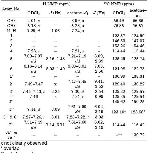
Table 1.1H and 13C NMR chemical shifts and coupling
constants of 3 in CDCl3 and acetone–d6
x not clearly observed * overlap.
H6`` 123.58 116.42, C7`` 123.58, C5``
123.58, C4``
139.72, C7a`` 139.72, C3a``
H7`` 116.42 123.58, C6``
139.72, C7a``
123.58, C5``
139.72, C3a`` 123.58, C4``
x: not observed.
While the doublet of a doublet observed in CDCl3 at δ =
7.09–7.07 ppm (J = 8.16 and 1.43 Hz) is assigned to H5
coupling to H4 in the trisubstituted ring. This proton was
shown doublet of a doublet and shifted a bit downfield in acetone–d6. It was also observed that in CDCl3 and
acetone–d6 H4 appeared as a singlet at δ = 7.28 and
7.21 ppm, respectively. Additionally, the proton of N–H observed as singlet and resonated at δ = 7.24 ppm in acetone–d6, and it appeared as doublet in CDCl3 to δ =
7.25 ppm, (J = 1.06 Hz). In acetone–d6, the coupling
between N–H and H6 was observed most probably due
to the interaction of the solvent with the compound. We suggested the triplet of signal in acetone–d6 at δ =
7.23–7.22 ppm, (J = 3.03 Hz) and at δ = 7.27–7.26 ppm, (J = 3.01 Hz) in CDCl3 to be assigned to H5`` and H6`` in
the benzimidazole ring due to coupling with H4`` and H7``.
The difference in chemical shift of H4`` and H7`` is also
apparent in both solvents. In CDCl3, H7`` and H4`` signal
were found to be separated; H7`` appeared as doublet of
a doublet at δ = 7.51–7.49 ppm, (J = 7.14 and 3.71 Hz), while H4`` appeared as doublet at δ = 7.44 ppm, J cannot
be determined exactly due to peaks overlapping. However, in acetone–d6 both protons shown signal as
doublet of a doublet at δ = 7.61–7.60 ppm (J = 6.02 and 3.19 Hz).
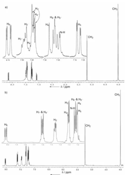
Fig 3. 1H NMR spectra of 3 in a) CDCl3 and b) in
acetone–d6.
From Figure 3, the signal of the benzyl ring protons H2` appeared as doublet of a doublet at δ =
7.47– 7.44 ppm, (J = 9.41 and 3.52 Hz) in acetone–d6
although the peaks were not clearly shown in CDCl3.
The H4` splitting patterns in CDCl3 was not clear due to
that overlapping of the peaks but it appeared as a triplet at δ = 7.31 ppm, (J = 0.99 Hz) in acetone–d6. On
the other hand, the protons of H3` displayed the signal
as a triplet at δ = 7.45–7.43 ppm, (J = 3.25 Hz) in CDCl3, and as doublet at δ = 7.30 ppm, (J = 2.54 Hz) in
acetone–d6. The methoxy OMe and methylene CH2
protons were observed in CDCl3 at δ = 4.01 and 5.18
ppm, and at δ = 3.99 and 5.23 ppm in acetone–d6,
respectively. Table 1 shows the chemical shift values of
3 in both solvents.
We found both spectra of the benzimidazole 3
were differed in splitting patterns for some of the aromatic protons, which may be due to the solubility of
3 in both solvents, including some effect from that six–
membered illusory ring, which was formed from the intramolecular hydrogen bonding between N–H in the benzimidazole ring and the oxygen atom in the benzyl ring (Figure 1). 1H–1H COSY and HMBC experiments
were performed to further confirm that assigned peaks.
13
C NMR
The 13C NMR spectra of
3 were obtained and
shown in Figure 4. The quaternary carbons in 3 were
identified and recognized by APT and or DEPT–135 NMR experiments in both solvents. The quaternary carbon signals for C3, C2``,C2, C1` and C1 in CDCl3 were
observed at δ = 153.26, 149.62, 146.53, 136.99 and 123.37 ppm, respectively. In acetone–d6 these values
were found to be shifted slightly downfield. The signals of the quaternary carbons C3a`` and C7a`` were absent in
CDCl3, but, APT NMR experiment showed the existing of
this peak at δ = 138.87 ppm in acetone–d6. A NMR solid
state study by Claramunt et al. showed that these signals appeared at δ = 136.9 ppm [37].The CH2 and
OMe signals in both solvents were found at about δ = 76.60 and 56.50 ppm, respectively.
The spectra showed that some of the carbons in the benzimidazole ring overlapped with the other carbons in trisubstituted ring. For example, in CDCl3 C7``
signal overlapped with C4 at δ = 114.44 ppm, while the
peaks were separated clearly in acetone–d6, (Figure 4).
Similarly, carbons C4``, C5`` and C6`` appeared at the
same chemical shift in both solvents. The carbons in trisubstituted ring C6 and C5 were resonated in CDCl3 at
δ = 121.98 and 125.29 ppm, respectively, while the benzyl ring carbons were observed at δ = 129.40, 129.35 and 129.32 ppm for C2`, C4` and C3` in CDCl3,
respectively. Table 1 summarizes the chemical shift values discussed.
To confirmed of the assignment signals and the overlapping between the aromatic carbons, the proton
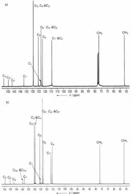
Fig 4. 13C NMR spectra of 3 in a) CDCl3 and b) in
acetone–d6.
coupled 13C NMR spectra of the benzimidazole 3 were
also conducted. Additionally, the HMQC and HMBC experiments done, further aid in assigning the peaks.
1
H–1H COSY
The signals of 3 are also assigned with an aid by
the COSY experiment. Figure 5 shows the 1H–1H
COSY NMR spectra of 3 in CDCl3 and acetone–d6. The
signal in both solvents at δ = 4.01-3.99 ppm, was assigned to the methoxy protons, OMe and it showed correlations with CH2 at δ = 5.18-5.23 ppm. As
expected, proton H5 was correlated with H4 and H6 in
CDCl3 at δ = 7.25 and 8.16–8.14 ppm, respectively.
Besides H5 and H4, proton H6 show correlations clearly
with H4 and N–H although this correlation is not
observed in acetone–d6. H5``, H6`` were observed to be
correlated with both protons H7`` and H4`` at δ = 7.51–
7.49 and 7.43 ppm in CDCl3 and at δ = 7.61–7.60 ppm
in acetone–d6.
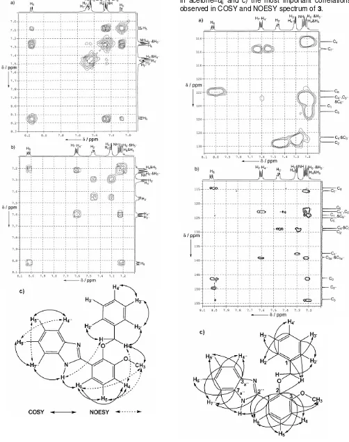
Fig 5. 1H–1H connectivities in the COSY a) in CDCl3, b)
in acetone–d6 and c) the most important correlations
observed in COSY and NOESY spectrum of 3.
Fig 6. 1H–13C connectivities of 3 in acetone–d6 a) in the
HMQC, b) in the HMBC and c) the most important correlations observed.
1
H–13C HMQC NMR
The 2D HMQC NMR spectrum was conducted to determine which hydrogens are connected to which carbons. The HMQC NMR spectrum for 3 was shown in
Figure 6a, and it confirms the attachments between all hydrogens and their corresponding carbons. In acetone– d6, the signals owing to C4, C7``, C6, C5, C4`, C3` and C2`
atoms are observed at the respective δ = 115.44, 116.42, 122.73, 125.74 and 129–130 ppm, while the sig-nals of the one bond 13C–1H connectivities are also well observed for OMe and CH2 atoms whereby the cross
peaks appear at δ = 56.85 and 76.57 ppm, respectively. Table 2 shows the summarized value for HMQC of 3.
1
H–13C HMBC spectra
The 2D HMBC NMR spectrum was conducted to examine the long–range 1H–13C connectivities. The HMBC NMR spectrum for 3 was shown in Figure 6b.
The aromatic quaternary carbons are established through the connectivities between the carbon and its neighboring proton by using a long–range correlated HMBC experiment. The signal in acetone–d6 of methoxy
protons showed 3J–correlation with C
3 at δ = 154.46 ppm
and 4J–correlation with C4 at δ = 115.44 ppm. While the
protons of CH2 were observed 2J–correlation with C1`
and 3J–correlation with C
2` and C2 at δ = 138.31, 130.22
and 147.07 ppm, respectively.
Additionally, the long–range HMBC cross peaks of the quaternary carbon C3 with H5 appeared at δ = 7.21–
7.19 ppm and was correlated with H6 at δ = 8.03–8.01
ppm. Proton H6 was demonstrated to correlate with C2``
and C2. H4 and H5 were also observed to be correlated
with C2. The spectrum also shows that C3a`` and C7a``
were correlated with N–H, H6`` and H5`` at δ = 7.24–7.22
ppm and with H7`` and H4`` at δ = 7.61–7.60 ppm. The
chemical shifts of the HMBC of 3 are summarized in
Table 2.
CONCLUSION
We have reported the complete assignments of the benzimidazole 3 using 1H, 13C, COSY, HMQC and
HMBC NMR in both CDCl3 and acetone-d6. Although the
APT and DEPT–135 spectra were not shown, that experiments were performed and the results were discussed. Combination of the information gathered from experiments done in both solvents helps in assigning peak splittings of compound 3. The differences in the
peak splittings can be seen in a few cases. For example, in CDCl3, H7`` displayed doublet of a doublet at δ =
7.51-7.49 ppm and H4`` signal was overlapped at δ = 7.44
ppm. While in acetone-d6 both protons shown the
signal as doublet of doublet at δ = 7.61-7.60 ppm. The quaternary carbon peaks of C3a`` and C7a`` were absent
in CDCl3, but they were observed clearly in acetone-d6
at δ = 139.72 ppm, and confirmed by APT NMR
experiment. The cause of these differences may be due to the solubility of 3 in both solvents, including
some effect from that six–membered illusory ring, which it was formed from the intramolecular hydrogen bonding between N-H in the benzimidazole ring and the oxygen atom in the benzyl ring. Overall, experiments done in acetone-d6 demonstrated clearer
peak splittings than in CDCl3. Further reactions using
the compound to synthesis biologically important compounds are in progress.
ACKNOWLEDGEMENTS
We thank the Malaysian Government and Universiti Sains Malaysia (USM) for IRPA short–term grant [304/PKIMIA/638007] to conduct this work. M.H. Al–Douh thanks Yemen Government and Hadhramout University of Science and Technology (HUST) for financial scholarship.
REFERENCES
1. Wright, J.B., 1951, Chem. Rev., 48, 397–541 2. Hofmann, K., 1953, The Chemistry of Heterocyclic
Compounds Imidazole and Its Derivatives Part I, Interscience, London, 247–324.
3. Craigo, W.A., LeSueur, B.W. and Skibo, E.B., 1999, J. Med. Chem., 42, 3324–3333
4. White, A.W., Curtin, N.J., Eastman, B.W., Golding, B.T., Hostomsky, Z., Kyle, S., Li, J., Maegley, K.A., Skalitzky, D.J., Webber, S.E., Yu, X.H. and Griffin, R.J., 2004, Bioorg. Med. Chem. Lett., 14, 2433– 2437.
5. Gudmundsson, K.S., Tidwell, J., Lippa, N., Koszalka, G.W., van Draanen, N., Ptak, R.G., Drach, J.C. and Townsend, L.B., 2000, J. Med.
11. Mekapati, S.B. and Hansch, C., 2001, Bioorg. Med. Chem., 9, 2885–2893.
12. Hong, S.Y., Chung, K.H., You, H.J., Choi, I.H., Chae, M.J., Han, J.Y., Jung, O.J., Kang, S.J. and Ryu, C.K., 2004, Bioorg. Med. Chem. Lett., 14, 2563–3566.
13. Kopanska (n. Zastapilo), K., Najda, A., Zebrowska, J., Chomicz, L., Piekarczyk, J., Myjak, P. and Bretner, M., 2004, Bioorg. Med. Chem., 12, 2617– 2624.
14. Ozden, S., Atabey, D., Yildiz, S. and Goker, H., 2005, Bioorg. Med. Chem., 13, 1587–1597
15. Nezhad, A.K., Rad, M.N.S., Mohabatkar, H., Asraria, Z. and Hemmateenejada, B., 2005, Bioorg. Med. Chem., 13, 1931–1938
16. Kazimierczuk, Z., Andrzejewska, M., Kaustova, J. and Klimesova, V., 2005, Eur. J. Med. Chem., 40, 203–208.
17. Mavrova, A.T., Anichina, K.K., Vuchev, D.I., Tsenov, J.A., Kondeva, M.S. and Micheva, M.K., 2005, Bioorg. Med. Chem., 13, 5550–5559.
18. Sondhi, S.M., Sharma, V.K., Verma, R.P., Singhal, N., Shukla, R., Raghubir, R. and Dubey, M.P., 1999, Synthesis, 5, 878–884
19. Sondhi, S.M., Singh, N., Kumar, A., Lozach, O. and Meijer, L., 2006, Bioorg. Med. Chem., 14, 3758– 3765.
20. Roth, T., Morningstar, M.L., Boyer, P.L., Hughes, S.H., Bukheit, R.W. and Michejda, C.J., 1997, J. Med. Chem., 40, 4199–4207
21. Porcari, A.R., Devivar, R.V., Kucera, L.S., Drach, J.C. and Townsend, L.B., 1999, J. Med. Chem., 41, 1251–1262
22. Rao, A., Chimirri, A., Clercq, E.D., Monforte, A.M., Monforte, P., Pannecouque, C. and Zappala, M., 2002, Il Farmaco, 57, 819–823
23. Rao, A., Chimirri, A., Ferro,S., Monforte, A.M., Monoforte, P. and Zappala, M., 2004, ARKIVOC, v, 147–155
24. Rida, S.M., El–Hawash, S.A.M., Fahmy, H.T.Y., Hazzaa, A.A. and El–Meligy, M.M.M., 2006, Arch. Pharm. Res., 29, 826–833.
25. Kumar, D., Jacob, M.R., Reynolds, M.B. and Kerwin, S.M., 2002, Bioorg. Med. Chem., 10, 3997–4004 26. Demirayak, S., Mohsen, U.A. and Karaburun, A.C.,
2002, Eur. J. Med. Chem., 37, 255–260
27. Andrzejewska, M., Mulia, L.Y., Rivera, R.C., Tapia, A., Vilpo, L., Vilpo, J. and Kazimierczuk, Z., 2002, Eur. J. Med. Chem., 37, 973–978
28. Kupchinsky, S., Centioni, S., Howard, T., Trzupek, J., Roller, S., Carnahan, V., Townes, H., Purnell, B., Price, C., Handl, H., Summerville, K., Johnson, K., Toth, J., Hudson, S., Kiakos, K., Hartley, J.A. and Lee, M., 2004, Bioorg. Med. Chem., 12, 6221–6236 29. Huang, S.T., Hsei, I.J. and Chen, C., 2006, Bioorg.
Med. Chem., 14, 6106–6119.
30. Soto, S.E., Molina, R.V., Crespo, F.A., Galicia, J.V., Diaz, H.M., Piedra, M.T. and Vazquez, G.N., 2006, Life Science, 79, 430–435.
31. Vazquez, G.N., Diaz, H.M., Crespo, F.A., Rivera, I.L., Molina, R.V., Muniz, O.M. and Soto, S.E., 2006, Bioorg. Med. Chem. Lett., 16, 4169–4173. 32. Al–Douh, M.H., Hamid, S.A., Osman, H., Ng, S.L. reflections were collected and 8446 are unique (Rint
= 0.0539), R = 0.0501, ωR = 0.1282 for 227 parameters and 8446 reflections (I > 2σ (I)). Residual electron density extremes were 0.613 and –0.326 e Å–3. The intensity data was collected at
297 K on SMART APEX2 CCD area–detector diffractometer with graphite–monochromated MoKa
radiation (0.71073 Å), θ range 2.21 to 37.50o [38].
All absorption corrections were performed by using SADABS the multiscan program [38]. The structure was solved and refined by SHELXTL against F2[39]. The H atoms were refined as riding and the
Uiso values were freely refined, they were placed in calculated positions, with C–H = 0.93–0.97 Å
and N–H = 0.89 Å. The software was used
SHELXTL [39] and PLATON [40]. These data can be obtained free of charge from International Union of Crystallography IUCr cv2101 or The Cambridge Crystallographic Data Centre CCDC 620951. Reference: (doi:10.1107/S160053680603251X). 35. Bruker program 1D WIN–NMR (release 6. 0) and
2D WIN–NMR (release 6.1).
36. Berger, S. and Braun, S., 2004, 200 and More NMR Experiments, A Practical Course, Wiley– VCH, Weinheim.
37. Claramunt, R.M., Lopez, C. and Elguero, J., 2006, ARKIVOC, v, 5–11.
38. APEX2 (Version 1.27), SAINT (Version 7.12A), and SADABS (Version 2004/1), 2005, Bruker AXS Inc., Madison, Wisconsin, USA.
39. Sheldrick, G.M., SHELXTL. (Version 5.1), 1998, Program for the Solution of Crystal Structures, Bruker AXS Inc., Madison, Wisconsin, USA. 40. Spek, A.L., 2003, J. Appl. Cryst., 36, 7–13.