A Thesis for the Degree of
Ph.D. in Engineering
Development of an in vitro neurovascular unit model
by on-chip tissue engineering
February 2019
Graduate School of Science and Technology
Keio University
Thesis Abstract
Neurovascular unit (NVU) is a conceptual unit in the brain, composed of neurons, glial cells such as astrocytes, microvasculatures covered by pericytes and extracellular matrix. This warns against focusing on only neurons and indicates the importance of investigating interactions between neurons and other brain cells for therapeutic approaches for neurodegenerative diseases. Microvasculatures in NVU have brain-specific functions called blood-brain barrier (BBB) and limit the transport of drugs to neurons. Although many studies have been performed for investigating BBB functions, an in vitro three-dimensional (3D) NVU model has not been established because of the gap between in vitro cell culture microenvironments and in vivo cellular conditions. Therefore, the aim of this study is to establish an in vitro 3D NVU model using a microfluidic device.
Chapter 1 summarizes the background, previous studies and objectives of this study. Chapter 2 summarizes materials and methods of this study.
Chapter 3 describes the establishment of an in vitro brain angiogenesis model focusing on microvasculatures in NVU. In particular, organ-specificity of endothelial cells (ECs) is focused on, and brain ECs and human umbilical vein endothelial cells (HUVECs) are cultured to construct microvasculatures. These organ-specific microvasculatures are compared in terms of microvascular formation processes, endothelial barrier functions and endothelial junction protein expressions. Consequently, an in vitro brain angiogenesis model is successfully established. In addition, it is found that endothelial barrier functions are greater in brain microvasculatures compared to microvasculatures formed by HUVECs.
Chapter 4 describes the integration of brain angiogenesis and neurogenesis models toward the construction of an in vitro NVU structure. First, brain ECs and mesenchymal stem cells are cocultured to investigate cell culture conditions for inducing angiogenesis. Next, the optimized culture condition for angiogenesis is applied to the culture of neural stem cells (NSCs), which succeeds in inducing 3D neurite extension. Finally, these angiogenesis and neurogenesis models are combined, which results in the construction of neurovascular tissues.
Chapter 5 describes investigation of long-term culture to construct NVU tissues including astrocytes. NSCs differentiate into astrocytes in long-term culture, which allows to construct a tissue composed of neurons, astrocytes and microvasculatures. However, further investigation is needed to construct more physiological NVU structures.
論
文
要
旨
【訳題】
オンチップ組織工学による生体外Neurovascular Unitモデルの開発
Neurovascular Unit(NVU)とは,主にニューロン,アストロサイトなどのグ リア細胞,ペリサイトを伴う微小血管,細胞外マトリクスから成る概念的な構 成単位である.このことは,神経変性疾患の治療に対する治療戦略として,ニ ューロンのみに着目するのではなく,周辺の細胞との相互作用の調査を行うこ との重要性を示唆している.NVUにおける微小血管には血液脳関門という脳血 管特有のバリア機能があり,ニューロンに対する薬剤の供給を制限する.これ まで血液脳関門の機能について多くの研究が行われてきたが,生体外の脳血管 モデルは生体内の環境との乖離があり,いまだにNVUの3次元生体外モデルは 実現していない.そこで本研究では,マイクロ流体デバイスという細胞の 3 次 元培養が可能なチップを用いて,神経幹細胞と脳の血管内皮細胞を培養し,生 体外NVUモデルを構築することを目的とした. 第1章に,本研究に関する基礎事項,従来の研究,本研究の目的を概説した. 第2章に,本研究で用いた試薬類や実験手法などについてまとめて述べた. 第3章では,まずNVUの中の微小血管のみに着目し,生体外血管新生モデル の確立のために行った実験について述べた.特に血管内皮細胞の臓器特異性に ついて着目し,脳血管内皮細胞およびヒト臍帯静脈内皮細胞を用いてそれぞれ 微小血管網の構築を行い,その構築プロセス,血管バリア機能の評価,血管内 皮細胞のジャンクションタンパク群の発現評価などを行った.その結果,生体 外脳血管新生モデルを確立するとともに,脳血管において血管バリア機能が優 れていることを見出した. 第4章では,生体外NVU構造の構築を目指し,血管新生モデルと神経新生モ デルの統合を試みた.まず,脳血管内皮細胞と間葉系幹細胞の共培養によって 血管新生を誘導できるような細胞培養条件を検討した.次に,その培養条件下 で神経幹細胞を培養し,3次元的な神経網形成を誘導することに成功した.さら に,血管新生と組み合わせることで,神経網および微小血管からなる神経・血 管組織の構築に成功した. 第 5 章では,第 4 章で述べた培養系を長期培養にすることにより,神経網と 微小血管に加えアストロサイトを含んだNVU様組織の構築に必要な培養条件を 検討した.長期培養によって神経幹細胞からアストロサイトへの分化誘導が確 認され,神経・血管・アストロサイトを含む組織の構築に成功したが,生体内 の模擬のため,さらなる検討が必要であることが示唆された. 第 6 章に,結論として各章の内容をまとめて研究成果を要約し,今後の展望 について述べた.
Contents
Chapter 1 General Introduction ... 1
1-1 Neurovascular unit (NVU) ... 1
1-2 Blood vessels and capillaries ... 1
1-2-1 Fundamental features of blood vessels and capillaries ... 1
1-2-2 Vasculogenesis and angiogenesis ... 4
1-3 Extracellular matrix (ECM) ... 10
1-4 Mesenchymal stem cell (MSC) ... 10
1-5 Blood-brain barrier (BBB) ... 11
1-5-1 Fundamental features of BBB ... 11
1-5-2 BBB molecules ... 13
1-5-3 BBB transport pathways ... 13
1-6 Neural stem cell (NSC) ... 14
1-7 Organ-specific ECs ... 16
1-8 In vitro vasculogenesis/angiogenesis models ... 16
1-9 In vitro BBB models ... 18
1-10 Application of in vitro BBB models to drug screening studies ... 22
1-11 Objectives ... 23
Chapter 2 Materials and methods ... 26
2-1 Cell culture ... 26
2-2 Microfluidic device preparation ... 27
2-3 Hydrogel filling and cell seeding in microfluidic devices ... 28
2-4 Immunocytochemistry ... 29
2-4-1 Immunofluorescence staining procedures ... 29
2-4-2 Antibodies ... 29
2-5 Quantitative analysis ... 31
2-5-1 Microvasculatures ... 31
2-5-3 Gel collapse in triculture of NSCs, BMECs and MSCs ... 31
2-5-4 Dextran perfusion and measurement of microvascular permeability coefficient ... 32
2-5-5 Statistical analysis... 34
Chapter 3 Comparison of organ-specific endothelial cells in terms of microvascular formation and endothelial barrier functions ... 35
3-1 Introduction ... 35
3-2 Experimental design ... 38
3-3 Results ... 42
3-3-1 Initial vascular sprout formation in BMEC/HUVEC-MSC cocultures ... 42
3-3-2 Microvascular network formation in BMEC/HUVEC-MSC cocultures ... 45
3-3-3 Basement membrane formation and pericyte coverage around microvasculatures ... 48
3-3-4 Permeability assay of microvascular walls using fluorescent dextran solution ... 51
3-3-5 Junction protein expression in microvascular networks ... 54
3-3-6 Inflammatory response of BMEC microvasculature induced by thrombin ... 57
3-4 Discussion ... 60
3-5 Summary ... 63
Chapter 4 Integration of neurogenesis and angiogenesis models for constructing a neurovascular tissue . 65 4-1 Introduction ... 65
4-2 Experimental design ... 67
4-3 Results ... 71
4-3-1 Neurite formation of neurons differentiated from NSCs in different gel scaffolds ... 71
4-3-2 Formation of microvasculature in BMEC-MSC coculture in different gel scaffolds ... 75
4-3-3 Optimization of the concentration of fibrin-Matrigel mixed gel ... 78
4-3-4 The process of neurogenesis and neurite extension in NSC culture ... 80
4-3-5 Triculture of NSCs, BMECs and MSCs for constructing neurovascular tissues ... 82
4-4 Discussion ... 89
4-5 Summary ... 92 Chapter 5 Investigation of triculture conditions of NSCs, BMECs and MSCs for the construction of
NVU-like structure ... 93
5-1 Introduction ... 93
5-2 Experimental design ... 94
5-3 Results ... 98
5-3-1 BMEC-MSC coculture in combination medium of NSC medium, EGM-2 and DMEM ... 98
5-3-2 NSC monoculture in the mixture of NSC medium and EGM-2 ... 101
5-3-3 Triculture of NSCs, BMECs and MSCs to construct a neuron-astrocyte-capillary tissue ... 104
5-4 Discussion ... 107
5-5 Summary ... 109
Chapter 6 Concluding remarks ... 111
6-1 Summary ... 111 6-2 Future perspectives ... 114 References... 117 Achievements ... 131 Publications ... 131 International conferences ... 131 Domestic conferences ... 132 Acknowledgments
List of abbreviations and symbols
2D: two-dimensional 3D: three-dimensional AD: Alzheimer’s disease Ang: angiopoietin AQP: aquaporin Aβ: amyloid-β
BBB: blood-brain barrier
BDNF: brain derived neurotrophic factor bFGF: basic fibroblast growth factor BM: basement membrane
BMEC: brain microvascular endothelial cell CNTF: ciliary neurotrophic factor
DAPI: 4′,6-diamidino-2-phenylindole Dll4: delta-like ligand 4
EC: endothelial cell ECM: extracellular matrix FBS: fetal bovine serum
FITC-dextran: fluorescein isothiocyanate-conjugated dextran GFAP: glial fibrillary acidic protein
Glut: glucose transporter
HIF-1α: hypoxia inducible factor-1α
HUVEC: human umbilical vein endothelial cell IGF-1: insulin-like growth factor-1
MAP2: microtubule associated protein 2 MMP: matrix metalloproteinases
NG-2: neuron-glial antigen-2 NSC: neural stem cell
NVU: neurovascular unit PBS: phosphate buffered saline PDGF: platelet-derived growth factor PDMS: polydimethylsiloxane
PECAM-1: platelet endothelial cell adhesion molecule SMC: smooth muscle cell
TEER: transendothelial electrical resistance VE-cadherin: vascular endothelial cadherin VEGF: vascular endothelial growth factor α-SMA: α-smooth muscle actin
Symbols used in chapters 2 and 3
cm: solute concentration inside cell membrane [mol/cm3]
c0: solute concentration difference across vessel wall [mol/cm3]
D: diffusion coefficient [m2/s]
I: fluorescence intensity
I0: initial intravascular fluorescence intensity
J: flux [mol/cm2/s]
l: vessel length [cm]
N: amount of dextran molecules [mol]
P: permeability coefficient [cm/s]
r: vessel radius [cm]
S: area of vessel wall [cm2]
V: volume of vessel [cm3]
Chapter 1 General Introduction
1-1 Neurovascular unit (NVU)
The brain consumes almost 20% of all nutrients present in the blood (Zlokovic, 2008). For the proper function of the brain, neurons always demand nutrients, mainly glucose, from closely-existing capillaries. Almost all neurons in human brain have capillaries in close proximity (Zlokovic, 2005). Astrocytes exist in close proximity between neurons and capillaries. This neuron-astrocyte-capillary organization is important for astrocytic transport of nutrients from blood to neurons, and this unique multicellular structure is called the neurovascular unit (NVU) defined as a structure composed of neurons, astrocytes, capillaries and extracellular matrix (ECM).
The concept of NVU was proposed in the Stroke Program Review Group held in 2001 by the National Institutes of Neurological Disorders and Stroke
(https://www.ninds.nih.gov/) (Lo et al., 2003). The concept of NVU implies the
importance of considering multicellular structure of NVU. The loss of neuronal functions causes neurodegenerative diseases such as Alzheimer’s disease and Parkinson’s disease. Neuronal functions are supported by surrounding other cells. It is important to consider interactions between neurons and other cell types in NVU for the further understanding of pathological states in neurodegenerative diseases (Iadecola, 2017).
1-2 Blood vessels and capillaries
1-2-1 Fundamental features of blood vessels and capillaries
Blood vessels are composed of endothelial cells (ECs), basement membrane (BM), mural cells and ECM. ECs form monolayer wall of blood vessels. BMs are various secreted proteins from ECs and mural cells, including type IV collagen, laminin, fibronectin, heparan sulfate proteoglycan and so on. Mural cells are smooth muscle cells
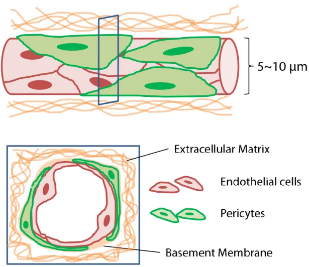
(SMCs) and pericytes. Mural cells exist closely around blood vessel wall and interact with endothelial monolayer. Pericytes exist around capillaries (Fig. 1-1) while SMCs exist around larger vessels. The average diameter of capillaries is 5–10 μm. ECM is composed of collagens, fibronectin, laminin, proteoglycans and so on (Frantz et al., 2010). ECM forms net fiber structure and almost all cells attach the fiber structure to live and form organs. Blood vessels are distributed throughout the body and mainly work as a conduit of blood. Blood vessels supply oxygen and nutrients to parenchymal cells. ECs are connected with each other through various junctions. Leakage of macromolecules and cells floating in blood into parenchyma is normally restricted by tight junctions and adherens junctions while oxygen and nutrients are properly transported to organ parenchyma through vascular walls.
Fig. 1-1 Schematic illustrations of capillary microenvironment.
Capillaries are small blood vessels whose diameter ranges from 5 to 10 μm. Capillaries are sparsely wrapped by pericytes. BM proteins localize at outer capillary surface.
1-2-2 Vasculogenesis and angiogenesis
Blood vessels constitute a circulatory system throughout human body. They transport oxygen and nutrients throughout our body to maintain functions of each organ and sustain our lives. In the generation of our body through embryogenesis, there are two main mechanisms of creating blood vessel networks.
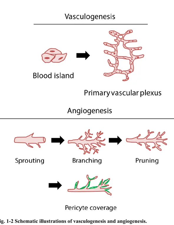
Vasculogenesis is the first step to build up vascular networks (Fig. 1-2). In the embryonic stage, hemangioblasts, which are stem cells of ECs, proliferate to create a group of cells called blood island. Hemangioblasts existing in the periphery of blood islands differentiate into ECs and create primary vascular plexus. The series of processes of creating primary vascular plexus is called vasculogenesis. As a next step, ECs sprout from the pre-existing vascular plexus and form more complex vascular networks through branching and pruning processes. These growing vascular networks are then wrapped by other cell types such as SMCs and pericytes through interactions among these cell types. Pericytes wrap around capillaries to stabilize their structures. Capillaries wrapped by pericytes become mature and stable. This morphogenesis from vascular plexus to mature vascular networks is called angiogenesis.
Fig. 1-2 Schematic illustrations of vasculogenesis and angiogenesis.
Vasculogenesis is the first step of vascular formation processes in which endothelial stem/progenitor cells expand and form primary vascular plexus. Angiogenesis is the following step for forming more complex vascular networks. Angiogenic processes are classified into sprouting, branching, pruning and pericyte coverage.
Angiogenesis is a multi-step morphogenesis of creating new blood vessels. In angiogenic processes, ECs show different morphologies and functions depending on the parts of growing vessels (Ribatti and Crivellato, 2012) (Fig. 1-3). As a first step, vascular sprouts are formed by leading ECs. these ECs are called tip cells (Gerhardt et al., 2003). Tip cells exist at the front edge of vascular sprouts and guide other ECs to elongate vascular sprouts. Tip cells extend filopodial structures, which sense guidance cues such as vascular endothelial growth factor (VEGF) (Ruhrberg et al., 2002). ECs following tip cells are called stalk cells, which proliferate and form vascular lumen structures. The selection of tip cells out of pre-existing vascular ECs is performed by signaling processes between VEGF and VEGF receptor 2 (VEGFR2), one of the receptors for VEGF (Bentley et al., 2014). ECs constituting vascular monolayer have VEGFR2. When VEGF bind VEGFR2 in ECs, these ECs activate Notch receptor in adjacent ECs by supplying delta-like ligand 4 (Dll4). ECs whose Notch receptors are activated by Dll4 behave like stalk cells while ECs which have high VEGF/VEGFR2 signaling level stay located at the leading edge of vascular sprouts as tip cells. Position changes between tip cells and stalk cells can happen depending on the balance between VEGF/VEGFR2 signaling and Dll4/Notch signaling. ECs residing in the pre-existing capillary with a stable morphology are called phalanx cells. Phalanx cells have VEGFR1, which acts as a suppressor of VEGF binding to VEGFR2. Phalanx cells contribute to stop VEGF/VEGFR2 signaling and finish angiogenic processes (Bautch, 2009).
Fig. 1-3 Types of ECs in processes of angiogenesis.
Tip cells are leading cells of growing vasculatures and extend filopodial structures sensing angiogenic factors. Stalk cells are proliferating cells in growing vasculatures. Stalk cells form vascular lumen structures. Phalanx cells exist in the root of growing vessels and contribute to finish angiogenic processes.
VEGF promotes EC proliferation, inhibits EC apoptosis and increase endothelial permeability. VEGF regulates vascular development throughout embryogenesis. The expression of VEGF is induced by hypoxic conditions. Especially, hypoxia inducible factor-1α (HIF-1α) promotes the expression of VEGF (Zimna and Kurpisz, 2015).
Growing capillaries become mature through interactions between ECs and pericytes. ECs secrete platelet-derived growth factor-B (PDGF-B) to recruit pericytes. Pericytes have PDGF receptor-β (PDGFR-β). Proliferation and migration of pericyte progenitor cells are promoted through PDGF/PDGFR-β signaling, resulting in pericyte covering around growing endothelial tubes.
Angiopoietin-1 (Ang-1) is a major angiogenic factor to promote vascular maturation (Ribatti et al., 2011) (Fig. 1-4). Ang-1 is secreted by pericytes and binds to endothelial Tie-2, a receptor for Ang-1. Angiopoietin-2 (Ang-2) is expressed in ECs and acts as a de-stabilizing factor of vessels. VEGF increases the production of Ang-2 by ECs, and Ang-2 binds to pericyte Tie-2 receptor, resulting in the dissociation of pericytes from vessels. In this state, VEGF induces angiogenesis by stimulating ECs without pericytes. ECs without the coverage of pericytes and stimulus of VEGF undergo apoptosis.
Fig. 1-4 Interactions between ECs and pericytes.
Ang-1 secreted by pericytes binds to endothelial Tie-2, which promotes pericyte association and capillary maturation. Endothelial Ang-2 binds to Tie-2 in pericytes, which generates pericyte dissociation. ECs without pericyte coverage can elongate vascular sprouts toward the region with abundant angiogenic factors such as VEGF.
1-3 Extracellular matrix (ECM)
ECM is a scaffold of organs and has fiber network structures. ECMs are essential for cells to live and function in every organ. Cells attach to ECM structure through integrins. Cell–ECM attachment through integrins is important for cell–cell or cell–ECM interactions. Main components of ECM are collagens, fibronectin, laminin, proteoglycans (Frantz et al., 2010). The components of ECM are different in every organ. In general, collagen I and fibronectin are abundant in many organs. However, brain ECM has much less collagen I and fibronectin (Bonneh-Barkay and Wiley, 2009; Zimmermann and Dours-Zimmermann, 2008). Hyaluronan is abundant in brain ECM and various proteoglycans bind to hyaluronan. Brain cells attach to these proteoglycans.
1-4 Mesenchymal stem cell (MSC)
Mesenchymal stem cells (MSCs) exist in many tissues, and are isolated from brain, spleen, liver, kidney, lung, thymus and pancreas (Lai et al., 2015). MSCs are originally defined as multipotent stem cells which have an ability to differentiate into adipocytes, chondrocytes and osteocytes (Lai et al., 2015). MSCs are now known to differentiate into stromal support cells and secrete factors to support other cells (Lai et al., 2015; Volarevic et al., 2011). In particular, MSCs are known to have some effects on angiogenesis (Bronckaers et al., 2014; Nassiri and Rahbarghazi, 2014; Volarevic et al., 2011). MSCs secrete VEGF, basic fibroblast growth factor (bFGF), insulin-like growth factor-1 (IGF-1) and PDGF to support EC survival and promote angiogenesis in a paracrine manner. It was reported that co-implantation of ECs and mesenchymal precursor cells leaded to the formation of long-lasting blood vessels in mice (Koike et al., 2004). The implanted mesenchymal precursor cells expressed mural cell marker, suggesting that MSCs can differentiate into pericytes. In addition to the proangiogenic effects of MSCs, MSCs are also known to promote endothelial junction protein expressions such as vascular endothelial cadherin (VE-cadherin) and Claudin-1 in a
juxtacrine manner (Menge et al., 2013; Nassiri and Rahbarghazi, 2014). MSCs can inhibit angiogenic processes by enhancing vascular integrity. Taken together, MSCs have both positive effects and negative effects on angiogenesis and play important roles throughout angiogenic processes. The relationship between MSCs and pericytes still remains to be elucidated. However, some previous studies reported that MSCs and pericytes have many common features (Caplan, 2017; de Souza et al., 2016).
1-5 Blood-brain barrier (BBB)
1-5-1 Fundamental features of BBBThe blood-brain barrier (BBB) is a brain-specific endothelial barrier. ECs constituting brain microvasculatures are connected with each other by tight junction proteins and strictly regulate the transport of oxygen and nutrients to brain parenchyma.
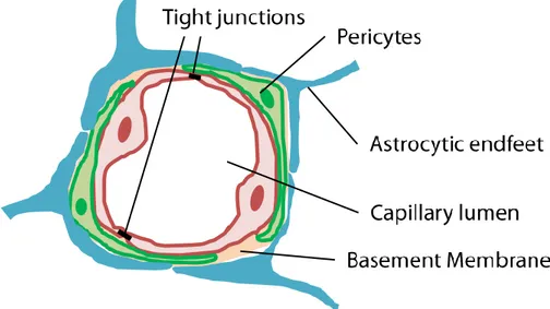
Brain microvasculatures are formed by ECs, pericytes and astrocytes (Fig. 1-5). Brain microvasculatures are wrapped by pericytes and both ECs and pericytes are surrounded by BM. Astrocytic endfeet ensheath ECs and pericytes. BMs are composed of different proteins including type IV collagen, laminin, nidogen and so on (Thomsen et al., 2017). These BM proteins are secreted by ECs and pericytes.
Fig. 1-5 BBB microenvironment.
ECs are sealed with each other by tight junction proteins and form capillary lumen. Pericytes wrap capillary lumen structures. Basement membranes ensheath both ECs and pericytes. Astrocytic endfeet wrap ECs and pericytes from further outside.
1-5-2 BBB molecules
Brain ECs are connected with each other by tight junction proteins. Tight junction proteins expressed in a brain are occludin, claudin 1, claudin 3, claudin 5 and claudin 12, and tight junction accessory proteins, ZO-1, ZO-2 and ZO-3, are also expressed in endothelial intercellular region (Reinhold and Rittner, 2017; Tietz and Engelhardt, 2015). In addition, adherens junction proteins such as platelet endothelial cell adhesion molecule (PECAM-1) and VE-cadherin are known to regulate BBB functions (Gavard and Gutkind, 2006).
1-5-3 BBB transport pathways
Previous studies investigated BBB-specific transport pathways (Abbott et al., 2010; Sweeney et al., 2018). ECs connected by tight junctions strictly prohibit the passage of substances through endothelial monolayer as a paracellular route. While ECs of brain microvasculatures have low rate of paracellular and transcellular transport, they have specific transporters and receptors for the entry of specific substances into brain parenchyma. BBB protects neurons from neurotoxins existing in blood circulation and nurture them by supplying nutrients. O2 and CO2 cross the BBB by simple diffusion.
Water is transported via water channels called aquaporin (AQP) receptors. Glucose, which is the main energy source of a brain, is transported by glucose transporter isotype-1 (Glut1). Brain ECs highly express Glut-1 protein. Glucose transport depends on the concentration gradient between blood and brain parenchyma. Peptides and proteins are transported via receptor-mediated transcytosis. The transport of amino acids, hormones and vitamins is performed by carrier-mediated transport.
1-6 Neural stem cell (NSC)
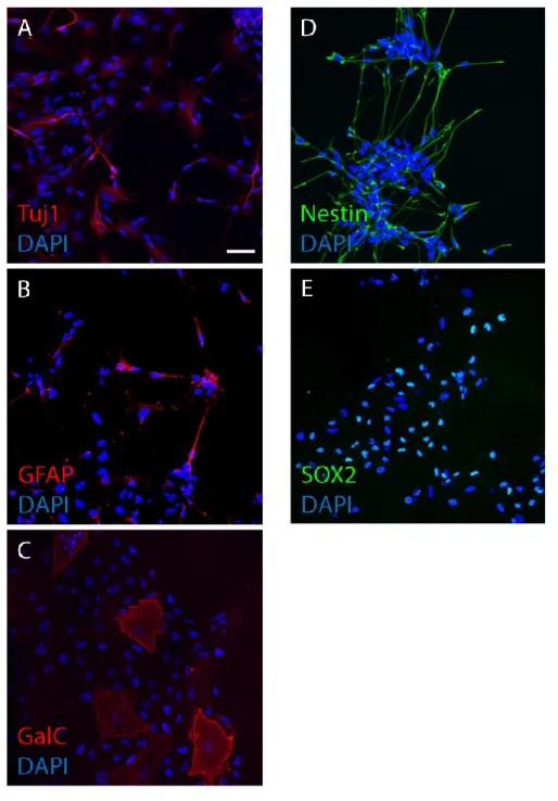
The brain is composed of neurons, glial cells including astrocytes and oligodendrocytes, blood vessels and ECM. Neurons, astrocytes and oligodendrocytes are generated by the same stem cell source called neural stem cell (NSC). While NSCs can generate neurons, astrocytes and oligodendrocytes, NSCs can divide symmetrically to generate the same stem cell (Ahmed, 2009; Temple, 2001). Undifferentiated cells which can self-renew and generate neurons, astrocytes and oligodendrocytes are defined as NSCs. Cultured NSCs also have an ability to generate differentiated cells (Fig. 1-6 A, B, C) while they can proliferate symmetrically (Fig. 1-6 D, E).
In terms of cultured NSC differentiation, previous in vitro studies investigated effects of soluble factors on the NSC differentiation into neuronal or glial lineages (Bhattacharya et al., 2008; Chen et al., 2013). For example, it was reported that brain-derived neurotrophic factor (BDNF) promoted NSC proliferation in a dose-dependent manner, and BDNF effectively promoted neuronal and oligodendrocytic differentiation (Chen et al., 2013). In terms of retinal stem cell differentiation, ciliary neurotrophic factor (CNTF) have concentration-dependent effects on neuronal or glial differentiation (Bhattacharya et al., 2008). It was reported that low concentration of CNTF induced neuronal differentiation while high concentration of CNTF induced glial differentiation. Fetal bovine serum (FBS), which is a widely-used essential supplement containing many factors required for cell growth and survival (Brunner et al., 2010) was also reported to have different effects on NSC differentiation (Hu et al., 2013; Hung and Young, 2006). These studies reported that FBS promoted NSC differentiation into astrocytes, not neurons. Taken together, previous in vitro studies suggested that some additional factors in NSC culture medium can promote neuronal or glial differentiation, further suggesting that specially-designed growth factor cocktail can be used for the desired expansion of neuronal and glial lineages in NSC culture.
Fig. 1-6 Neural stem cell culture. (A: neurons, B: astrocytes, C: oligodendrocytes, D, E: neural stem cells) (Scale bar, 50 μm)
NSCs were cultured using culture dishes for 14 days. Immunofluorescence staining was performed for differentiated cells (A, B, C) and undifferentiated stem cells (D, E). NSCs have an ability to generate differentiated cells in addition to the ability of self-renewal. Antibodies used here are described in chapter 2.
1-7 Organ-specific ECs
Every organ has its own vascular characteristics and patterning that are generated through interactions between ECs and parenchymal cells (Géraud et al., 2014). ECs in different organs are heterogeneous depending on organ-specific functions. Differences of ECs were previously reported (Géraud et al., 2014; Rafii et al., 2016). ECs are classified into three groups; continuous non-fenestrated ECs, continuous fenestrated ECs and discontinuous ECs. For example, brain ECs are continuous non-fenestrated ECs. ECs in choroid plexus are continuous and fenestrated. Liver sinusoidal ECs are discontinuous. Brain ECs strictly restrain the passage of molecules over 1 nm in size, while liver sinusoidal ECs allow permeation of molecules of 60 nm in size (Géraud et al., 2014).
ECs always interact with parenchymal cells. Organ-specific ECs are different not only in terms of fenestrae but also in terms of abilities to secrete angiocrine factors. Angiocrine factors are secreted by ECs and can stimulate organ-repair of damaged or diseased organs. A previous study showed that expression profiles of angiocrine factors in ECs were different among various organs (Nolan et al., 2013).
These differences of organ-specific ECs are not negligible when engineering multicellular structure such as BBB. To mimic microvascular environment by a tissue engineering approach, in which ECs interact with parenchymal cells, organ-specific ECs derived from the target organ need to be used for the recapitulation of the multicellular organization and its formation processes.
1-8 In vitro vasculogenesis/angiogenesis models
Previous studies reported in vitro vasculogenesis and angiogenesis models based on microfluidic cell culture systems (Kim et al., 2015, 2013; Lim et al., 2013; Moya et al., 2013). Microfluidic devices are made of polydimethylsiloxane (PDMS) and have designed microchannels. Microfluidic devices enabled us to culture cells
three-dimensionally within injected ECM scaffolds in the devices. By culturing ECs in ECM scaffolds such as collagen and fibrin and supplying growth factors, vascular formation can be induced through vasculogenesis or angiogenesis processes.
In previous studies, vascular formation was demonstrated by ECs, and most of these studies used human umbilical vein endothelial cells (HUVECs) (Kim et al., 2015, 2013; Lim et al., 2013). Vascular formation was induced within a week. Engineered vascular networks expressed not only endothelial junction markers such as VE cadherin, but also expressed basement membrane proteins, which were signs for the maturation of engineered vascular networks.
Functions of engineered vasculatures were also investigated. Especially, endothelial barrier function was analyzed. To analyze the barrier function of engineered vasculatures, it is needed to perfuse fluorescent dextran solution into the vasculatures. Previous studies successfully perfused the dextran solution into the engineered vasculature, and furthermore, the passage of the solution through the vascular wall was analyzed to measure the endothelial barrier function (Bang et al., 2017; Bichsel et al., 2015; Lee et al., 2014; Sobrino et al., 2016).
The construction of vascular networks and the perfusion of solution into the networks were of great importance for fundamental studies of vascular biology and drug screening studies. However, engineering of capillary-level microvascular networks with the coverage of pericytes has not been realized. In addition, as far as I know, in vitro
BBB model of functional brain microvascular networks has not been reported while most previous studies were performed with HUVECs. Until now, previous in vitro BBB models have investigated endothelial barrier functions with/without pericytes and astrocytes in 2D-based cell culture systems.
1-9 In vitro BBB models
The main components of BBB are ECs, astrocytes, pericytes and BMs. ECs in BBB express tight junction proteins, and pericytes and astrocytes closely exist around ECs. Previous studies focused on this characteristic morphology of brain microvasculatures and demonstrated BBB microenvironment by culturing these cell types.
Transwell cell culture system has been mostly used as an in vitro BBB model (Banerjee et al., 2016; Bicker et al., 2014) (Fig. 1-7). Transwell system is a combination of well plates and semipermeable membrane inserts. Cells can be cultured on the top surface of the insert, the bottom surface of the insert and the bottom of wells. To mimic the in vivo microenvironment, ECs are basically cultured on the top surface of the insert. When ECs are cocultured with pericyte or astrocyte, pericytes or astrocytes are cultured on the bottom surface of the insert or the bottom of the well. Tri-culture system is realized by culturing ECs on the top surface of the insert and culturing other cell types on the bottom surface of the insert or the bottom of the well, which is a mimicry of in vivo BBB microvascular environment. BM proteins such as laminin or collagen IV can be coated on the top surface of the insert before seeding ECs on the insert. Transwell cell culture system not only mimics the in vivo brain microvascular environment, but also is useful for analyzing interactions among these cell types in paracrine manner.
Using Transwell cell culture system, endothelial barrier functions have been investigated. Transendothelial electrical resistance (TEER), which represents the resistance towards ion diffusion through endothelial monolayer, has been analyzed in the Transwell cell culture system to estimate endothelial barrier functions. This analysis is realized by employing two electrodes in apical side and basal side of EC monolayer respectively. Previous studies demonstrated that astrocytes or pericytes augmented the endothelial barrier functions estimated by TEER analysis in Transwell coculture and tri-culture systems.
interactions among ECs, astrocytes and pericytes in terms of endothelial barrier functions, there is still a gap between in vitro BBB models and native EC conditions in vivo (Bicker et al., 2014). Native in vivo capillaries are three-dimensional, while ECs in previous in vitro BBB models formed 2D monolayer on a culture substrate. In addition, ECs are covered by pericytes and astrocytic endfeet and the percentage of pericyte coverage around capillaries are the highest in the brain compared to the other organs (Daneman et al., 2010; Keaney and Campbell, 2015; Thomsen et al., 2017). These fundamental features of brain capillaries were not included in previous in vitro studies, highlighting the disparity between in vitro BBB models and native EC conditions in vivo.
Instead of Transwell cell culture system, a three-dimensional in vitro BBB model has been reported using a microfluidic cell culture system (Adriani et al., 2017). This study focused on the organization of the in vivo NVU structure in which astrocytes lie between neurons and microvasculatures. To mimic this organization, neurons and astrocytes were seeded within two adjacent collagen gel channels and ECs were seeded in medium channel to form EC monolayer. This study demonstrated that cultured neurons showed neurite elongation and exhibited neuronal activity. Endothelial barrier functions were analyzed by introducing fluorescent dextran solution into the EC-coated microchannel. However, this study implied some difficulties in engineering functional NVU structures. One was the difficulty in the mimicry of the NVU organization. This study indeed mimicked the cell arrangement composed of neurons, astrocytes and microvasculatures by seeding these cells in adjacent microchannels. However, these cell types were cultured within compartmentalized gel scaffolds respectively while astrocytes closely exist between neurons and microvasculatures in vivo. Another difficulty was the analysis of endothelial barrier functions. Permeability coefficients were calculated based on the fluorescence intensity of introduced fluorescent dextran into the EC-coated microchannel. However, cultured ECs formed monolayer, not lumen structures, and consequently had relatively large permeability coefficients compared to
previous studies. In addition, permeability coefficients were smaller in EC monoculture condition than coculture condition with neurons and astrocytes. This result contradicted previous reports that astrocytes augmented endothelial barrier functions performed by Transwell cell culture system and TEER approach. It is still challenging to construct a functional 3D NVU structure in vitro.
Fig. 1-7 Previous in vitro BBB model using Transwell cell culture system.
ECs were cultured in Transwell system and endothelial barrier functions were estimated by TEER analysis, which is the measurement of electrical resistance across an endothelial monolayer. Endothelial barrier function is augmented by culturing ECs with astrocytes and pericytes.
1-10 Application of in vitro BBB models to drug screening studies
One of the main objectives of establishing an in vitro BBB model is the technical development to investigate the drug delivery to brain parenchyma. BBB excludes almost 100% of large molecules (>500 Da) from the brain parenchyma and more than 98% of all small molecules (Pardridge W., 2005). Only 7% of drugs for brain diseases in clinical development reach the marketplace (Booth and Kim, 2012). The difficulty in the prediction of BBB passage makes it quite difficult to develop a new drug for neurodegenerative diseases. To overcome the difficulty, an in vitro BBB model as a tool of testing drug efficacy is needed.In vitro disease models were reported previously. For example, pathological states of Alzheimer’s disease (AD) were mimicked in an in vitro manner (Park et al., 2015). Neural progenitor cells were cultured in a microfluidic device which has a series of hemisphere wells on the bottom of a microfluidic channel. These cells were cultured in hemisphere wells as spheroid culture. A shear flow was applied in the channel. As a result, the shear flow enhanced differentiation of neural progenitor cells into neurons and the formation of neuronal networks among hemisphere wells. In pathological states of AD, the accumulation of amyloid-β (Aβ) is widely recognized. When Aβ was added to the cell culture medium, viabilities and connectivity of neurons were decreased especially in flow condition compared to static condition. This previous study investigated the effects of fluid flow and Aβ protein toxicity on neurospheroids and suggested the potential of the in vitro model as a drug screening model.
Another previous study established an in vitro BBB model focusing on the transport of solutes through endothelial monolayer cultured in a microfluidic channel, in which fluid flow can be applied (Booth and Kim, 2012). The fluid flow can be applied by the connection of a pump system to the microfluidic cell culture system. This previous study reported that fluid flow augmented the endothelial barrier function measured by TEER approach. In addition, ECs were exposed to histamine, known as an
inflammatory mediator for BBB disruption, resulting in the temporal disruption of the endothelial barrier function. This in vitro BBB model can be used as a tool for testing effects of barrier-enhancing or barrier-opening drugs.
Previous in vitro drug screening studies recapitulated some characteristics of pathological states or brain endothelial barrier functions. However, previous in vitro
disease models and BBB models have some limitations, which prevents the mimicry of
in vivo 3D characteristics of the BBB. Previous in vitro disease models have been performed using only neural cells, and mimicking the BBB has not been realized (Yi et al., 2015). In addition, there are many BBB studies performed using rodent cells or in vivo rodent models. However, these models reflect only some aspects of human neurodegenerative diseases because of species differences. Therefore, to fully understand the pathology of human diseases and the prediction of drug efficacy, human cell-based in vitro BBB model is needed, which will potentially be of great use for drug screening studies.
1-11 Objectives
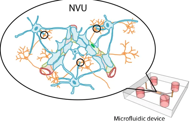
The final goal of this thesis is the engineering of an NVU structure (Fig. 1-8). Main cellular components of NVU are ECs, pericytes, neurons and astrocytes. Morphological features of NVU include microvascular networks, coverage of pericytes and astrocytic endfeet around microvasculatures and neighboring neural networks, which generate neuron-astrocyte-microvascular system. If we can establish a cell culture system of NVU, we can possibly apply the system to studies for neurodegenerative diseases such as Alzheimer’s disease and Parkinson’s disease by a drug screening approach or by a recapitulation of pathological processes and the investigation of the processes.
Previous in vitro NVU models have mainly focused on endothelial barrier functions specific for brain microvasculatures using Transwell cell culture system. In these studies, cellular microenvironment of BBB was mimicked, and endothelial barrier functions
were estimated by TEER approach. However, there is still a gap between the cellular microenvironment in the Transwell system and in vivo BBB microenvironment. In vivo
complex microvascular structure has neither been considered in previous in vitro cell culture systems nor has been engineered by a tissue engineering approach.
One of the potential approaches of the engineering NVU structure in vitro is a microfluidic device. Microfluidic device has a set of microchannels designed to realize specific features of a target tissue or to investigate interactions among different cell types. Tissue engineering approaches can be performed using microfluidic devices because of the capability of culturing cells three-dimensionally within ECM scaffolds to investigate formation processes of a target tissue or to recapitulate morphological features of a target tissue.
To achieve the engineering of functional NVU organization, it is quite important to construct the characteristic structure of NVU including the features of BBB. In this organization, capillary networks can be engineered through the recapitulation of angiogenic processes. As well as capillary networks, neural and glial networks can be engineered by inducing the differentiation of NSCs and morphogenesis of differentiated cells. Therefore, in vitro NVU model can be considered as the combination of angiogenesis model and neurogenesis model. Hence, cell culture conditions for neural cells and ECs were investigated to induce neurogenesis and angiogenesis respectively.
In this thesis, effects of organ-specific ECs on angiogenic microvascular formation and its function were first investigated by culturing brain microvascular ECs (BMECs) or HUVECs with MSCs in chapter 3, which was an essential milestone in the construction of an in vitro BBB model. Next, an in vitro neurogenesis-angiogenesis model was established in chapter 4. In chapter 5, culture conditions for a long-term triculture of NSCs, BMECs and MSCs were investigated to construct a tissue composed of neurons, astrocytes and microvasculatures. Finally, summary and future perspectives were described in chapter 6.
Fig. 1-8 Schematic illustration of an in vitro NVU model in a microfluidic device. The purpose of this study is the construction of an in vitro NVU model which is
composed of neurons (orange), astrocytes (blue) and microvasculatures (red) covered by pericytes (green). Especially, two characteristics of NVU are focused on; coverage of astrocytic endfeet around microvasculatures and close contact between astrocytes and neurons (circles).
Chapter 2 Materials and methods
2-1 Cell culture
(chapter 3, 4, 5)Cells used in this thesis were human brain microvascular endothelial cells (BMECs), human umbilical vein endothelial cells (HUVECs), human H9-ESC derived neural stem cells (NSCs) and human mesenchymal stem cells (MSCs).
BMECs were commercially obtained (Cell Systems Corporation, Kirkland, WA, USA). BMECs were cultured in Endothelial Cell Growth Medium-2 BulletKit (EGM-2, Lonza, Basel, Switzerland) or CSC complete medium (Cell Systems Corporation), depending on experiments. BMECs were expanded in fibronectin-coated culture dishes for no more than 7 passages.
HUVECs were commercially obtained (Lonza). HUVECs were cultured in EGM-2. HUVECs were expanded in fibronectin-coated culture dishes for no more than 7 passages.
NSCs were commercially obtained (GIBCO, Gaithersburg, MD, USA) and expanded in fibronectin-coated culture dishes. NSCs were cultured in NSC medium kit (StemProⓇ NSC SFM, GIBCO): Knockout DMEM/F12 with 2% StemProⓇ Neural Supplement, 1% GlutaMAX, 20 ng/mL basic fibroblast growth factor (bFGF), 20 ng/mL epidermal growth factor and 1% Antibiotic-Antimycotic (GIBCO). NSCs were expanded for no more than 5 passages.
Isolation of MSCs was described in a previous study (Yamamoto et al., 2013). Briefly, MSCs were isolated from human bone marrow using LNGFR (CD271) and Thy-1 (CD90) surface markers. First, bone marrow mononuclear cells (PoieticsTM;
Lonza) were suspended at 1 to 5 × 10⁷ cells/mL in ice-cold Hank’s balanced salt
solution supplemented with 2% fetal bovine serum (FBS), 10 mM HEPES and 1% penicillin/streptomycin. Next, the cells were stained for 30 minutes on ice with the monoclonal antibodies. To eliminate dead cells from the flow cytometric cell sorting
(FACS) system, propodium iodide (2 μg/mL) was used. A triple-laser MoFlo (Beckman Coulter, CA, USA) or FACSVantage SE (Becton Dickinson, Heidelberg, Germany) were used for FACS analysis.
MSCs were used to promote vascular formation and stabilize vascular structures (Yamamoto et al., 2018, 2013). MSCs were cultured in the MSC growth medium: Dulbecco’s Modified Eagle Medium with low glucose (DMEM, Invitrogen, Carlsbad, CA, USA) supplemented with 20% FBS and 1% Antibiotic-Antimycotic. MSCs were expanded for no more than 8 passages.
2-2 Microfluidic device preparation
(chapter 3, 4, 5)Fabrication processes of the microfluidic device used in this thesis were described in detail in a previous study (Shin et al., 2012). Briefly, the microfluidic device was made of poly-dimethylsiloxane (PDMS; Silgard184, Dow Corning, Midland, MI, USA) and was produced by soft lithography with SU-8 patterned wafers. After peeling the cured PDMS from the SU-8 mold, it was cut into pieces and punched out to make media inlets and gel inlets. To form microchannels, each device was plasma-bonded with a coverglass. Microchannels were filled with 1 mg/mL poly-d-lysine solution (Sigma-Aldrich, St. Louis, MO, USA), and the devices were placed in a humidified 5% CO2 incubator at 37°C for overnight. The devices were then rinsed twice with sterile
deionized water and dried at 60°C for 24 hours.
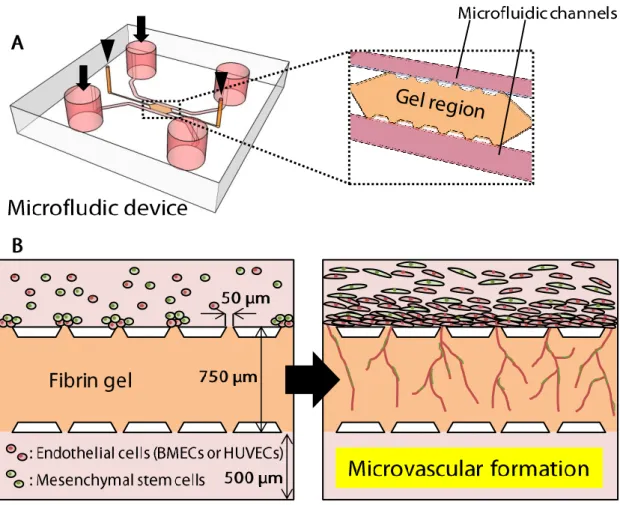
This microfluidic device has three channels. The central channel, which is filled with hydrogel, is sandwiched by two parallel microfluidic channels. The central channel has two gel-inlets which are 1.2 mm in diameter, and two microfluidic channels have two media-inlets which are 4.0 mm in diameter. The height of each channel is 135 μm. The width of the central channel is 750 μm or 1300 μm, which were used depending on experiments, and the width of microfluidic channels is 500 μm.
2-3 Hydrogel filling and cell seeding in microfluidic devices
(chapter 3, 4, 5)Fibrinogen, CorningTM MatrigelTM GFR (Growth Factor Reduced) Membrane
Matrix and hyaluronan (HyStemⓇ Cell Culture Scaffold Kit, Sigma-Aldrich) were used as gel scaffolds for cell culture in microfluidic devices.
Fibrinogen was dissolved in phosphate buffered saline (PBS) to prepare pre-gel solution. The final concentration of fibrinogen solution was 4 mg/mL in chapter 3, and 2 mg/mL in chapter 4 and chapter 5. In chapter 4 and chapter 5, Matrigel and hyaluronan were additionally used with fibrinogen. The final concentration of Matrigel was 2, 4 or 8 mg/mL, depending on experiments. The final concentration of hyaluronan was 2 mg/mL. Pre-gel solutions of Matrigel and hyaluronan were mixed with fibrin pre-gel solution to make fibrin-Matrigel solution and fibrin-hyaluronan solution before injection into microfluidic devices. Pre-gel solution was injected through the two gel inlets immediately after thrombin (10 units/mL, Sigma-Aldrich) was added to the solution to start gelation. After the gel injection, devices were placed in a humidified 5% CO2
incubator at 37 °C for 30 minutes to complete polymerization of the pre-gel solution. When injecting pre-gel solution, it was easily confined within the central channel because of surface tension between PDMS posts (Shin et al., 2012). After gelation, microfluidic channels were filled with culture medium from media inlets. The microfluidic devices were kept in an incubator until use.
BMECs, HUVECs, NSCs or MSCs were suspended in their own culture medium after these cells were dissociated from culture dishes using TrypLE (GIBCO). The cell concentration was 1 × 10⁶ cells/mL for all cell types. After aspirating culture medium
from reservoirs of a microfluidic device, 1 × 10⁴ cells were seeded in the microfluidic
device through media reservoirs. When seeding cells, devices were tilted by 90° to allow cells to adhere to the gel surface between trapezoid posts. After cell seeding, devices were incubated for 30 minutes in a humidified 5% CO2 incubator at 37 °C to
Culture medium was exchanged every day. Phase-contrast images were taken every day to monitor the process of cell migration and morphogenesis.
2-4 Immunocytochemistry
2-4-1 Immunofluorescence staining procedures(chapter 3, 4, 5)
For the imaging of constructed neurites and microvasculatures, cells were fixed with 4% paraformaldehyde for 15 minutes at room temperature and permeabilized with 0.1% Triton X-100 (Sigma-Aldrich) for 5 minutes. After permeabilization, the cells were treated with BlockAce (Dainippon Pharmaceutical, Japan) for 1 hour to block non-specific staining. The cells were then incubated at 4°C overnight with primary antibodies. Thereafter, the cells were incubated with secondary antibodies at room temperature for 2 hours. The cells were finally incubated with 4′,6-diamidino-2-phenylindole (DAPI; Invitrogen) for staining cell nuclei. The cells were rinsed with PBS between each step.
Z-stack fluorescence images were taken by a confocal laser-scanning microscope (LSM 700, Carl Zeiss, Germany). Two-dimensional projection immunofluorescence images were generated with the z-stack images using ImageJ (National Institutes of Health, Bethesda, MD, USA).
2-4-2 Antibodies(chapter 3, 4, 5)
Primary antibodies used in this thesis are as follows: (product name; source; catalog number; company; dilution)
BMECs and HUVECs
Human CD31/PECAM-1 Antibody; sheep; AF806; R&D Systems; 1:100.
VE-Cadherin (D87F2) XPⓇ Rabbit mAb; rabbit; #2500; Cell Signaling Technology; 1:100.
ZO-1 Monoclonal Antibody (ZO-1-1A12); mouse; 33-9100; Thermo Fisher Scientific; 1:50.
Occludin Polyclonal Antibody; rabbit; 71-1500; Thermo Fisher Scientific; 1:100.
Pericytes differentiated from MSCs
Monoclonal Anti-Actin, α-Smooth Muscle antibody produced in mouse; mouse; A2547; Sigma-Aldrich; 1:200.
Anti-PDGFR beta antibody [Y92] – C-terminal; rabbit; ab32570; abcam; 1:100.
Human NG2/MCSP PE-conjugated Antibody; mouse; FAB2585P; R&D Systems; 1:50.
Basement membranes
Anti (mouse) Laminin; mouse; LB-1013; LSL; 1:200.
Anti Collagen IV α5(IV) Chain, Human (Mono); rat; C-452-EX; Chigei Medical Research Institute; 1:50.
NSCs
Purified anti-Tubulin β3 (TUBB3) [Tuj1]; mouse; 801202; BioLegend; 1:500.
Microtubule associated protein 2 (MAP2) Antibody (M13); mouse; 13-1500; Thermo Fisher Scientific; 1:100.
GFAP Polyclonal Antibody; rabbit; PA5-16291; Thermo Fisher Scientific; 1:100.
Anti-Galactocerebroside Antibody, clone mGalC; mouse; MAB342; Merck Millipore; 1:100.
Anti-Human/Mouse SOX2 Monoclonal Antibody; mouse; MAB2018; R&D Systems; 1:100.
Anti-Nestin antibody – Neural Stem Cell Marker; rabbit; ab92391; abcam; 1:200.
Secondary antibodies (1:100 dilution) and the antibody for staining cell nuclei (DAPI, 1:250 dilution) used in this thesis were purchased from Invitrogen.
2-5 Quantitative analysis
2-5-1 Microvasculatures(chapter 3, 4)Quantitative analyses were performed for sprout and microvascular length, sprout number, branch points, vessel outer diameter and pericyte number. Microvascular length was quantified using z-stack immunofluorescence images of engineered microvasculatures. After 2D projection images were generated with z-stack images of microvasculatures, sprout length and microvascular length in the 2D projection images were traced using the freehand line tool in ImageJ. The length of the traced lines was measured to quantify sprout length and microvascular length. Sprout number and branch point were quantified by manually counting the sprout number and branch points in each image. Vessel diameter was measured at 1/3 and 2/3 of the width of the central channel along the Y-axis. Pericyte number was quantified by counting α-SMA-expressing cell nuclei on the outer surface of microvascular configurations.
Quantitative analyses of junction proteins were also performed using ImageJ. After thresholding each immunofluorescence image, image calculator, a plugin of ImageJ, was used to calculate fluorescence intensity of junction proteins in ECs constituting microvasculatures.
2-5-2 Neurites(chapter 4)
Neurite length was measured by the same method of measuring microvascular length. To quantify the number of three-dimensionally migrating cells, cell nuclei which located at least 15-μm far from the top or bottom surfaces of a 3D hydrogel scaffold were counted using ImageJ. This counted cell number was divided by the total cell number in the 3D hydrogel scaffold to calculate the percentage of cells in 3D region.
2-5-3 Gel collapse in triculture of NSCs, BMECs and MSCs(chapter 4)
gel collapse was performed using ImageJ by surrounding the devoid area in each z-projection image.
2-5-4 Dextran perfusion and measurement of microvascular permeability coefficient(chapter 3)
To analyze microvascular permeability, fluorescein isothiocyanate-conjugated dextran (FITC-dextran, 2000 kDa) or Texas Red-conjugated dextran (70 kDa) were introduced into engineered microvasculatures. After aspirating culture medium from reservoirs of a microfluidic device, 10 μL of 50 μg/mL FITC-dextran or Texas Red-dextran solution was added to a microchannel containing seeded ECs and MSCs. Fluorescence images were obtained every 10 seconds using the 20 × objective lens of a confocal laser-scanning microscope (LSM 700).
Permeability coefficients (P) of BMEC/HUVEC microvasculatures were calculated using Fick’s First Law. In general, Fick’s First Law is represented by the following equation:
𝐽 = −𝐷𝑑𝑐m
𝑑𝑥 (1)
where J is the flux of solute across microvascular wall, D is the diffusion coefficient of solutes in endothelial monolayer constituting microvascular wall and cm is the
concentration of the solute inside EC membrane. When the width of the cell membrane is given by d, concentrations of solutes at the intravascular border (cm (0)) and
extravascular border (cm (d)) are represented using a partition coefficient β as:
𝑐m(0) = β𝑐1 (2) 𝑐m(𝑑) = β𝑐2 (3)
where c1 is the concentration of solutes in microvascular lumen and c2 is the
concentration of solutes in extravascular region. Substitution of equations (2) and (3) into equation (1) yields:
𝐽 = −𝐷𝑑𝑐m 𝑑𝑥 = −𝐷 𝑐m(𝑑) − 𝑐m(0) 𝑑 = 𝐷β(𝑐1− 𝑐2) 𝑑 (4)
Equation (4) shows that the flux is in proportion to the concentration difference between intravascular region and extravascular region. It is difficult to determine each proportionality constant of Dβ/d individually. Therefore, these constants are expressed in a single constant P as a permeability coefficient, yielding the following equation.
𝐽 = 𝑃(𝑐1− 𝑐2) (5)
Permeability coefficient is calculated from the flux of solute for a concentration difference (c0) across an area of microvascular wall (S) using Fick’s First Law
(Adamson et al., 1994; Bichsel et al., 2015; Kim et al., 2013).
𝑃 = 𝐽 × 1
𝑐0 (6)
The flux J is calculated from the rate of the transport of dextran molecules (N) across the microvascular wall after the microvasculature was filled with the dextran solution.
𝐽 =𝑑𝑁 𝑑𝑡 ×
1
𝑆 (7)
The concentration c0 is represented using the amount of dextran molecules that initially
fill the microvasculature.
𝑐0 =𝑁0
𝑉 (8)
where V is the volume of the microvasculature. It is assumed that the number of solute molecules was proportional to the fluorescence intensity (I), substitution of equations (7) and (8) into equation (6) yields
𝑃 =𝑉 𝑆×
𝑑𝐼 𝑑𝑡⁄
𝐼0 (9)
where I0 is the initial intravascular fluorescence intensity, and dI/dt is the change in fluorescence intensity per unit time in the extravascular region. It was assumed that the microvascular lumens were circular.
𝑆 = 2π𝑟𝑙 (10) 𝑉 = π𝑟2𝑙 (11)
where r is the microvascular radius and l is microvascular length. Substitution of equations (10) and (11) into equation (9) yields
𝑃 =𝑟2×𝑑𝐼 𝑑𝑡𝐼⁄
0 (12)
Permeability coefficient P was calculated using equation (12). 2-5-5 Statistical analysis
Experiments were repeated three times to confirm the reproducibility of results. Data were represented as means ± SEM. Welch’s t-test was used to test for differences between the two groups, which were considered statistically significant at p < 0.05.
Chapter 3 Comparison of organ-specific endothelial
cells in terms of microvascular formation and
endothelial barrier functions
3-1 Introduction
Microvascular networks are essential for correct organ functioning. Each organ has its own vascular characteristics and functions that are maintained through interactions between ECs and parenchymal cells within vascular niches (Géraud et al., 2014). Angiocrine and paracrine factors secreted by ECs regulate organ homeostasis and function (Rafii et al., 2016), and the fundamental function of microvascular networks is to deliver oxygen and nutrients throughout organs. ECs that coat microvascular walls adhere to each other through junction proteins such as VE-cadherin, occludin, and ZO-1 (Abbott et al., 2010; Greene and Campbell, 2016). While the transport of macromolecules and undesired substances such as neurotoxins out of the microvasculature is strongly inhibited by endothelial barrier functions, ECs regulate the active transport of nutrients and oxygen into the organ parenchyma by paracellular and transcellular pathways (Abbott et al., 2010; Barar et al., 2016; Greene and Campbell, 2016). In addition to this universal function, ECs display slightly different features depending on the organ in which they reside. Notably, brain ECs cooperate with astrocytes and pericytes to form a characteristic vascular microenvironment called BBB (Zhao et al., 2015). Brain microvascular networks strictly prohibit the transport of toxic substances to the brain parenchyma and effectively transfer compounds essential for brain homeostasis. On the other hand, ECs of the kidney glomeruli and liver effectively maintain active transport of blood-borne molecules to the parenchyma (Ben-Zvi et al., 2014; Géraud et al., 2014). In addition to these morphological features of organ-specific
ECs, ECs from different organs exhibit tissue-specific expression profiles of transcription factors, angiocrine growth factors, adhesion molecules, and chemokines (Nolan et al., 2013). Such differences in both the structural organization and function of organ-specific ECs suggest that organ specificity should be considered when investigating interactions between ECs and other cell types constituting a target organ.
The brain has organ-specific ECs that play important roles in the functioning of the BBB. Many studies have investigated BBB development and functions because the regulation of the BBB is important for both drug delivery and drug screening (Banerjee et al., 2016; Bicker et al., 2014). Transport properties of the brain capillary endothelium and interactions between ECs and perivascular cells, such as pericytes and astrocytes, have also been the focus of previous studies (Banerjee et al., 2016; Helm et al., 2016; Keaney and Campbell, 2015; Sweeney et al., 2018). By culturing ECs with pericytes and/or astrocytes, cell culture systems have been observed to mimic in vivo
microvascular environments. In some studies, a Transwell cell culture system was used to obtain TEER measurements that were further analyzed to estimate endothelial barrier functions. These studies established that endothelial barrier function is augmented when ECs are cultured with astrocytes and pericytes. However, the ECs in these studies formed a two-dimensional (2D) monolayer rather than a native in vivo
three-dimensional (3D) capillary structure. It has also been reported that ECs isolated from brain capillaries tend to lose their barrier properties (Banerjee et al., 2016). In addition, ECs are covered by pericytes and astrocytic endfeet and the percentage of pericyte coverage around capillaries are the highest in the brain compared to the other organs (Armulik et al., 2010; Daneman et al., 2010; Keaney and Campbell, 2015; Thomsen et al., 2017; Trost et al., 2016; Zlokovic, 2008). These fundamental features of brain capillaries were not included in previous in vitro studies, highlighting the disparity between in vitro BBB models and native EC conditions in vivo.
To bridge this gap, it is imperative that a successful 3D brain microvascular network be constructed. An in vitro BBB model requires two key features: the construction of
functional capillary-level microvascular networks and the coverage of pericytes and astrocytic endfeet around the microvasculatures. The construction of capillary-level microvasculatures is particularly challenging, especially in terms of capillary diameter: indeed, most previously reported microvasculatures have been between 20 and 100 μm in diameter (Kim et al., 2015, 2013; Nguyen et al., 2013), much larger than that of real capillaries. Toward the establishment of an in vitro BBB model, construction of capillary-level microvascular networks with pericyte coverage was first focused on. Previous studies that have constructed in vitro microvascular networks have used microfluidic cell culture platforms in which cells were cultured in 3D gel scaffolds mimicking an in vivo ECM (Kim et al., 2015; Kim et al., 2013). We created an angiogenesis model using ECs and MSCs (Yamamoto et al., 2018). HUVECs were used in these studies.
In this chapter, a microfluidic cell culture platform was developed by coculturing BMECs and MSCs, an essential milestone in the construction of an in vitro BBB model. To compare the characteristics of organ-specific ECs, HUVECs were also cultured with MSCs as opposed to BMECs. MSCs were used in the coculture because of their ability to differentiate into pericytes both in vivo (Koike et al., 2004) and in vitro (Yamamoto et al., 2018, 2013). Whether organ-specific ECs would affect vascular formation processes and whether microvascular networks constructed using such ECs would exhibit their own characteristics in terms of endothelial barrier functions and fundamental morphology were assessed in this chapter.
3-2 Experimental design
Detailed methods for the experiments described in this chapter are summarized in chapter 2. Here, experimental settings are described below.
Microfluidic device:
Schematic illustrations of the microfluidic device used in this chapter are shown in Fig. 3-1. This microfluidic device has three microchannels. The central channel is used as cell culture channel by injecting gel scaffolds. Two parallel microfluidic channels are used as media channels.
The width of the central channel is 750 μm, The width was determined by preliminary experiments. When ECs and MSCs are seeded in one microfluidic channel in the 750-μm device, they elongate vascular structures and these vasculatures reach the opposite side of the channel about 5-7 days. The penetration of microvasculatures across the central channel is needed to perfuse dextran solutions into the microvasculatures to investigate endothelial barrier functions. Therefore, we used the 750-μm device for EC-MSC cocultures.
The width between trapezoidal posts is 50 μm, which is determined by following reasons. First one is to prevent the leakage of gel solutions out of the central channel when injected. Gel solutions are easily fixed in the central channel because of the surface tension between these posts (Huang et al., 2009; Shin et al., 2012). Second is to minimize the undesired leakage of fluorescent dextran solution from microfluidic channel to the central channel. When constructing microvasculatures, some spaces exist between microvasculatures and trapezoidal posts. Some of the solutes of fluorescent dextran solution can pass through the space, which may diffuse throughout the central channel. To avoid this, the width between each post was set to be 50 μm. Microfluidic devices whose width between posts was 30 μm was tried to make, only to find it difficult to produce these devices stably.
Cells:
BMECs, HUVECs and MSCs were used in this chapter. The amount of all cell types seeded in each device was 1.0 × 10⁴ cells. The ratio of ECs and MSCs was 1:1, which
was optimized in a previous study (Yamamoto et al., 2013).
Angiogenesis is the growth of new blood vessels from pre-existing ones. Endothelial vascular sprouts are formed by degrading surrounding ECM around blood vessels through production of matrix metalloproteinases (MMPs). ECs and MSCs were seeded on the gel surface between each post in order to mimic angiogenic processes in a microfluidic device. Cultured ECs proliferate and form vascular-like structure inside the microfluidic channel where cells are seeded. Some ECs migrate into the gel scaffold and form vascular sprouts. These sprouts are elongated and become mature by the coverage of pericytes which are differentiated from MSCs. It was reported that the differentiation of MSCs into pericytes was induced by culturing MSCs with ECs (Koike et al., 2004; Yamamoto et al., 2018).
Medium:
For all experiments in this chapter, 1:1 mixture of EGM-2 and MSC growth medium was used.
Additional supplement to medium:
The medium was supplemented with 10 ng/mL VEGF and 10 ng/mL bFGF to promote cell proliferation and microvascular formation.
Gel scaffold:
Fibrin (4 mg/mL) was used for all experiments in this chapter because fibrin has been widely used as a gel scaffold for EC culture to induce angiogenic processes in vitro