Research Journal of Pharmaceutical, Biological and Chemical
Sciences
Antiviral Activity of Marine Sponges Homaxinella tanitai and Microxina
Subtilis against Hepatitis C Virus.
Suciati
1*,
Irsyad Abdillah
1, Muhammad Ihsan Uddin
1, Achmad Fauzi
1, Myrna Adianti
2, and
Achmad Fuad
1,2.
1Department of Pharmacognosy and Phytochemistry, Faculty of Pharmacy Universitas Airlangga, Jl. Dharmawangsa Dalam
Surabaya 60286, East Java, Indonesia;
2Institute of Tropical Diseases, Universitas Airlangga, Jl. Mulyorejo, Surabaya 60115, East Java Indonesia
ABSTRACT
The aim of the study was to screen antiviral activity of marine sponges extracts and fractions against hepatitis C virus, as well as to investigate the mode of action of the extract. The results showed that extracts of marine sponges Homaxinella tanitai dan Microxina subtilis inhibited the growth of HCV at IC50 of 27.1 and 40.5
µg/mL, respectively. Three fractions from H. tanitai and two fractions from M. subtilis gave more than 50% inhibiton againts HCV at concentration 100 µg/mL. The time of addition experiment revealed that H. tanitai
and M. subtilis extracts both act at enty and post entry steps. These results suggest that H. Tanitai and M. subtilis extracts could serve as potential candidates for antiviral agents against hepatitis C virus.
Keywords: antiviral, hepatitis C virus, marine sponges, Homaxinella tanitai, Microxina subtilis
INTRODUCTION
Hepatitis C virus (HCV) is a positive-stranded RNA virus in the genus Hepacivirus of the family Flaviviridae. HCV has been considered as causatif agent of both acute and chronic hepatitis leading to the development of hepatic cirrhosis and hepatocellular carcinoma (1,2). Approximately 130-150 million people globally infected by hepatitis C, and estimated 700.000 people die each year from hepatitis C (World Health Organization, 2016). Based on the heterogeneity of the viral genome, HCV is currently classified into seven genotypes (3–9) and more than 67 subtypes (1a,1b, 2a, 2b etc.) (3, 4). The viral genome, which is a singlestranded open reading encoding a polyprotein precursor consisting of 3000 amino acid residues that is processed by virus-encoded and host cellular proteases into structural and nonstructural proteins. The HCV proteins also play essential roles in the pathological processes associated with HCV infection, such as carcinogenesis as well as glucose and lipid metabolic disorders (4, 5). Current standar therapy for hepatitis C is using triple combination of pegylated interferon, ribavirin and specific NS3 serine protease inhibitor, such as telaprevir and boceprevir. The treatment is considered effective with viral clearance rate to >70% (10,11,12). However, it cause severe side effect, such as anemia and skin rashes from the use of telaprevir and boceprevir, and the presence of drug-resistence virus. Therefore the search for effective and safer therapy for hepatitis C by using natural resources, such as marine sponges is still needed.
The oceans, which occopies over 70% of the Earth’s surface, have been the habitat of various living creatures, including algae, sponges, cnidarians, molluscs, bryozoans, ascidians and echinoderms as well as microorganisms. This species diversity makes the marine environment one of the most prolific sources of natural products. Many of these marine creatures produce unique and biologically active compounds which may not be found in the terrestrial ecosystem. These metabolites may be produced as a means of self-defence against predation, since many marine organisms have no spine or protective shell (13). Amongst marine resources, sponges have been the focus of study for many years. Marine sponges are simple, multicellular, and sessile invertebrates with approximately 15.000 species have been discovered WorldWide. More than 5300 metabolites have been reported from this organism (14). Many of these metabolites showed pronounced bioactivity including antiviral.
In this study we investigated anti HCV activity of ethyl acetate extracts of Homaxinella tanitai and
Microxina subtilis collected from Barrang Lompo Island, South Sulawesi, Indonesia. Flash column fractions of the sponge extracts were also subjected to anti HCV assay. In order to determine the mode of action of the extracts time of addition experiments were conducted.
MATERIAL AND METHODS
Biological Material
Sponges were collected by using SCUBA at a depth of 8-10 m from around Barrang Lompo Island, Makassar, South Sulawesi on May 17th 2014. Samples were kept in plastic pack in ice boxes immediately after collection. Sponge specimens were then freezed at -20°C until analysis. Identification of sponges were conducted by Ecology Laboratory, Department of Biology, Faculty of Mathematic and Sciences, Institut Teknologi Sepuluh November, Surabaya. Voucher specimens were kept in etanol 70% at the Faculty of Pharmacy, Universitas Airlangga under the accession number 17-5-14-3 and 17-5-14-4.
Extraction and fractionation
Anti HCV Assay
The protocol as has been described in (15,16,17). Huh7it cells were seeded in 48-well plates (5×10⁴
cells/well) a day before infection. The same volume of JFH1a, with multiplication of infection (MOI) of 0.1 focus-forming units (ffu)/cell,was mixed with serial dilutions of the extracts (100, 50, 25, 12.5, 6.25 and 3.1
μg/ml) and inoculated to the cells. Single concentration of 100 μg/ml samples were used for flash column fractions, as well as for time of addition experiments. The mixture was incubated at 37°C for 2 h, and the cells were rinsed twice with serum-free medium to remove residual virus, followed by incubation for an additional 46 h with the same medium. At 48 h p.i., the culture supernatants were collected and used for virus titration.
Virus titration was conducted by placing Huh7it-1 cells (2.4 × 10⁴ cells/well) in a 96-well plate 1 day
prior to virus infection. Culture supernatants obtained from HCV-infected cells were serially diluted 25-fold in culture medium and inoculated to the cells. The virus was adsorbed to the cells at 37°C for 2 h, followed by incubation for 46 h with a medium containing 0.4% methylcellulose (Sigma-Aldrich). HCV titers were conducted using a focus formation assay. HCV antigen-positive cells were stained with HCV-infected patient’s serum and horseradish peroxidase-conjugated goat anti-human IgG (MBL, Tokyo, Japan). A metal enhanced DAB substrate kit (Thermo Fisher Scientific Inc., Rockford, IL, USA) was used to detect the infectious foci, which were then imaged and counted using the katikati counter software.
The mode of action of the extracts were examined by conducting time of addition experiments. Two sets of experiments were done in parallel: (i) to assess the antiviral effect at the entry step, a mixture of HCV and sample was inoculated into the cells. After virus adsorption for 2 hours, the residual virus and the sample were removed. The cells were then refed with fresh medium without sample for 46 hours; (ii) to assess the antiviral effect at the post entry step, HCV was inoculated to the cells in the absence of the sample. After virus adsorption for 2 hours, medium containing samples were then added followed by incubation at 46 hours; (iii) as a positive control, HCV mixed with the sample was inoculated to the cells. After virus adsorption for 2 hours, the residual virus and the sample were removed, and cells were refed with fresh medium containing the sample for 46 hours. Culture supernatants were obtained at 1 and 2 days post-infection (dpi) and titrated for virus infectivity. Virus and cells treated with medium containing 0.1% DMSO served as a control. The percentage of inhibition of the samples against the virus were then calculated by comparing to the control.
Cytotoxicity Assay
The cytotoxicity of the samples was determined by MTT method as has been described previously (17). The method was based on colorimetric reaction of 3-(4,5-dimethylthiazol-2-yl)-2,5-diphenyltetrazolium bromide with enzyme dehydogenase inside living cells to form a coloured formazan dye, which corresponded to the number of viable cells. In this assay Huh7it cells were mixed serial dillution of the samples or control in 96 well plates. After 46 hours incubation condition of the cells were observed under microscope. The medium was removed from 96 well plates and then 150 μl/well of MTT 10% was added, followed by incubation for 4
hours at 37°C. The MTT solution was removed from 96 well plates and 100 μl/well of DMSO 100% was then
added. Reaction mixtures were homogenize by shaking for 0.5 min before measurement of absorbance at 560 nm and 750 nm.
RESULTS
Anti HCV activities of Homaxinela tanitai and Microxina subtilis extracts
The ethyl acetate extracts of H.tanitai and M. subtilis showed anti-HCV activities with 50%-inhibitory concentration (IC50) of 27.1 μg/ml and 40.5 μg/ml, respectively (Table 1). Cytotoxicity of the crude extract
against Huh7it-1 cells was determined using MTT assay. Severe cytotoxicity was not observed at the concentrations tested and the CC50 value was > 100 μg/ml. The selectivity index was evaluated (SI; CC50/IC50);
Table 1. IC50 and CC50 values of H. tanitai and M. subtilis extracts by using Sepacore® flash column chromatography, employing gradient elution of n-hexane, ethyl acetate and methanol in order of increasing polarity to obtain 7 fractions. Each fractions were then submitted to anti HCV assay using single concentration of 100 μg/ml. The percentage of inhibition of each fractions was calculated (Table 2).
Table 2. Anti HCV activities of H. tanitai and M. subtilis fractions
Time of addition experiments were performed to determine the possible mode of action of the extracts. Single concentration of extracts were used in this experiment. Tested samples were added to the cell only during viral inoculation or only after viral inoculation for the remaining culture period until virus harvest. The results (Table 3) suggested that both extracts inhibited HCV activities at both entry and post-entry steps.
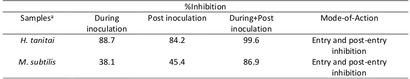
Table 3. Time of addition analysis of H tanitai and M. subtilis extracts
%Inhibition
Marine sponges have been the fruitful source of many bioactive metabolites, including antiviral. Several marine sponges have been reported to have anti HCV activity, such as Amphimedon sp.(18) and undescribed species of the genus Latrunculia (19). The ethyl acetate extract of Amphimedon sp. showed inhibition of both protease and helicase activities of hepatitis C virus NS3. Anti HCV metabolites have also been succesfully isolated from marine sponges, such as halisulfate 3 and suvanine both obtained from unidentified Demospongiae spong derived from Okinawa Japan. Both compounds inhibited NS3 helicase-activity in a dose dependent manner with IC50 values of 4 and 3 µM, respectively (12).Another sponge metabolite reported to
have anti HCV activity is a series of alkaloid discorhabdins, i.e discorhabdins A, C and dihydrodiscorhabdin C
In this study ethyl acetate extracts of H. tanitai and M. subtilis exhibited anti HCV activity with IC50
values of 27.1 and 40.5 µg/mL, respectively. In addition, both extract did not dispaly cytotoxicity at the highest concentration tested 100 µg/mL. Flash column chromatography on the extracts were carried out, and obtained seven fractions, which then subjected to anti-HCV assay. The results (Table 2) showed that three fractions from
H. tanitai and two fractions from M. subtilis gave > 50% inhibitions against HCV at concentration of 100 µg/mL. In addition, these results also indicated that the possible anti HCV metabolites of H. tanitai and M. subtilis are semi polar or polar compounds, in which eluted in combination of ethyl acetate and methanol from flash column chromatography.
In order to determine the mode of action of extracts, whether it exerted the effect on the entry or post entry steps, time of addition experiments were carried out. To examine the HCV effect at the entry step, sample was only added at the first 2 h of experiment, followed by incubation of cells with fresh media without sample for 46 h. Meanwhile, to determine whether sample act the post entry step, virus was inoculated into the cell in the absence of sample, after virus adsorption for 2 h, sample was added and incubated for 46h. The results (Table 3) showed that extract of H. tanitai exhibited 88.7 and 84.2 % inhibition against HCV at 99.6 µg/mL during inoculation and post inoculation, respectively. However, better inhibition (100%) of extract was obtained when it was added through out experiment. This result suggested that H. tanitai extract act at both entry and post entry steps. At the same concentration extract of M. subtilis showed lower inhibition againts HCV compare to extract of H. tanitai at 38.1 and 45.4 % during inoculation and post inoculation, respectively. However, the extract gave higher inhibition at 86.9%, when it was added in both steps (during and after inoculation), which indicated that M. subtilis extract exerted the activity at entry and post entry steps.
CONCLUSION
The results of this study suggest that the ethyl acetate extracts and fractions of marine sponges
Homaxinella tanitai and Microxina subtilis exhibit antiviral activity against hepatitis C virus. Both extracts exerted the effect at entry and post entry steps.
ACKNOWLEDGEMENT
Authors acknowledge the Ministry of Research, Technology and Higher Education of the Republic of Indonesia for research grant PUPT under contract number 018/SP2H/LT/DRPM/II/2016.
REFERENCES
[1] Baldo V, Baldovin T, Trivello R, Floreani A. Curr Pharm Des 2008; 14: 1646–1654. [2] Seeff LB. Hepatology 2002; 36: S35–46.
[3] Gottwein JM, Scheel TK, Jensen TB, Lademann JB, Prentoe JC, Knudsen ML, Hoegh AM, Bukh J. Hepatology 2009; 49: 364–77.
[4] Smith DB, Bukh J, Kuiken C, Muerhoff AS, Rice CM, Stapleton JT, Simmonds P. Hepatology 2013; 59: 318–27.
[5] Moradpour D, Penin F, Rice CM Nat Rev Microbiol 2007; 5: 453–6. [6] Shoji I, Deng L, Hotta H. Front Microbiol 2011; 2: 278.
[7] Arzumanyan A, Reis HM, Feitelson MA. Nat Rev Cancer 2013; 13: 123–35. [8] Shepard CW, Finelli L, Alter MJ. Lancet Infect Dis 2005; 5: 558–67.
[9] Mohd Hanafiah K, Groeger J, Flaxman AD, Wiersma ST. Hepatology 2013; 57: 1333–42 [10] Ghany MG, Nelson DR, Strader DB, Thomas DL, Seeff LB. Hepatology 2011; 54: 1433–1444. [11] Liang TJ, Ghany MG. N Engl J Med 2013; 368: 1907 – 1917.
[12] Furuta A, Salam KA, Hermawan I, Akimitsu N, Tanaka J, Tani H, Yamasita A, Moriishi K, Nakoshi M, Tsubuki M, Peng PW, Suzuki Y, Yammoto N, Sekiguchi Y, Tsuneda S, Noda N. Mar Drugs 2014; 12: 462
– 476.
[13] Whitehead R. Annu. Rep. Prog. Chem. Sect. B 1999; 95: 183-205.
[14] Ye J, Zhou F, Al-Kareef AMQ, Wang H. J Asian Nat Prod Res 2015; 17: 1 – 23.
[16] Aoki C, Hartati S, Santi MR, Lydwina, Firdaus R, Hanafi M, Kardono LBS, Shimizu Y, Sudarmono P, Hotta H. Int J Pharm Pharm Sci 2014; 6: 211 – 215.
[17] Tumewu L, Apryani E, Santi MR, Wahyuni TS, Permanasari AA, Adianti M, Aoki C, Widyawaruyanti A, Hafid AF, Lusida MI, Soetjipto, Hotta H. Procedia Chemistry 2016; 18: 169 – 173.
[18] Fujimoto Y, Salam KA, Furuta A, Matsuda Y, Fujita O, Tani H, Ikeda M, Kato N, Sakamoto N, Maekawa S, Enomoto N, de Voogd NJ, Nakakoshi M, Tsubuki M, Sekiguchi Y, Tsuneda S, Akimitsu N, Noda N, Yamashita A, Tanaka J, Moriishi K. Plos One 2012; 7: 1 – 12.