University of Zagreb
FACULTY OF SCIENCE
DEPARTMENT OF BIOLOGY
Maja Mucko
MORPHOLOGY AND PHYLOGENY OF
PICOEUKARYOTES AND PLANKTONIC
PENNATE DIATOMS IN THE MIDDLE AND
SOUTH ADRIATIC SEA
DOCTORAL THESIS
Sveučilište u Zagrebu
PRIRODOSLOVNO-MATEMATIČKI FAKULTET
BIOLOŠKI ODSJEK
Maja Mucko
MORFOLOGIJA I FILOGENIJA
PIKOEUKARIOTA I PENATNIH
PLANKTONSKIH DIJATOMEJA U
SREDNJEM I JUŽNOM JADRANU
DOKTORSKI RAD
This doctoral dissertation was carried out as a part of the postgraduate programme at University of Zagreb, Faculty of Science, Department of Biology – Botany, under the supervision of dr.sc. Zrinka Ljubešić. The research was performed in the frame of the Bio-tracing Adriatic Water Masses (BIOTA) project, supported by the Croatian Science Foundation (project number UIP-2013-11-6433); project leader dr.sc. Zrinka Ljubešić). The experimental part of the research was carried out in part at Ruđer Bošković Institute, Zagreb, Croatia, while part of the bioinformatic analyses was carried out at University of Arkansas, Fayeteville, USA.
Acknowledgments
Firstly, I would like to express my sincere gratitude to my supervisor Dr Zrinka Ljubešić for the continuous support of my Ph.D. study and related research, for her patience, guidance, motivation, and immense knowledge. She helped me in all phases of research and writing of this thesis. I could not have imagined having a better supervisor for my Ph.D. study.
In addition to my supervisor, I would like to thank my thesis committee: Dr Daniela Marić Pfannkuchen, Dr Sunčica Bosak and Dr Regine Jahn, for their insightful comments and encouragement, but also for the hard questions, which motivated me to widen my research from various perspectives.
My sincere thanks also goes to Dr Sandi Orlić and Dr Petra Peharec Štefanić, who enabled me access to the laboratory and research facilities without which I would not be able to examine my samples. I would also like to thank my ‘over the sea’ colleagues and co-authors Dr Teofil Nakov, Dr Elizabeth Ruck, Dr Daniel Vaulot, Dr Romain Gastineau, Dr David G Mann and Dr Rosa Trobajo for their sincere guidance and fruitful discussions. Without they precious support it would not be possible to conduct this research.
I thank my fellow lab mates, current and previous, colleagues from abroad, students, interns and
colleagues passing through the lab or cafeteria☺. Thank you Sunčica, Maja, Anamarija, Pero, Kora,
Tonka, Mirela, Ana, Tihana, Tanja, Mišel, Dorotea, Zuzanna, Chetan, Solenn, Karoline, Mirta, Veronika, Nikola; for the stimulating discussions, for help, support and encouragement, and for all the fun we have had in the last four years. Additionally, I would like to thank the rest of co-authors on research publications involved in this thesis.
I would also like to thank my girls, my best friends, who were, are, or will be on the same life journey, professionally and privately, for their help, patience, listening, encouraging, laughing, partying, crying, and experiencing all the other feelings in between… Thank you Petra, thank you Marina, thank you Sany, thank you Franka, thank you Zrinka! Priatel’ce 4ever ☺
I would like to thank my family: my parents Pero and Ljiljanka, for their irreplaceable love and support, to my brother Danijel and my sisters Martina and Petra for supporting me spiritually throughout writing this thesis and my life in general.
In the end, I want to thank my husband Antonio, for his overwhelming love, support, for being my soulmate and my rock, for giving me much needed advice, for being just the opposite to my headstrong soul in unpredictable situations, for giving me higher purpose in my life, for being just my love…
University of Zagreb Doctoral thesis
Faculty of Science
Department of Biology
MORPHOLOGY AND PHYLOGENY OF PICOEUKARYOTES AND PLANKTONIC PENNATE DIATOMS IN THE MIDDLE AND SOUTH ADRIATIC SEA
MAJA MUCKO
Faculty of Science, Department of Biology
The smallest protists, generally called picoeukaryotes, and planktonic pennate diatoms have received limited attention in world oceans, although they are numerous and important for primary production, biogeochemical cycles and for carbon injection into the deep ocean. In this thesis, represented through seven scientific publications, cultivated planktonic pennate diatoms and picoeukaryotes were investigated with traditional methods (light and electron microscopy, morphometry) and with molecular analyses, including multi-gene phylogenies, and were identified as genera Entomoneis, Pseudo-nitzschia, Haslea and Picochlorum. Detailed investigations of genus Entomoneis resulted with description of seven new species. General picoeukaryotic diversity was addressed by eDNA amplicon sequencing V4 variable region of nuclear 18S rRNA gene. Obtained results identified 95% of the picoeukaryotic community as hetero- or mixotrophic, mostly belonging to parasitic dinoflagellates and radiolarians, while only 5% of the community was represented with photoautotrophic picoeukaryotes belonging to classes of green algae, haptophytes, stramenopiles and cryptophytes. One cultivated Picochlorum sp. strain was subjected to growth rate experiment and pigment and lipid analyses, resulting in interesting and promising data for future biotechnological studies.
(52 pages, 3 figures, 232 references, original in English)
Keywords: picoeukaryotes, planktonic pennate diatoms, morphology, phylogeny
Supervisor: Dr Zrinka Ljubešić, Associated Professor
Reviewers: Dr Daniela Marić Pfannkuchen, Scientific collaborator
Dr Sunčica Bosak, Assistant Professor
Sveučilište u Zagrebu Doktorski rad Prirodoslovno-matematički fakultet
Biološki odsjek
MORFOLOGIJA I FILOGENIJA PIKOEUKARIOTA I PENATNIH PLANKTONSKIH DIJATOMEJA U SREDNJEM I JUŽNOM JADRANU
MAJA MUCKO
Prirodoslovno-matematički fakultet, Biološki odsjek
Najmanji protisti, poznati kao pikoeukarioti i penatne planktonske diatomeje često su zanemareni u istraživanjima svjetskih oceana, iako predstavljaju brojnu i važnu komponentu za primarne proizvodnje, biogeokemijskih ciklusa i značajni su za protok ugljika u duboki ocean. U sklopu ove disertacije kroz sedam znanstvenih publikacija navedene skupine organizama istraživane su tradicionalnim metodama (svjetlosna i elektronska mikroskopija, morfometrija) i molekularnim analizama koje uključuju filogenetske anlize sa više različitih genskih markera te su identificirane kao rodovi Entomoneis, Pseudo-nitzschia, Haslea i Picochlorum. Detaljna analiza roda Entomoneis rezultirala je opisom sedam novih vrsta za znanost. Bioraznolikost ukupne pikoeukariotske zajednice u južnom Jadranu odredila se sekvenciranjem amplikona okolišne DNA i to prema varijabilnoj V4 regiji nuklearnog 18S rRNA gena. Dobiveni rezultati identificirali su 95% pikoeukariotske zajednice kao hetero- ili miksotrofne organizme, većim dijelom pripadajući parazitskim dinoflagelatima i radiolarijama, dok je samo 5% zajednice zastupljeno fotoautotrofnim pikoeukariotima koji pripadaju razredima zelenih algi, haptofita, stramenopila i kriptofita. Jedan kultivirani soj alge Picochlorum sp. istražen je kroz eksperiment određivanja brzine rasta te analizama pigmenta i lipida, što je rezultiralo zanimljivim i obećavajućim rezultatima koji mogu poslužiti kao okosnica za buduće biotehnološke studije. (52 stranice, 3 slike, 232 literaturna navoda, jezik izvornika: engleski)
Ključne riječi: pikoeukarioti, penatne planktonske dijatomeje, morfologija, filogenija Mentor: Izv. prof. dr. sc. Zrinka Ljubešić
Ocjenjivači: Dr. sc. Daniela Marić Pfannkuchen, znanstveni suradnik Doc. dr. sc. Sunčica Bosak
Table of content
LIST OF PUBLICATIONS ... I
THESIS SUMMARY ...II
PROŠIRENI SAŽETAK... V
INTRODUCTION ... VIII
The unseen diversity of marine microbial protists ...1
Picoeukaryotes: significance and diversity ...6
Diatoms: significance and diversity ...8
Species concepts in protistology ... 12
Adriatic Sea: dynamic and oligotrophic ecosystem and a natural laboratory ... 16
THESIS OUTLINE ... 19
INDIVIDUAL PUBLICATIONS ...IX Publication I ... X Publication II ...XI Publication III ...XII Publication IV ... XIII Publication V... XIV Publication VI ... XV Publication VII ... XVI DISCUSSION ... XVII The Adriatic Sea - a 'hotspot' of biodiversity ... 20
Species concept of this thesis ... 25
CONCLUSIONS ... 30
LITERATURE ... 31
I
LIST OF PUBLICATIONS
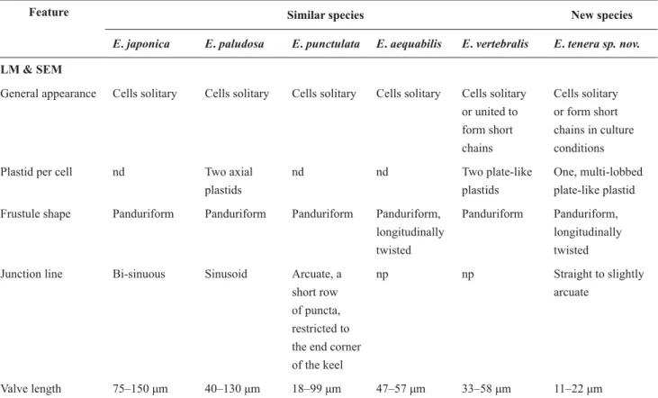
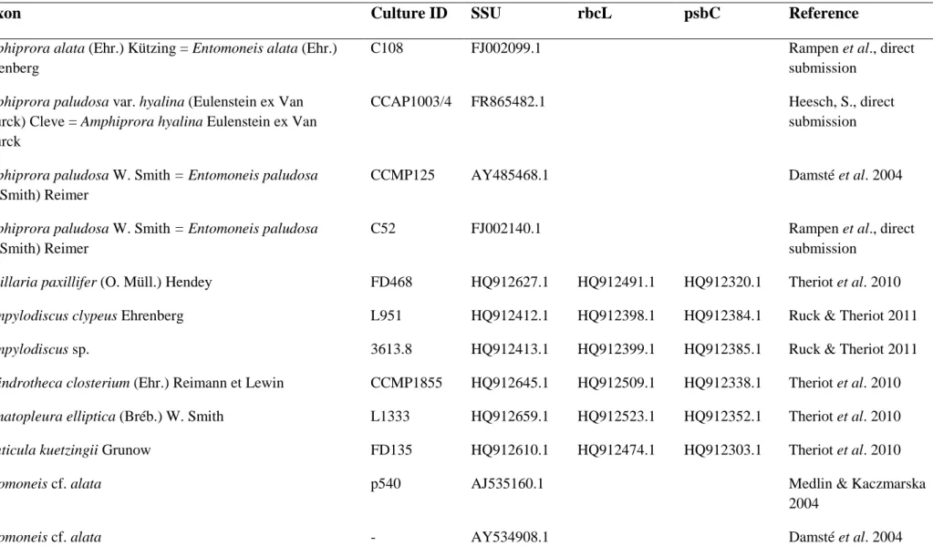
I. Mejdandžić M, Bosak S, Orlić S, Gligora Udovič M, Peharec Štefanić P, Špoljarić I, Mršić G, Ljubešić Z (2017) Entomoneis tenera sp. nov., a new marine planktonic diatom (Entomoneidaceae, Bacillariophyta) from the Adriatic Sea. Phytotaxa 292: 1-18. II. Mejdandžić M, Bosak S, Nakov T, Ruck E, Orlić S, Gligora Udovič M, Peharec
Štefanić P, Špoljarić I, Mršić G, Ljubešić Z (2018) Morphological diversity and phylogeny of the diatom genus Entomoneis (Bacillariophyta) in marine plankton: six new species from the Adriatic Sea. Journal of Phycology 54: 275-298.
III. Grbin D, Pfannkuchen M, Babić I, Mejdandžić M, Mihanović H, Marić Pfannkuchen D, Godrijan J, Peharec Štefanić P, Olujić G, Ljubešić Z (2017) Multigene phylogeny and morphology of newly isolated strain of Pseudo-nitzschia mannii Amato & Montresor (Adriatic Sea). Diatom Research 32: 127-131.
IV. Mejdandžić M, Bosak S, Ljubešić Z (2017) Blue Diatoms: Global Phenomenon of ˝Greening˝ in Shellfish and Record of Planktonic Haslea Species in the South Adriatic Sea. Naše more, Znanstveno-stručni časopis za more i pomorstvo 64: 38-44.
V. Ljubešić Z, Mejdandžić M, Bošnjak I, Bosak S (2016) Comparing methods in picoplankton abundance estimation. In: Rapp. Comm. int. Mer Médit. 41.
VI. Mucko M, Novak T, Medić N, Gašparović B, Peharec Štefanić P, Orlić S, Ljubešić Z (2018) Characterization of newly isolated photosynthetic marine pico green algae (Picochlorum, Trebouxiophyceae) from the Adriatic Sea. Submitted to Acta Botanica Croatica
VII. Mucko M, Bosak S, Casotti R, Balestra C, Ljubešić Z (2018) Picoplankton winter diversity in an oligotrophic marginal sea. Submitted to Marine Genomics
II
THESIS SUMMARY
Free-living microbial populations in pelagic parts of oceans are represented as plankton, diverse collection of organisms passively driven by water currents, which includes organisms belonging to Bacteria, Archaea, Eukarya (protists and fungi) and viruses. The most abundant Bacteria belong to classes Proteobacteria, mostly Alphaproteobacteria and Gammaproteobacteria and phototrophic Cyanobacteria (predominantly genera Prochlorococcus and Synechococcus) while Archeal diversity strongly dominates with classes Euryarcheota and Crenarchaeota. It is important to emphasize that the newest tree of life published by Hug et al. (2016) shows Bacteria as the most diverse domain with new classes discovered every day, while Archaea and Eukarya, at first sight, in comparison to Bacteria are less diverse. Nevertheless, microbial eukaryotes represent a large range of phototrophic, mixotrophic and heterotrophic organisms with huge morphological and phylogenetic diversity. The most dominant marine microbial eukaryotes belong to super groups Archaeplastida, phyla Chlorophyta and Prasinophyta; Chromalveolata, phyla Dinophyta, Bacillariophyta and MAST (Marine Stramenopiles), classes Pelagophyceae, Cryptophyceae and Prymnesiophyceae; Excavata, phylum Euglenozoa; Opistokontha, phyla Choanoflagellata and Fungi; Rhizaria, phyla Cercozoa and Radiolaria (Massana and Pedrós-Alió 2008). Picoeukaryotes (PEs) are organisms with cell size ≤ 3µm, including phototrophs, heterotrophs, and mixotrophs, which can be involved in mutualistic relationships such as parasitism or symbiosis with larger organisms and are of great importance for biogeochemical cycles in oceans. The diatoms are microscopic eukaryotic algae with unique silicified shell called frustule, and, based on their symmetry and phylogeny can be divided in radial centrics (Coscinodiscophyceae, radial symmetry), bi- or multipolar centrics (Mediophyceae, radial to irregular symmetry), araphid pennates and raphid pennates (Bacillariophyceae, bilateral symmetry), all divided into 9 major phylogenetic clades. In contrast to PEs who have few or non-diagnosable morphological parameters to be identified with, diatoms have a large number of specific morphological features on their frustules and traditionally have been identified primarily with light and electron microscopy. Both PEs and pennate diatoms in marine plankton have often been neglected in microscopical examination of field samples: PEs mostly because of their size, and planktonic pennate diatoms mostly because of their difficult identification using light microscopy, light silification and small abundance. Therefore, the need for the introduction of various molecular methods in their detection and correct identification was acknowledged in last few decades. The diatoms are today commonly identified using nuclear 18S rRNA gene
III
and chloroplast encoded rbcL and psbC genes, while PEs, depending on lineages, using nuclear 18S rRNA, plastid 16S rRNA and ITS genes and chloroplast or mitochondrial encoded rbcL or COI. However, multi-gene phylogenies are often not possible to obtain since a lot of PEs or pennate diatoms cannot be cultivated. Therefore, next generation sequencing (NGS) methods in examining environmental DNA (eDNA) are introduced in marine phytoplankton research which rely on barcode regions used for species identification and diversity estimations. Additionally, from the biotechnological aspect, some members of pennate diatoms and especially PEs can be interesting and promising.
The aim of this thesis was to obtain a more complete picture of the diversity of marine PEs and pennate planktonic diatoms in the middle and south Adriatic Sea using traditional microscopy, molecular identification and defining physiological attributes, and to try to answer the following important questions during that process: i) Where is the phylogenetical border between species and genus in picoeukaryotes and planktonic pennate diatoms? Does the current knowledge of species/genus border adequately reflect the use and availability of new genetic markers in picoeukaryotes and planktonic pennate diatom research and microscopy in classical morphology? ii) Is the Adriatic Sea a good model for studying shifts in diversity in the plankton communities due to ongoing climate changes? iii) What is the potential for isolation and cultivation of new strains of PEs and planktonic pennate diatoms with the possible biotechnological application?
In this thesis, represented through seven publications, newly isolated and cultivated planktonic pennate diatoms were identified as belonging to the genera Entomoneis Ehrenberg, Pseudo-nitzschia H. Peragallo and Haslea Simonsen while PEs as genus Picochlorum W.J.Henley, J.L.Hironaka, L.Guillou, M.A.Buchheim, J.A.Buchheim, M.W.Fawley & K.P.Fawley. Within the genus Entomoneis, seven new species were described: E. tenera, E. pusilla, E. gracilis, E. vilicicii, E. infula, E. adriatica and E. umbratica (publications I and II). Further on, Adriatic strain of known species Pseudo-nitzschia mannii was characterized (publication III), and unknown species Haslea sp. and her biotechnologically important pigment marennine was detected (publication IV). Additionally, unknown species of Picochlorum sp., from genus with yet unknown new species number and great biotechnological potential, was characterized (publications V and VI). General PEs diversity was addressed by eDNA amplicon sequencing of nuclear 18S rRNA gene, V4 variable region and obtained results identified 95% of the PEs community as hetero- or mixotrophic, while only 5% of the
IV
community was represented with photoautotrophic PEs belonging to classes of green algae, haptophytes, stramenopiles and cryptophytes (publication VII).
The scientific contribution of this thesis is increasing the general scientific knowledge about the analyzed genera of PEs and diatoms and the descriptions of up to now unknown organisms. Additionally, this thesis provides first NGS analysis regarding PEs in the Adriatic Sea, filling the crucial knowledge gap in plankton studies in this oligotrophic ecosystem. One cultivated Picochlorum sp. strain was subjected to growth rate experiments and pigment and lipid analyses, resulting in interesting and promising data for future experimental approaches and biotechnological applications for this highly resilient pico-photoautotroph. Biotechnology in algae, a challenging and promising field of research is with this thesis enriched with green algae strains full of potential for upcoming and evolving varieties of green technologies. With ongoing climate change, studies like this are of the utmost importance for efforts in preserving the ocean and the world’s largest biome.
V
PROŠIRENI SAŽETAK
Slobodno živuće mikrobne populacije u pelagičkim dijelovima oceana zajednički se nazivaju planktonom - raznovrsnom skupinom organizama pasivno pokretanima vodenim strujama, koji uključuju organizme iz triju domena živog svijeta: Bacteria, Archaea, Eukarya (protisti i gljive), te viruse. Najbrojnije bakterije pripadaju koljenu Proteobacteria, najviše razredima Alphaproteobacteria i Gammaproteobacteria te autotrofnim cijanobakterijama (rodovi Prochlorococcus i Synechococcus), dok najdominantnije arheje pripadaju razredima Euryarcheota i Crenarchaeota. Važno je naglasiti da u najnovijem stablu života kojeg su prikazali Hug i suradnici (2016) najveću raznolikost pokazuje upravo domena Bacteria, sa svakodnevno otkrivanim novim razredima, dok je raznolikost arheja i eukariota naizgled puno manja. Usprkos tome, mikrobni eukarioti ipak predstavljaju široki raspon fotoautotrofnih, miksotrofnih i heterotrofnih organizama koji se iznimno morfološki i filogenetski razlikuju. Najbrojniji oceanski mikrobni eukarioti pripadaju supergrupama Archaeplastida, koljenima Chlorophyta i Prasinophyta; Chromalveolata, koljenima Dinophyta, Bacillariophyta, MAST (Marine Stramenopiles), razredima Pelagophyceae, Cryptophyceae i Prymnesiophyceae; Excavata, koljenu Euglenozoa; Opistokontha, koljenima Choanoflagellata i Fungi; Rhizaria, koljenima Cercozoa i Radiolaria (Massana and Pedrós-Alió 2008). Pikoeukarioti su organizmi s veličinom stanica ≤ 3 μm u promjeru, uključuju fotoautotrofe, heterotrofe i miksotrofe koji mogu biti uključeni u mutualističke odnose kao što su parazitizam i simbioza s većim organizmima, a od velike su važnosti za biogeokemijske cikluse u oceanima. Dijatomeje su mikroskopske eukariotske alge s jedinstvenom silificiranom ljušturom zvanom frustula, koje na temelju njihove simetrije i filogenije razlikujemo kao radijalne centrice (Coscinodiscophyceae, radijalna simetrija), bi- ili multipolarne centrice (Mediophyceae, radijalna do nepravilna simetrija) ili arafidne i rafidne penate (Bacillariophyceae, bilateralna simetrija), koje se zajedno dijele unutar 9 filogenetski različitih skupina. Za razliku od pikoeukariota koji ili nemaju uopće, ili imaju samo nekoliko morfoloških parametara, dijatomejske frustule posjeduju velik broj specifičnih morfoloških svojstava te se tradicionalno se identificiraju pomoću svjetlosnog i elektronskog mikroskopa. Pikoeukarioti i penatne planktonske dijatomeje se često predviđaju i zanemaruju u morskim okolišnim uzorcima prilikom klasičnih analiza mikroskopijom: pikoeukarioti uglavnom zbog svoje veličine, a penatne planktonske dijatomeje uglavnom zbog teške identifikacije putem svjetlosne mikroskopije, slabe silificiranosti te male brojnosti u planktonu. Stoga je uvođenje različitih molekularnih metoda u njihovo otkrivanje i ispravnu identifikaciju u posljednjih nekoliko
VI
desetljeća postalo obavezno. Diatomeje se danas obično identificiraju pomoću više različitih gena kao što su nuklearni 18S rRNA gen te kloroplastni rbcL i psbC geni, dok pikoeukarioti, ovisno o taksonomskim razredima, korištenjem nuklearnog 18S rRNA gena, plastidnog 16S rRNA gena, varijabilne ITS regije te kloroplastnog ili mitohondrijskog gena kao što su rbcL ili COI. Ipak, nerijetko identifikacija putem filogenije nije moguća jer se velika većina pikoeukariota i dijatomeja ne mogu uspješno uzgajati u laboratorijskim uvjetima pa se danas uvode i metode sekvenciranja sljedeće generacije (NGS) u različite analize okolišne DNA (eDNA), kako bi se otkrile i identificirale teško uzgojive i rijetke svojte. Osim toga, s biotehnološkog aspekta, neke penatne planktonske dijatomeje, a osobito pikoeukarioti, mogu biti zanimljivi i obećavajući.
Cilj ove disertacije je dobiti potpuniju sliku raznolikosti morskih pikoeukariota i planktonskih penatnih dijatomeja u Jadranu koristeći tradicionalnu mikroskopiju, suvremenu molekularnu identifikaciju i karakterizaciju fizioloških parametara organizama, te u tom procesu pokušati odgovoriti na sljedeća važna pitanja: i) Gdje je granica vrste i roda kod pikoeukariota i penatnih planktonskih dijatomeja? Da li su dosadašnje spoznaje o granicama vrste i roda adekvatne obzirom na korištenje i dostupnost novih genskih markera u istraživanjima pikoeukariota i penatnih planktonskih dijatomeja kao i mikroskopije u klasičnoj morfologiji? ii) Zbog klimatskih promjena dolazi do oligotrofikacije oceana te promjena u planktonskim zajednicama. Da li je Jadransko more idealan model za proučavanje tih promjena zbog svoje oligotrofije? iii) Kolika je mogućnost izolacije novih klonova sa potencijalnom biotehnološkom primjenom?
U ovoj disertaciji, predstavljenoj kroz sedam znanstvenih publikacija, zabilježeni su i istraženi kultivirani dijatomejski rodovi Entomoneis Ehrenberg, Pseudo-nitzschia H. Peragallo i Haslea Simonsen te pikoeukariotski rod Picochlorum W.J.Henley, J.L.Hironaka, L.Guillou, M.A.Buchheim, J.A.Buchheim, M.W.Fawley & K.P.Fawley. Rod Entomoneis zastupljen je sa sedam novih vrsta: E. tenera, E. pusilla, E. gracilis, E. vilicicii, E. infula, E. adriatica i E. umbratica (publikacije I i II). Nadalje, rod Pseudo-nitzschia istražen je s karakterizacijom novog kultiviranog soja poznate vrste P. mannii (publikacija III), a rod Haslea s još nepoznatom ‘plavom’ dijatomejom Haslea sp. čiji je biotehnološki važan pigment marenin zabilježen (publikacija IV). Dodatno, rod Picochlorum okarakteriziran je sa jednim novoizoliranim sojem velikog biotehnološkog potencijala, dok je broj novih vrsta još uvijek nepoznat (publikacije V i VI). Opća raznolikost pikoeukariota istražena je sekvenciranjem amplikona eDNA koristeći varijabilnu V4 regiju nuklearnog 18S rRNA gena, a dobiveni su
VII
rezultati identificirali 95% zajednice kao heterotrofnu i/ili miksotrofnu, dok je samo 5% zajednice predstavljeno fotoautotrofnim pikoeukariotima koji pripadaju razredima zelenih algi, haptofitima, stramenopilima i kriptofitima (publikacija VII).
Znanstveni doprinos ove disertacije je obogaćivanje općeg znanstvenog znanja spomenutih rodova pikoeukariota i penatnih planktonskih dijatomeja s opisima do sada nepoznatih vrsta te karakterizaciji novo izoliranih sojeva poznatih vrsta. Osim toga, ova disertacija daje prvi skup podataka prikupljenih sekvenciranjem sljedeće generacije na Illumina platrofmi koji se odnosi na pikoeukariote u Jadranskome moru, dajući prijeko potrebno znanje planktonskim istraživanjima u ovom oligotrofnom ekosustavu. Biotehnološki potencijal jednog kultiviranog soja roda Picochlorum dodatno je analiziran u eksperimentu rasta te analizama pigmenta i lipida, što je rezultiralo zanimljivim i obećavajućim podacima za buduće eksperimentalne pristupe na ovom plastičnom i otpornom piko-fotoautotrofu. Biotehnologija primjenjiva na algama, izazovno i obećavajuće područje istraživanja je s ovom disertacijom dobilo uvid u nove mogućnosti i nove sojeve primjenjive u nadolazećim i rastućim zelenim tehnologijama. Obzirom na prisutne klimatske promjene, studije poput ove su od najveće važnosti za očuvanje oceana, najvećeg svjetskog bioma.
VIII
INTRODUCTION
„To many people, 'biodiversity' is almost synonymous with the word 'nature,' and 'nature' brings to mind steamy forests and the big creatures that dwell there. Fair enough. But biodiversity is much more than that, for it encompasses not only the diversity of species, but also the diversity within species.“ - Cary Fowler
INTRODUCTION
1
The unseen diversity of marine microbial protists
Aquatic ecosystems account for 70% of the Earth's surface (excluding ice and groundwater ecosystems) and preservation of this vast ecosystem is of the utmost importance to humankind (Costanza et al., 1997). Marine ecosystems comprise 97% of aquatic ecosystems in a form of a continuous body of seawater that holds about 320 million cubic miles (1.35 billion cubic kilometres) (NatGeoEd.org). The marine ecosystems are threatened by global changes, such as climate change, overfishing, and pollution, but most of us see just the small picture in form of a decreased number of commercially important fish, marine mammals or increasing amount of plastics and other pollutants in aquatic ecosystems. Most of the world's oceans are unreachable, harboring small amounts of primary producers, consequently called ˝blue deserts˝, but on the other hand, marine ecosystems harbor a large diversity of microbial populations that ensure their functioning and sustainability.
Free-living microbial populations in pelagic parts of oceans are represented as plankton, diverse collection of organisms passively driven by water currents, which include autotrophic, heterotrophic and mixotrophic organisms: Bacteria, Archaea, members of Eukarya (protists and fungi), and viruses. On average, a liter of seawater contains ~106 eukaryotic cells (Brown et al., 2009), ~108 prokaryotic cells and (Whitman et al., 1998) and ~109-1011 virus-like particles (Wilhelm and Matteson, 2008). Plankton organisms dominate marine ecosystem in terms of both abundance and biomass (Zinger et al., 2012), and can be divided depending on their feeding preferences into phytoplankton (plant-like plankton composed of phototrophic protists; cells can perform photosynthesis), zooplankton (animals feeding and grazing on phytoplankton; heterotrophs), bacterioplankton (bacteria, cyanobacteria and archaea) and virioplankton (viruses). Microbial community drives every one of the major biogeochemical cycles that make the ocean processes crucial for all other ecosystems on earth (Worden et al., 2015). Marine microorganisms are a diverse pool of species; for instance, Bacteria within the global ocean are estimated to consist of more than ~2×10⁶ taxa (Curtis et al., 2002), Archaea ~2×10⁴ taxa
(Massana et al., 2000) and 2.2×10⁶ Eukarya taxa (Mora et al., 2011).
Protists are diverse eukaryotic organisms mostly found as single cells, although many species form colonies formed of several to numerous cells (Caron et al., 2012). They are distributed throughout all branches of the eukaryotic tree of life (Baldauf, 2008; Burki et al., 2014; Figure 1). Together with virioplankton and heterotrophic bacterioplankton, protists form the ˝microbial loop˝, contributing predominantly to organic matter and nutrient recycling (Azam et al., 1983; Pernthaler, 2005; Pomeroy et al., 2007).
INTRODUCTION
2
Figure 1. Global tree of eukaryotes from a consensus of phylogenetic evidence (in particular, phylogenomics), rare genomic signatures, and morphological characteristics. Numerous eukaryotic groups are shown (not exhaustively), regardless of their taxonomic rank. Cartoons illustrate the diversity constituting the largest assemblages (colored boxes). The branching pattern does not necessarily represent the inferred relationships between the lineages. Dotted lines denote uncertain relationships, including conflicting positions. Note the solid branch leading to haptophytes and rappemonads. Adapted from Burki et al., 2014.
Major lineages inside photosynthetic protists are Dinophyta (dinoflagellates), Ochrophyta (Stramenopiles, diatoms, golden algae and brown algae), Chlorophyta (Archaeplastida, marine green algae), Haptophyta (mainly coccolithophorids) and Cryptophyta (flagellated microalgae containing phycoerythrin accessory pigments) (Anderson et al., 2011; Wang et al., 2016; Tragin et al., 2017). Generally, protists can be divided into size fractions: pico- (0.2 to 2(3) µm), nano- (2(3) to 20 µm) and micro- (20 to 200 µm) fraction (Sieburth et al. 1978). Smallest protists, generally called picoeukaryotes (PEs), and pennate planktonic diatoms (size fractions nano and micro; 3-200 µm) received limited attention in world oceans, although they are numerous and important for both primary production, as well as for carbon injection into deep ocean (Worden et al., 2004; Agusti et al., 2015). PEs have especially important roles in oligotrophic ecosystems,
INTRODUCTION
3
where together with picocyanobacteria (such as Prochlorococcus and Synechococcus) are the driving force of primary production, but they are even more important at high latitudes, where marine cyanobacteria are less numerous (Lovejoy et al., 2007; Balzano et al., 2012; Flombaum et al., 2013).
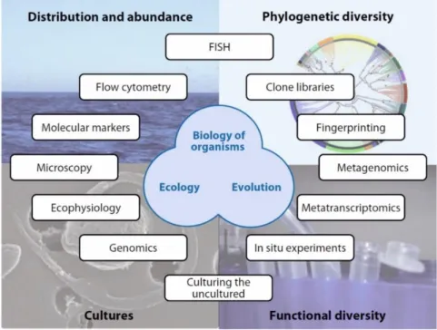
Marine protists can be investigated with traditional techniques such as light, electron and epifluorescence microscopy, pigment analysis with High-Performance Liquid Chromatography (HPLC), flow cytometry, etc. Additionally, other tools can be applied in protist studies, depending on the area of research (abundance, phylogenetic diversity, cultures and functional diversity, Figure 2). Morphology, as a study of external and internal structure, shape and form of organisms with exhaustive observations of various details specific to each taxon, and phylogeny as a study of evolutionary relationships between organisms are the most used ways of investigating living organisms, among them protists.
Fundamentally, there is widespread acceptance that identification of protist species using light microscopy alone is no longer sufficient or adequate. Light microscopy has very limited use to assess picoplankton diversity in the field, but in cultures it can provide valuable information on their size, shape of the cells, number of plastids, swimming or sliding behavior, consequently allowing identification of the species. Additionally, specific fixatives, such as Lugol or osmium can make certain cellular features (flagella) more visible or the fixatives can destroy the samples. Descriptions based on microscopy and holotypes deposited in designated institutes as fixed slide preparations or photographs can help to describe a morphotype but fail to identify species (Adl et al. 2007). More improved electron microscopy (transmission or scanning electron microscopy) allows to describe to a certain extent in morphological diversity of larger protist cells (for example dinoflagellates or diatoms), but it is limited when applied to small protists (Moestrup and Throndsen, 1988). Electron microscopy can allow us to determine specific ornamentation of cells, as was shown for minute Bathycoccus prasinos W. Eikrem & J. Throndsen or Imantonia rotunda N. Reynolds (Eikrem and Throndsen, 1990). However, usage of electron microscopy has some limitations. Cells need to be fixed and concentrated before embedding or mounting, which can result in the loss of small or delicate forms. Microscopes are usually too big to carry to field and demand specific environment (certain temperature and light conditions) to be adequately placed. Epifluorescence microscopy relies on the emission of light by cellular compounds (e.g. pigments) or by strains specific for certain components such as DNA. The vast diversity of heterotrophic protists is difficult to examine
INTRODUCTION
4
under light and electron microscopy, so staining and epifluorescence microscopy has proven to be a better tool in investigations of these cells (Sherr and Sherr, 1993).
Pigmented protists (i.e. phytoplankton) possesses a collection of pigments which are somewhat specific to certain lineages, such as classes (for example fucoxanthin and diatoxanthin in diatoms, peridinin and diadinoxanthin in dinoflagellates or prasinoxanthin in prasinophytes) (Wright and Jeffrey, 2006). Pigment analysis expanded rapidly in the 1980s with the development of automated HPLC methods for pigment separations, useful for routine shipboard or shore-based determinations (e.g. Wright and Shearer, 1984; Roy, 1987). HPLC method allows separation of up to 40 pigments - chlorophylls, carotenoids and their derivatives. Types of microalgae that had previously been missed in microscopic analysis of field samples through filtering loss or preservation damage could now be recognized from their pigment signatures (e.g. Gieskes and Kraay, 1983; Guillard et al., 1985). However, HPLC as a method has some disadvantages: (i) due to the liability and rapid degradation of phytoplankton pigments, special conditions must be employed to preserve samples, and samples must contain certain concentration of pigments so they can be detected (ii) these pigments are particularly sensitive to light, heat, oxygen, acids and alkalis; (iii) whilst a number of pigments is specific to particular classes or genera, a number of other pigments is spread across many algal classes making interpretation of the data difficult (iv) expression of the pigments is quite variable with the contents of a particular cell varying with a number of environmental factors such as irradiance and nutrients. Despite these disadvantages, HPLC is still the best technique for mapping phytoplankton populations and monitoring their abundance and composition.
Pigmented cells can be quantified using flow cytometry, a technique able to separate cells based on their size and auto-fluorescence (Marie et al., 1997). Flow cytometry is an analytical technique, based on optical properties and/or specific fluorescence of particles (cells) and their constant flow through the flow chamber/counting chamber within a sheat fluid. Flow-cytometry measures and enumerates small cells and particles such as phytoplankton, bacteria, and viruses. Light scattered by each individual cell (a function of cell size and refractive index) and fluorescence from pigments such as chlorophyll or phycoerythrin are recorded in real time (Marie, 1999). Major advantages of flow cytometry include speed, accuracy, and absence of sample preparation, at least to analyze photosynthetic pigments. This revolutionary method led to the discovery of very important picoplanktonic organisms such as Prochlorococcus (Chisholm, 1988) and Ostreococcus C. Courties & M.-J. Chrétiennot-Dinet (Courties et al., 1994). Still, scattering and fluorescence properties are not sufficient to discriminate taxa within
INTRODUCTION
5
picoeukaryotes, with the exception of cryptophytes that contain phycoerythrin (Li and Dickie, 2001). Therefore, potential applications of flow cytometry are considerably enhanced when samples are stained with fluorescent markers binding to specific cell compounds.
The tiny proportion of cultivable marine protists, our inability to identify them, interpret their diversity and to study them in detail all contribute to huge knowledge gaps, which are often difficultly bridged (Massana, 2011; del Campo et al., 2014).
Figure 2. Overview of approaches to investigate cell biology, ecology, and evolution of marine protists, treating four main study areas: distribution and abundance, phylogenetic diversity, functional diversity and culture studies. Abbreviation: FISH, fluorescence in situ hybridization. Adapted from Massana, 2011.
One universal method to access diversity among all organisms is applicable to genes and proteins which present a certain degree of variability (Anne, 2006). Molecular markers can be divided into three categories (Schlötterer, 2004): the protein variants (i.e. allozymes), the DNA (Deoxyribo Nucleic Acid) sequence variations (i.e. polymorphisms) and the DNA repeat variation. Remarkable progress in molecular biology and science altogether brought revolutionary methods such as the Sanger method (Sanger and Coulson, 1975) and Polymerase chain reaction (PCR; Saiki et al., 1985). Later on, next-generation sequencing (NGS) or high-throughput sequencing (HTS) methods such as 454 pyrosequencing (based on Ronaghi et al., 1998) in 2005 (Margulies et al., 2005), and then Illumina sequencing (based on Canard and Sarfati, 1994) in 2007. HTS methods allowed the transition between clone libraries sequenced
INTRODUCTION
6
by Sanger method and the large metabarcoding datasets, where Sanger sequencing provides a relatively low number of long high-quality sequences, while HTS provides a large amount of medium-quality sequences and allows only small fragments to be sequenced. Several different gene markers to assess the protist diversity have been used in HTS methods: V4 region of 18S rRNA gene (Massana et al., 2014; Zimmermann et al. 2011); V9 region of 18S rRNA gene (De Vargas et al., 2015); rbcL (large subunit of the ribulose-1,5-biphosphate carboxylase-oxygenase) encoded in plastid genomes; and cox1 (cytochrome c oxidase subunit I) gene encoded in mitochondrial genomes (Kermarrec et al., 2013); 16S rRNA plastid gene (Lepère et al., 2009; Choi et al., 2017); or more species specific hypervariable region such as ITS (internally transcribed spacer of the rRNA operon) (Coleman, 2003; Rodríguez-Martínez et al., 2013). These sophisticated sequencing methods, called metabarcoding led us to ambitious projects such as Tara Oceans ( https://oceans.taraexpeditions.org/en/m/about-tara/les-expeditions/tara-oceans/) or Ocean Sampling Day project (https://www.microb3.eu/osd.html), which together obtained large and useful datasets of marine eukaryotic diversity. Along with metabarcoding, metagenomics can allow us to investigate community gene repertoires and metabolic potential, whereas metatranscriptomics can provide insights into realized functions (Dinsdale et al., 2008; Frias-Lopez et al., 2008).
Picoeukaryotes: significance and diversity
Picoeukaryotes (PEs) are single-celled ubiquitous organisms that possess a nucleus and a minimal number of organelles (mitochondrion, Golgi apparatus, optionally flagellum, and chloroplasts if photosynthetic; then called photosynthetic picoeukaryotes (PPEs)), and like the rest of eukaryotes, their cellular components are the evolutionary products of endosymbiosis which occurred once or several times in their evolution (Delwiche, 1999). These organisms are the smallest organisms among eukaryotes, having cells between 0.2 to 3µm in diameter, including phototrophs, heterotrophs, and mixotrophs, which can be involved in mutualistic relationships with larger organisms such as parasitism or symbiosis (Acosta et al., 2013). PEs are an important constituent of the ocean's microbiota and perform essential roles in biogeochemical cycles, and form, together with prokaryotes, an ocean’s veil above which larger protists and metazoans might bloom (Massana, 2011).
PPEs account for a significant fraction of primary production, especially in oligotrophic conditions (Li, 1995; Worden and Not, 2008). Although numerically less abundant than marine cyanobacteria, PPEs constitute a third active group of the marine picophytoplankton which was reported several times: in the North Atlantic (Li et al., 1992); Arabian Sea (Shalapyonok et al.,
INTRODUCTION
7
2001); equatorial Pacific (Mackey et al., 2002); Sargasso Sea and Mediterranean Sea (DuRand et al., 2001; Brunet et al., 2007). PPEs reach 1-3×10³ cells mL-1 in oligotrophic systems and up to 105 cells mL-1 in nutrient rich coastal zones (Sanders et al., 2000; Li, 2009). Together with cyanobacteria Prochlorococcus and Synechococcus they form the picophytplankton, which accounts for a high fraction (80-90%) of phytoplankton biomass in the oceans (Latasa and Bidigare, 1998; Not et al., 2008). They can be extremely abundant in some areas, such as California, USA, coastal site where PPEs were responsible for up to 76% of the net picoplanktonic production (Worden et al., 2004). Most dominant PPEs in world oceans proved to be Prymnesiophyceae, Prasinophyceae (Mamiellophyceae), Cryptophyceae, Pelagophyceae, Chrysophyceae, and Dictyochophyceae (Shi et al., 2009). Their role in the food chain can be of most importance, as some studies showed that PPEs are subjected to grazing more than cyanobacteria (for example Synechococcus), profoundly because of their size (cells larger than coccoid cyanobacteria) which can be substantial for carbon transfer to higher trophic levels (Stockner, 1988). This has direct consequences in packaging carbon in larger particles, which contributes to biological carbon pump whereby organic carbon is transferred to the deep ocean.
Heterotrophic PEs (HPEs) are generally considered bacterial grazers (Jürgens et al., 2008). They keep bacterial stocks stable, transfer dissolved organic matter to higher trophic levels and recycle nutrients that sustain regenerated primary production (Massana, 2011). Although considered less abundant than PPEs, HPEs proved to be very abundant in the oceans, defined by high operational taxonomic unit (OTU) numbers (De Vargas et al., 2015). Many studies showed great prevalence of HPEs over PPEs in oligotrophic ocean ecosystems, where was previously considered that PPEs are of an extreme importance for primary production (Shi et al., 2009, Acosta et al., 2013, De Vargas et al., 2015, Pernice et al., 2015, Pearman et al., 2017). Interactions between HPEs and prokaryotes have ecological implications, as bacterial abundances and community composition are strongly influenced by the predation pressure of HPEs (Jardillier et al., 2005). Besides primary production and bacterivory, HPEs can also influence different trophic levels through parasitic and mutualistic symbiotic associations (Worden and Not, 2008).
General PE diversity is still unexplored, due to various causes: i) small eukaryotic cells cannot be easily identified with traditional methods; ii) many are not easily cultivated in artificial media and laboratory conditions; iii) due to their uneven cellular properties, some taxonomic groups preserve better than others (Vaulot et al., 2008). Most of the cultured PPEs belong to Prasinophyceae, Pelagophyceae, Bolidophyceae, and Pinguiphyceae, while HPEs to
INTRODUCTION
8
Bicosoecida and Chrysophyceae (Vaulot et al., 2008; Jürgens et al., 2008). Most of picoeukaryotic taxonomic groups do not possess easily discriminated characteristics visible under light microscopy at the current resolution levels (Johnson and Sieburth, 1982; Andersen et al., 1996). With the development of electron microscopy, such as TEM, important diagnostic features were better detected (presence and shape of flagellar hairs or body scales, the presence of pyrenoids and starch inclusions, chloroplast organization and membrane configuration (Eikrem and Edvardsen, 1999)). Culturing through enrichment cultures, pre-filtered cultures, flow-cytometry sorted or manually isolated and serially diluted techniques allowed scientists to describe and culture various picoplankton species (e.g. Micromonas pusilla (Butcher) Manton & Parke (Butcher, 1952)) that in the end drastically improved our world collections of PPEs (Vaulot et al., 2004; Andersen and Kawachi, 2005). At the simplest level, photosynthetic pigments (as a key taxonomic diagnostic feature for microalgae) allows us to distinguish green, brown and red algae, but HPLC signature is often indicative of the class (e.g. prasinoxanthin is only present in Prasinophyceae) (Guillou et al., 1999). Due to inherent limitations of the above methods, for decades PEs were treated as a ˝black box˝ of difficult access (Massana, 2011). Molecular tools gave new insights into the microbial world and revolutionized microbial ecology (Massana, 2011). As mentioned in the previous section ˝The unseen diversity of marine microbial protists˝, HTS methods greatly improved general knowledge on PEs diversity in last decade, allowing us to identify uncultivable representatives and rare taxa.
Diatoms: significance and diversity
Diatoms (Bacillariophyta) are unicellular, mostly photoautotrophic heterokonts (a group of algae with golden-brown to brown chloroplasts originating from a red algal endosymbiont), often forming colonies, with cell sizes roughly between 10 and 200 µm. Some diatoms can live heterotrophically in the dark if supplied with a suitable source of organic carbon, while less than ten species are obligatory heterotrophs (from the genera Nitzschia Hassall and Hantzschia Grunow) (Round et al., 1990). Their unique hallmark is the ornamented compound silica cell wall called a frustule built of an amorphus hydrated silica (SiO2 × nH2O) and organic material
(proteins, polysaccharides). The frustule consists of two halves, unequal in size: smaller hypotheca and larger epitheca, each containing its valve – hypovalve and epivalve, which are held in place by silicified girdle bands called hypocingulum and epicingulum. The typical valve looks like a Petri dish with a flat area called the valve face and a rim called mantle (both cingulum together called mantellum). The flux of material across the frustule takes place via multiple sorts of pores or slits, while the cytoplasm is fully protected (Round et al., 1990). With
INTRODUCTION
9
their almost indestructible shells, conveniently small size, great variety and beauty, diatoms were perfectly ‘pre-adapted’ to their role as the objects of scientific fashion. There are a significant number of morphologic characters to be considered on each diatom frustule, most of them being in common for all diatoms (e.g. areolae, striae, virgae, raphe, fultoportulae or rimoportulae, central and apical pores, girdle bands areolae, etc.); but a lot of characters are genus, and/or species-specific, and need to be carefully investigated when observing a diatom frustule and identifying a species or discriminating against two similar species. Diatoms’ photosynthesis apparatus holds primary chlorophylls (Chl a and c) and protective xanthophyll (fucoxanthin, diadinoxanthin, diatoxanthin) and carotene (β-carotene).
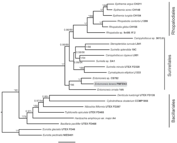
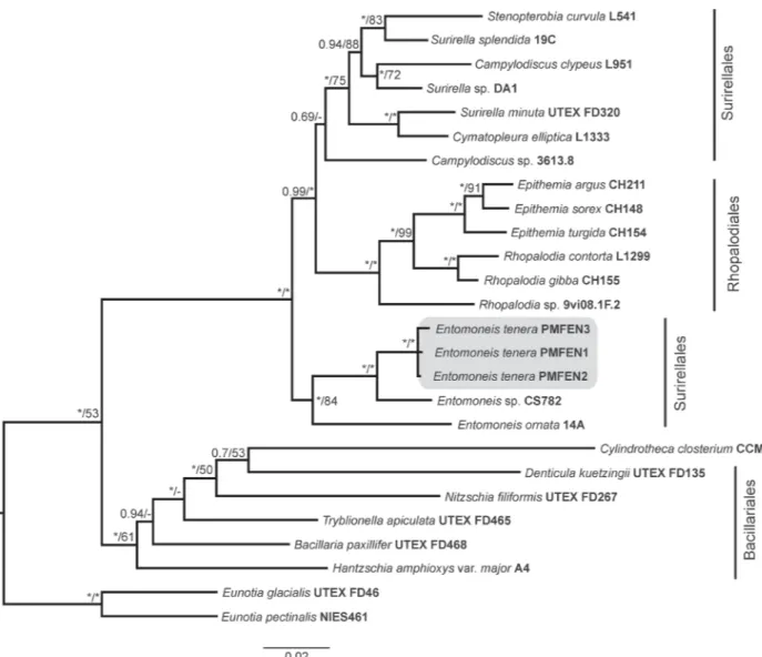
Historically, diatoms were divided into two major groups: the centrics and the pennates, which can be distinguished by their symmetry (centrics having radial symmetry, while pennates having bilateral symmetry), mode of sexual reproduction (oogamous centrics, while isogamous pennates; Figure 3) and plastid number and structure (discoid plastids in centrics, while plate-like plastids in pennates) (Round et al., 1990). Morphologically, pennates can be further divided into two groups by the presence or absence of a slit (raphe) in the valve for movement, i.e. motile pennates possessing raphes are ‘raphid’, whereas immotile pennates lacking raphes are ‘araphid’ diatoms. Today, the most accepted classification system presents diatoms in three classes: Coscinodiscophyceae (radial centric diatoms), Mediophyceae (bi- or multi- polar centric and some radial centric diatoms) and Bacillariophyceae (pennate diatoms) (Medlin and Kaczmarska, 2004). However, newest phylogenetical classification resolves evolution of diatoms as radial centrics grading into polar centrics, which grade into araphid pennates, which themselves grade into the monophyletic raphid pennates (Theriot et al. 2015). According to the phylogenetical division, diatoms are encompassed in 9 major phylogenetic clades: R1 (Leptocylindrus, Tenuicylindrus, Corethron), R2 (Ellerbeckia, Proboscia, Melosira, Aulacoseira, Paralia, Endictya, Stephanopyxis, Podosira), R3 (Rhizosolenia, Guinardia, Coscinodiscus, Actinocyclus, Actinoptychus, Aulacodiscus), P1 (Thalassiosira. Cyclotella, Triceatirum, Odontella, Biddulphia, Attheya, Lithodesmium, Ditylum, Eunotogramma), P2 (Ceratulina, Eucampia, Hemiaulus, Chaetoceros, Bacteriastrum, Acanthoceros, Urosolenia), P3 (Trigonium, Lampriscus, Stictocyclus, Isthmia, Climacosphenia, Chrysanthemodiscus, Toxarium, Ardissonea), A1 (Striatella, Asterionellopsis. Bleakeleya, Delphineis, Rhaphoneis, Plagiogramma), A2 (Fragilaria, Synedra, Staurosira, Licmophora, Diatoma, Opephora, Tabularia, Asterionella, Grammatophora, Thalassionema), Raphid pennates (Eunotia, Nitzschia, Pseudo-nitzschia, Fragilariopsis, etc.) (Theriot et al. 2015). Genera Entomoneis,
INTRODUCTION
10
Pseudo-nitzschia and Haslea, which are investigated in this thesis belong to the monophyletic raphid pennate diatoms, the youngest evolved diatoms in general (Theriot et al. 2015). The genus Entomoneis belongs to the specific canal-raphe diatoms from the lineage Surirellales (Ruck and Theriot 2011), while genus Pseudo-nitzschia which is sister to Fragilariopsis, anchored into the paraphyletic genera Nitzschia and Haslea, which are sisters to diverse genus Navicula, belonging to the canal-raphe lineage Bacillariales (Li et al. 2017; Ruck and Theriot 2011). From the evolutionary perspective, Pseudo-nitzschia evolved first, Haslea followed, and Entomoneis evolved most recently (Ruck and Theriot 2011). Pseudo-nitzschia and Haslea have a common ancestor in their canal raphe evolution, while taxa from the genus Entomoneis have a different ancestor, which is responsible for the evolution of its raphe canal on elevated keel, common for the orders Surirellales and Rhopalodiales (Ruck and Theriot 2011, Ruck et al. 2016).
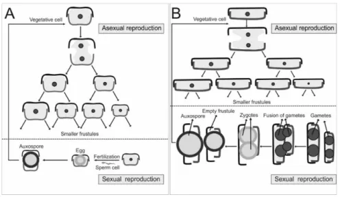
Diatom reproduction is particularly interesting, usually by mitosis, but with the reduction in cell sizes (Figure 3). When diatom cell undergoes mitosis, each daughter cell receives one of the two halves of the frustule, leaving daughter cell to synthesize its own hypotheca. After sub-sequential mitosis, diatoms diminish, leading to the critical point for sexual reproduction. In centrics, this process is oogamous, while in pennates is isogamous and includes ˝+˝ and ˝–˝ cells (as ˝female˝ and ˝male˝ cells) which need to get close together to exchange gametes. A zygote then buds into auxospore that expands to the initial vegetative diatom cell size.
Figure 3. Reproduction in diatoms. A: Oogamous centrics; B: Isogamous pennates
Diatoms commonly live in various terrestrial and aquatic habitats with enough water (or moisture), nutrients, sun energy and CO2 allowing them to perform photosynthesis and to create
INTRODUCTION
11
planktonic or benthic life, where in the plankton are found most commonly radial and bi- or multipolar centrics and a small amount of pennates, whereas pennates predominantly inhabit benthos (on various types of substrates: rocks, plant material, sediment, animals, etc.). Many diatoms secrete kinds of polysaccharides which encapsulate the cells, so they can use it to create all sorts of pads or stalks to attach to various surfaces, or to form colonies. Additionally, to endure in extreme environmental conditions, many diatom species can form resting stages, either in forms of auxospores or dormant vegetative cells (Round et al., 1990). Planktonic diatoms are the most successful group in phytoplankton, obtaining more than 20% of world’s carbon dioxide fixation, which in total exceeds carbon uptake by rain forests. Furthermore, they contribute approximately between 20% (Mann, 1999) and 45% (Yool and Tyrrell, 2003) of the global net primary production. Diatoms are abundant in nutrient-rich coastal ecosystems and at high latitudes and their diversity is currently estimated to ca. 200,000 species (Mann and Droop, 1996), but it is still debated among phycologists. Some estimations made by Guiry (2012) gave a conservative figure of 12,000 described species of diatoms and 8,000 yet to be discovered. However, Mann and Vanormelingen (2013) estimated at least 30,000 but possibly up to 100,000 species. Regardless of which of these estimates are more accurate, there is no doubt that large fraction of diatom diversity is yet to be described or even collected.
Generally, marine planktonic centric diatoms received greater scientific attention historically and today, taking into account their more noticeable morphology and abundance in light microscopy, while tender planktonic pennate diatoms (although they can have large cells) are often neglected in field samples. Additionally, centrics (both radial and multipolar) had been in the scientific focus because of their adaptations to the planktonic lifestyle in form of various colony formation (chains, common in genera Chaetoceros Ehrenberg, Bacteriastrum Shadbolt, Thalasiossira Cleve, Skeletonema Greville) and girdle elements expansion (e.g. Rhizosolenia Brightwell, Proboscia B.G. Sundstrom). Shape and length of chains in centric diatoms seem to be a compromise between various environmental factors, such as the effectiveness of grazer defense, adjustment of buoyancy or optimization for nutrient uptake (Reynolds, 1984; Karp-Boss et al., 1996). A small number of pennate planktonic diatoms adapted to planktonic lifestyle also form colonies, such as the genera Asterionellopsis Round, Asteroplanus C. Gardner & R.M. Crawford in R.M. Crawford & C. Gardner (helical winding colonies), Thalassionema Grunow ex Mereschkowsky (zigzag or stellate colonies), Thallasiothrix Cleve & Grunow (radiate colonies), Pseudo-nitzschia (needle-like chain colonies) and Fragilariopsis Hustedt (ribbon-like colonies) (Kooistra et al., 2007). The last two genera have been investigated the most
INTRODUCTION
12
among planktonic pennate diatoms, but even so, this amount of investigations is still scarce in comparison to centric diatoms (Kang and Fryxell, 1992; Lundholm et al., 2003; Trainer et al., 2012; Lim et al., 2018; Aslam et al., 2018). A possible reason why so few pennate diatoms are found in the plankton may have to do with their isogamous sexual reproduction, where isogametes are amoeboid and need to crawl to one another, or because single-celled pennates have been neglected in plankton due to their low numbers, light silification, small size and morphological characters only visible under electron microscopy. Nevertheless, pennates in plankton deserve more investigations globally.
Characterization of diatom diversity requires accurate and consistent taxon identification. Where morphological analyses alone often fail to provide a complete description, complementary approaches are performed to provide a uniform means of standardization in estimations of global diatom diversity (Logares et al., 2014). With the introduction of HTS methods in diatom research, biodiversity studies such as Tara Oceans, Malaspina and others, are beginning to bridge the knowledge gaps by generating large amounts of genetic and phenotypic data and uncovering biodiversity patterns at global scales (Nealson and Venter, 2007; De Vargas et al., 2015; Agusti et al., 2015). These studies have discovered novel diversity around species and genera which historically have been considered relatively species poor (e.g., Planktoniella F. Schütt; Malviya et al., 2016) and suggest that there are likely many other groups of planktonic diatoms that are similarly understudied. Most abundant species in marine plankton according to HTS results of big surveys belong to the genus Chaetoceros, followed by Fragilariopsis, Thalassiosira and Corethron Castracane (Malviya et al., 2016).
Species concepts in protistology
Nonexistence of a generally accepted concept for delimiting protists species has many unfortunate consequences (Boenigk et al., 2011). The first is a lack of basic communicability about fundamental biological units, with obvious negative implications for barcoding; the second is a lack of clarity regarding their evolutionary and ecological significance, while third is a drastic underestimation of protist diversity and importance in more general biodiversity papers (Boenigk et al., 2011). Looking back on the beginnings of taxonomical investigations of protists, scientists were faced with limitations of light, then scanning and transmission electron microscopy, but molecular methods at the beginning of the 1990’s have brought new light into protistology, allowing many new lineages to be discovered. A recent approach for overcoming the drawbacks of insufficient taxonomic coverage of the studied diversity is the use of operational taxonomic units (OTUs) (Green et al., 2004). In many cases, OTUs are treated
INTRODUCTION
13
synonymous to species and are used for estimating species richness (Boenigk et al., 2011). Nevertheless, we are often faced with the inconsistencies between molecular phylogenies on the one hand and morphological species denominations and traditional classification concepts on the other. Due to different methodology in species identification, protist diversity can often be misinterpreted. For instance, the existence of cryptic species (two or more genetically distinct species classified as a single species due to morphological identity) can result in increased biodiversity estimations; or by contrast, taxon-independent (OTU-based) diversity studies (often applied to microorganisms) can result in underestimation of natural biodiversity (Boenigk et al., 2011). Some of the key issues in alpha diversity among protist are: i) confusion arising from high levels of evolutionary convergence and morphological conservation; ii) morphological plasticity which can lead to unreliable species diagnoses; iii) different rates of phenotypic and genotypic divergence, meaning that a single genetic marker will provide inconsistent taxonomic signal; iv) uncertainty about diagnosable morphological characters for defining a genus/species; v) high levels of molecular diversity that are not correlated with known cells (from natural or cultivated material) leading to much higher genotypic than morphological diversity; vi) limited genetic information about many protist lineages (Boenigk et al., 2011).
Today, there are ca. 24 existing species concepts that can be considered, which is futile and ignores the biological realities (Boenigk et al., 2011). Among them, the most often discussed and applied concepts are the biological species concept (BSC), morphological species concept (MSC), phylogenetical species concept (PSC) and ecological species concept (ESC).
The BSC is a complex set of different processes important for delimiting species. In the essence, BSC defines a biological species as groups of interbreeding natural populations that are reproductively isolated from other such groups. The emphasis of this definition is not on the degree of morphological differences, but genetic ones. For example, some scientist agree that each species should be isolated reproductively, while others agree to allow a certain percent of species hybridization (with at least one other species, not necessarily being sister species) (Mallet, 2008). Additionally, we certainly cannot rule out horizontal gene transfer as a ˝invasion˝ into a genome, a problem in species delimitation when a single gene marker is used (but more on that in the section about PSC). When adapted to PEs, BSC considers that two populations belong to the same species if they can interbreed and their descendants are fertile. This can be particularly difficult to assess as most of PEs are asexual clonal organisms, with short periods of specific sexual reproduction, happening either when conditions in the
INTRODUCTION
14
environment are unfavorable, or in particular seasons (Grimsley et al., 2009 and references therein). Likewise, the BSC when applied to diatoms, where it is also based on separation of species according to their inability to interbreed, this concept is also problematic due to mostly unknown sexual reproduction (Medlin, 2018). Three genera, which are well studied regarding sexual reproduction (Pseudo-nitzschia, Melosira Agardh, and Haslea), represent indeed just a tip of an iceberg in the diatom world (Mouget et al., 2005; Mizuno, 2006, 2008; Kaczmarska and Ehrman, 2015). The examples are Haslea karadagensis, H. provincialis, H. ostrearia and Pseudo-nitzschia multiseries and P. pseudodelicatissima (Mouget et al. 2005; Gastineau et al. 2012; Gastineau et al. 2016). It is particularly complicated to interbreed Pseudo-nitzschia or Haslea species in cultures, as both ˝+˝ and ˝-˝ cells are needed to reproduce, normally present in dioecious species (Davidovich and Bates 1998). In Pseudo-nitzschia, allogamy is common mode of sexual reproduction where gamete production starts 2-3 days after mixing cells from different clonal cultures. Two types of sexes (˝+˝ and ˝-˝) cross and exchange gametes (2 per cell). Rearrangement of gametangia is not synchronous and may start first in either the larger or the smaller cell. Afterwards, plasmogamy happens and two large axospores form. Finally, two initial cells form within the auxospore (Davidovich and Bates 1998). In Haslea, sexual reproduction begins with two gametes per cell and then syngamy occurs, resulting in zygotes. Zygotes then grow and form tubular auxospores. Initial vegetative cells (one per auxospore) form within the auxospores (Gastineau et al. 2016).
The MSC relies on unique and discernible morphological characters that can be observed in a cell which can lead to species identification. However, in marine PEs, most of the unknown diversity is a direct consequence of the impossibility to get a pure culture, which is often necessary to define a species. Observations with light and electron microscopy can provide discernible morphological characters only in a small number of PEs, therefore, the MSC is in most cases inapplicable to the smallest eukaryotes (Potter et al., 1997; Rosselló-Mora and Amann, 2001). In diatoms, probably the most used and the applicable concept is morphological because of their intricate cell walls (Williams and Kociolek, 2007; Cox, 2009). However, this concept is not ‘foolproof’ because of diminishing cell size in cell division and morphological changes from the initial cell to the smallest one (more pronounced in pennate diatoms, subtler in centrics). Additionally, some diatom species can express different morphologies when living under different conditions (seasons or habitats) (Klee and Houk, 1996; Rose and Cox, 2014; Wetzel et al., 2015).
INTRODUCTION
15
As phenotypic (morphological) characters of species, discernible by light or electron microscopy are difficult to distinguish or are highly plastic within a species, it is important to combine phenotypic with genotypic data to delineate species, mostly using several gene markers (Boenigk et al., 2011). This is a base of the PSC, which states that separate species or higher taxonomical levels must be monophyletic. Monophyletic groupings are clusters of individuals that are diagnosable distinctly from other clusters and should display a parental pattern of ancestry and descent, independent of the marker used (Cracraft, 1989; Vanderlaan et al., 2013). In marine PEs, PSC can also be difficult to assess, since many PEs cannot be cultivated and are analyzed with different molecular markers. Therefore, during the last decade, PSC is sometimes transformed into a concept based on OTUs (Pedrós-Alió, 2006). PEs are more and more treated as prokaryotes in estimations of diversity, making the definition of a species less essential. Many studies investigating PEs diversity use OTUs as ‘species’ and delineate them on criteria of minimal similarity thresholds and number of clones (more clones, more accurate taxonomical delineation) representing an OTU (Pedrós-Alió, 2005). In diatoms, PSC is more thoroughly investigated, and it was estimated that all diatom species are monophyletic. Additionally, thanks to fossil records, we can apply molecular clocks to divergences and get an estimation of species separation and evolution (Medlin, 2018; Souffreau et al., 2011). Species can be estimated in the phylogenetic concept through cladistics analysis of their frustule components (Kooistra et al., 2010; Edgar et al., 2015; Pennesi et al., 2016); through geometric morphometric analyses (Beszteri et al., 2005a; Edgar et al., 2015) or through sequence analysis of one or more genes (Theriot et al., 2015). Problems arise when some authors accept paraphyletic groupings (emergence of a new taxa from different parent taxa, splitting the parent taxa into minimally two, or more new taxa; e.g. Pinnularia borealis Ehrenbeg complex, Pinseel et al., 2017), and others do not, resulting in loss of well known taxa (e.g. Hemidiscus Wallich, Gómez et al., 2017). Therefore, diatomists often cannot emphasize enough how important it is to combine molecular data with other species concepts (morphological, biological or ecological) to estimate a ‘species’. As a powerful tool, molecular data can identify multi-species complexes (cryptic species) and help to define species limits (Medlin, 2018). Additionally, molecular data can provide an objective framework to be used in determining physiological and morphological differences in taxonomic identification, and can also interpret gene flow and dispersal mechanisms, allowing us to better understand the biogeography of a certain species (Medlin, 2018).
INTRODUCTION
16
Finally, the ESC also provides useful information about strain occupation of certain niche, where two strains that occupy different niches are considered to belong to different species (Dykhuizen, 1998). On the other hand, the ecological species concept is very complicated to define, as an ecological niche of an organism relies on several physical-chemical and biological parameters that need to be considered simultaneously.
Combination of mentioned species concepts is popular, probably mostly the phylo-phenetic species definition, more applicable in prokaryotes, but also used for small eukaryotes, relies on information from 18S rRNA gene. It is accepted that two strains must show a similarity >97% to be considered part of the same species (Rosselló-Mora and Amann, 2001); but on the other hand, the same authors highlighted that 18S rRNA gene does not have enough resolution to delineate species. Such a rigid definition of species in marine PEs certainly cannot be easily accepted, so we must take into consideration both concepts – the morphological and phylogenetical. Nevertheless, every species concept has its benefits and drawbacks, leaving scientists to use each of them with caution and apply them as best they can to their organisms (Pedrós-Alió, 2006).
Adriatic Sea: dynamic and oligotrophic ecosystem and a natural laboratory
The Adriatic Sea, situated as the northernmost part of the Mediterranean Sea, with the length of 800 km extending from the Strait of Otranto to the northwest Po River valley, the width of 200-250 km and an average depth of 252 m, is a semi-enclosed oligotrophic basin bathymetrically divided into three areas. In the north, the Adriatic Sea is shallow (maximum depth 50 m), the middle Adriatic basin is deeper with a depth up to 280 m, while the southern part is characterized by a deep Southern Adriatic Pit (SAP) (maximum depth 1234 m). This partition also identifies distinct physical (Artegiani et al., 1997) and biological (Zavatarelli et al., 1998) oceanographic characteristics. The average temperature of the Adriatic Sea ranges between 22 and 24 °C in summer and from 12 to 14 °C in winter, while the salinity generally varies between 38 and 39 (Cushman-Roisin et al., 2013).
The Adriatic Sea is defined with two main currents – East Adriatic Current (EAC) and Western Adriatic Current (WAC). EAC brings highly saline and low-nutrient waters from Ionian and Levantine Seas, while WAC carries large amounts of high-nutrient freshwater from the Po River, which makes the Adriatic Sea a quite heterogeneous marine system with across-shelf and longitudinal trophic gradients (Polimene et al., 2006). Contributing to the dense water formation for the eastern Mediterranean deep circulation cell, southern Adriatic presents the entering point for water masses originating from the Ionian Sea: Ionian surface water (ISW)