Response to balloon injury is vascular bed specific
A consequence of de novo vessel structure?
Michael R. Ward
a,b,*, Peter Kanellakis
a, Debbie Ramsey
a, Garry L. Jennings
a,
Alex Bobik
aaBaker Medical Research Institute,Melbourne, Australia
bDepartment Cardio6ascular Medicine,Stanford Uni6ersity Medical Center,300 Pasteur Dri6e,Stanford CA,94305-5218 USA Received 17 February 1999; received in revised form 1 September 1999; accepted 28 September 1999
Abstract
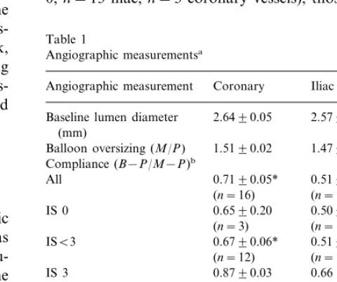
Relative contributions of remodelling and neointimal hyperplasia to restenosis after coronary angioplasty have been inferred from studies using iliofemoral arteries, despite differences in structure/function and smooth muscle cell lineage. We compared the response to balloon overstretch injury of coronary arteries (C,n=16) and similar sized branches of the iliac arteries (I,n=18) using preinjury vessel diameter (P), inflated balloon size in vivo (B) and the manufacturer predicted inflated size (M) to examine arterial compliance, as well as resulting injury and morphology in perfusion fixed vessels. Despite similar degrees of oversizing (M/P) in the coronary and iliac arteries (C, 1.4490.04; I, 1.5190.02), the compliance to overstretch (B−P/M−P) was significantly greater in the coronary than the iliac arteries (C, 0.7190.05; I, 0.5190.03) (PB0.05) and was associated with a higher injury score (C, 1.6490.31; I, 0.3990.18PB0.05) — only 5/18 iliac vessels had rupture of the IEL compared with 13/16 in the coronary bed. In a subgroup of animals whose vessels (C:n=7; I:n=8) were perfusion fixed 28 days after injury, coronary arteries had greater intimal area (C:1.0390.42; I:0.1090.03 mm2,PB0.05) but larger luminal area (C:1.6190.71; I:0.7690.51,
PB0.05) due to greater area within EEL (C:3.3890.49;I:1.4990.54,PB0.05) or less inward remodelling. The injuries resulting from similar strategies of balloon overstretch in the coronary and the iliac arteries are different and affect healing responses-iliac arteries remodel more while coronary arteries develop more intimal hyperplasia. These results indicate that caution is warranted when extrapolating results from the iliac to the coronary artery when investigating restenosis after angioplasty. © 2000 Elsevier Science Ireland Ltd. All rights reserved.
Keywords:Balloon overstrech; Coronory arteries; Iliac arteries
www.elsevier.com/locate/atherosclerosis
1. Introduction
Restenosis, which remains a significant limitation of percutaneous coronary revascularisation, results from a combination of negative remodelling (reduction in area within the external elastic lamina) and intimal growth (by smooth muscle cell proliferation and extracellular matrix deposition) [1]. Many experimental studies evalu-ating mechanisms of and therapeutic strategies for restenosis have been performed in the iliofemoral arter-ies [2,3] despite potential differences from the coronary arteries regarding both of these responses to injury.
The coronary arteries are morphologically distinct from peripheral arteries of similar size and this may affect the extent of injury and intimal hyperplasia after angioplasty. As the coronary vessels form by vasculoge-nesis (in situ formation by colocation of cellular ele-ments) rather than angiogenesis (axial growth from a vessel bud regulated by haemodynamics) [4], the internal elastic lamina is more fenestrated and incomplete and is more prone to forming intima [5]. In addition, as the coronary arteries are perfused during diastole, optimisa-tion of the pressure-flow relaoptimisa-tionship does not depend on the propagation of the systolic pressure wave, which requires vessel wall elasticity. Consequently the external elastic lamina is thinner [6], making rupture of the elastic laminae more likely, which has been strongly linked to neointima formation after injury [7,8].
* Corresponding author. Tel.:+1-650-723-1237; fax:+ 1-650-725-6766.
E-mail address:[email protected] (M.R. Ward)
Remodeling responses may also be different to those observed in the coronary arteries. Indeed, negative re-modelling is more prevalent in primary atherosclerotic lesions in the iliofemoral bed than in the coronary arteries [9], it is quite possible that negative remodelling would also be more frequent in the iliofemoral arteries after balloon dilatation.
Despite these potential differences a direct compari-son of coronary and iliac injury patterns and healing responses has not been performed. We therefore com-pared the response to balloon overstretch injury of iliofemoral and coronary vessels, by assessing compli-ance to balloon inflation, and the relative contributions of remodelling and intimal hyperplasia to lumen loss within the arteries during the healing response. We found that iliac arteries were less compliant than coro-nary vessels to overstretch, had less rupture of the elastic laminae and formed much less neointima, but had reduced lumen size 28 days after injury due to accentuated negative remodelling.
2. Methods
2.1. Animals, study design and drug administration
Mature male Boston mini-pigs (26 – 40 weeks old, 40 – 60 kg), were obtained from a colony at Monash University, Clayton, Australia. Injury of four arteries was attempted in each of the nine pigs: two coronary and two iliac branches. In all cases the iliac arteries injured were the right and left recurrent circumflex branch of the external iliac artery which are similar in size to the coronary arteries (2.5 – 3.0 mm diameter). Two of the three main coronary branches (right nary, left anterior descending and left circumflex coro-nary arteries) were dilated in each pig, except in two cases where only one was dilated due to difficulty with catheter engagement or arrhythmias. Angiographic measurements and injury scores were assessed for all vessels (see below). Seven coronary and eight iliac vessels (four pigs) were perfusion fixed in vivo 28 days after initial injury, and embedded in paraffin for mor-phometric assessment of vessel areas. Ten iliac and nine coronary vessels (five pigs) were harvested fresh at 5, 14 and 28 days, and were embedded in OCT (‘Tissue Tek’, Miles, Elkhart, IN), frozen using isopentane (Unilab, Australia) in liquid nitrogen and then stored at −70°C for immunohistochemical analysis as part of a separate study.
All animals were administered 300 mg Aspirin (Reckitt & Colman, West Ryde, Australia) per day orally, from 7 days prior to the initial procedures until the day of sacrifice, as well as 120 mg slow-release Verapamil (half a scored 240 mg tablet, Knoll, Lane Cove, Australia) within 12 h before each surgery.
Im-mediately prior to surgery the pigs were premedicated with intramuscular Acepromazine (0.1 mg/kg, Delta, Hornsby, Australia), Droleptan (10 mg, Delta, Hornsby, Australia) and Atropine sulphate (1.2 mg, Delta West, Bentley, Australia), anaesthesia induced with intravenous Propofol (150 – 200 mg, ICI, Mel-bourne, Australia), and then maintained with inhaled isoflourane (Abbott, Kurnell, Australia).
2.2. Angiography and angioplasty
All procedures were performed using an 8F JL4 guiding catheter through a sheath inserted into the common carotid artery (initial procedure right carotid, follow-up left carotid) after heparinisation (15 000 units, Fisons, Thornleigh, Australia). Angiography was performed after intraarterial glyceryl trinitrate 200 mg
(Fisons, Thornleigh, Australia) using Ioxaglate (Hexabrix, Mallinckrodt, Notting Hill, Australia) and was recorded for later analysis (Super VHS tape, Fuji, Germany) in the left anterior oblique view (25°) for the coronary arteries and in straight anteroposterior view for the iliac vessels. To easily identify the injured seg-ment at vessel harvest, the most proximal segseg-ment of each artery was injured. Arteries were balloon injured using standard human angioplasty catheters (semicom-pliant, 20 mm length) which were oversized according tothe manufacturer-specified inflated balloon size with balloon:artery ratio of 1.3 – 1.5:1. The balloon catheter was inflated to 10 atm for 30 s with three separate inflations separated by 1 min reperfusion periods. All measurements of vessels were made using handheld digital calipers (Mitutoyo, Japan) from the cine frame at end diastole at the point where the middle marker of the balloon catheter was sited, and using the guiding catheter as a reference. Vessel compliance (VC) was calculated by the following formula:
VC=(B−P)/(M−P)
whereB is inflated balloon size in vivo,Mis manufac-turer predicted inflated balloon size and P is preinjury vessel diameter.
2.3. Perfusion fixation and 6essel har6est
inferior vena cava. To fix the heart the aorta was cross-clamped and the right and left atrial ap-pendages incised, simultaneous to commencement of fixation through an aortic root cannula. After 5 min in vivo fixation, the vessels were then excised and care-fully debrided of the perivascular tissues, stored in 4% formalin in PBS for 24 h prior to paraffin embed-ding.
2.4. Morphometry and injury score
All vessels were cut into segments of 3 mm length resulting in six to seven sampling sites per balloon injured vessel. The region of maximal injury and in-tima formation were used for injury score and mor-phometry respectively (usually the same site).
Injury scores were assessed from 4mm sections (both
frozen and perfusion fixed vessels) in which the elastic laminae were highlighted using Masson’s Trichrome with Orcein staining or Van Giessen – Verhoff’s stain [10] for perfusion fixed and frozen vessels respectively. Injury score was assigned as previously described [7]: 0, Internal elastic lamina (IEL) not broken
1, IEL broken but media and external elastic laminae (EEL) intact
2, IEL broken with deep medial injury and partial rupture of EEL (EEL damaged but structurally intact)
3, EEL ruptured.
Intimal fracture length ratio (IELf/c) was calculated as the proportion of the IEL circumference which had been disrupted as previously described [8]. Medial, neointimal and vessel areas were determined for the perfusion fixed vessels only by planimetry using cus-tomised software (Capricorn Scientific, Woori Yallock, Australia) from sections magnified with a projecting microscope (NeoPromar Leitz Germany) onto a digitis-ing tablet (Complot series 7000 digitiser, Bausch and Lomb, Austin, TX, USA).
2.5. Calculation of late loss due to intima formation
and remodelling
An estimate of the proportion of late angiographic loss which could be attributed to intima formation was calculated as follows: lumen was assumed to be circu-lar immediately post dilatation and at follow-up; the cross-sectional area of the lumen at these two time points was then calculated aspr2 orp (minimal lumen diameter/2)2. The difference between these two areas was then calculated as late area loss. The proportion of late area loss due to intima formation was calculated by dividing intimal area on histology by late area loss. The remainder was then assumed to be due to inward remodelling.
2.6. Statistics
All data are presented as mean9SEM. For com-parison of two categorical variablesx2test was used or Fisher’s exact test where appropriate. For comparison of a continuous variable in coronary and iliac vessels Students t-test was used. For comparison of vessel compliance and balloon oversizing in multiple cate-gories (injury score) analysis of variance was used with post-hoc pairwise testing by Student’s t-test. For uni-variate regression analysis, simple linear regression was used. For multivariate analysis stepwise linear regres-sion was used.
3. Results
3.1. Relati6e compliance of the coronary and iliac arteries to o6erstretch injury and relationship to se6erity of injury sustained
The mean diameter on baseline angiography (P) and degree of balloon oversizing (M/P) of coronary and iliac vessels were similar. However, despite identical inflation pressures and times, expansion of the balloon (or compliance, B−P/M−P) within the coronary ar-teries was significantly greater than that seen within the iliac vessels (Table 1). As resistance to expansion may be significantly reduced if the elastic laminae were ruptured we also performed exploratory comparative analysis of compliance in the subgroups of iliac and coronary vessels where the IEL was intact (injury score 0,n=13 iliac,n=3 coronary vessels), those where the
Table 1
Balloon oversizing (M/P) 1.4790.06 Compliance (B−P/M−P)b
Lumen diameter at 2.1490.18* 1.4790.12 follow-up (mm)
Late lumen loss (mm) 0.8590.09* 1.2290.10 aAll values are mean9SEM. *PB0.05 versus iliac.
Fig. 1. Compliance and extent of vessel injury. Relationship between vessel compliance (B−P/M−P) and extent of rupture of the inter-nal elastic lamina (IELf/c) (see methods for definitions) for coronary (C, left,n=16) and iliac (I, right,n=18) vessels.
3.3. Se6erity of injury predicts extent of neointima formation but not remodelling
For the group which were perfusion fixed and quanti-tative morphometry performed the preinjury vessel size (C: 2.6790.08, I: 2.6490.05 mm) and balloon oversiz-ing (C: 1.4890.03, I: 1.5190.03) were similar between groups and representative of the whole study popula-tion. Angiographic follow-up diameter (C: 2.0590.13, I: 1.3890.12) and late loss (C: 0.9890.15, I: 1.349 0.14) in the fixed group were also representative of the group as a whole. The site used in each vessel for morphometric analysis was the site of greatest injury and intima formation. In all vessels this was also the site of smallest lumen area.
The coronary vessels developed significantly more intima than the iliac arteries (Fig. 2). The amount of intima formed after balloon overstretch injury of porcine vessels was highly dependent on the depth and circumferential extent of rupture of the elastic laminae consistent with observations from previous studies [7,8]. The intimal area for the vessels was strongly correlated with severity of injury (Fig. 2), and was predicted by the following formulae:
Intimal area (mm2)=2.53×IELf/c+0.0739
or
Intimal area (mm2)=0.451×Injury score+0.147
Due to the widely contrasting injury scores it was not possible to determine whether vessel type had any statistically significant effect on neointimal volume in-dependent of extent of injury. The mean (9SEM) percentage of angiographic late area loss which could be attributed to intima formation in the coronary ves-sels was 3398%, while in the iliac vessels it was 893%.
Despite much lower injury score, lumen size 28 days after injury in the iliac vessels was much smaller due to reduction in total vessel area (Fig. 2). Neither histolog-ical lumen size nor total vessel area were significantly correlated with injury score in either group or in all vessels. Similarly neither angiographic late loss or the percentage of late area loss which could be attributed to inward remodelling was significantly related to injury score.
Masson’s Trichrome staining with Orcein of coro-nary vessels showed that extensive rupture of the elastic laminae resulted in deposition of collagen interspersed with smooth muscle cells to breach the arc of rupture. Adventitial collagen accumulation was only moderate and predominantly confined to the region of most intimal and medial damage. In contrast in the iliac vessels moderate adventitial collagen was present even in vessels without rupture of the elastic lamina (Fig. 3). EEL was intact (injury scoreB3, n=17 iliac and n=
12 coronary vessels) and those in which the EEL was ruptured. This demonstrated that compliance within a given vascular bed was similar for injury scores 0 – 2 (Table 1): that is that the internal elastic lamina offered minimal resistance to balloon expansion. However, compliance measurements were increased if the external elastic lamina was ruptured (Table 1). In addition, the compliance measurements were significantly larger where the EEL was intact in the coronary vessels than in the iliac vessels.
Rupture of the internal elastic lamina (IEL) was observed in only five of 18 iliac vessels but in 13 of 16 coronary vessels (PB0.05), and mean injury score was 0.3990.18 in the iliac vessels and 1.6390.27 in the coronary vessels (PB0.05). The mean intimal fracture length ratio (IELf/c) in the all coronary and iliac vessels was 0.2490.04 and 0.0590.02 respectively (PB0.05). Exploratory analysis in only those vessels where the IEL had been broken revealed that IELf/c was still significantly greater in coronary (0.2990.04) than in iliac (0.1990.02) vessels (PB0.05). In coronary ves-sels, intimal fracture length was significantly predicted by vessel compliance ((B−P)/(M−P)) (Fig. 1). Due to the low number of iliac vessels which had a fractured IEL (5/18) this relationship was not statistically signifi-cant for the iliac vessels (P=0.45, 1−b=0.11). Im-portantly, the amount of balloon oversizing (M/P) did not predict either injury score or intimal fracture length (data not shown).
3.2. Angiographic acute gain and late lumen loss
4. Discussion
In this study we have demonstrated that the pattern of healing responses after balloon injury in the coro-nary bed differs significantly from that seen in the iliac vasculature. These differences should be taken into account when considering the applicability of studies
done in the iliac vessels to human coronary restenosis. When compared to the iliac vessels, the coronary arteries demonstrated increased angiographic compli-ance, sustained more extensive injury and developed more neointima. The greater balloon expansion (com-pliance) observed in the coronary vessels resulted from a combination of more frequent rupture of the elastic laminae and less elastic resistance. Even in vessels where the external elastic lamina remained intact bal-loon expansion was significantly greater in the coronary bed. The reduced elastic resistance and higher injury scores in the coronary vessels can be at least partly attributed to differences in the underlying vessel struc-ture, consequent to their unique origins and haemody-namic environment. More extensive injury in the coronary arteries was associated with a ten-fold in-crease in the size of neointima formed. While neointima formation has been previously shown to depend on the depth and extent of vessel injury [7,8] it is uncertain whether this can account for all of the observed differ-ence between coronary and iliac responses or whether known regional variability in smooth muscle cell biol-ogy may contribute [11]. Others have recently reported greater injury and neointima formation in coronary than in carotid vessels in response to similar balloon overstretch injury [12]. Our study has extended these findings by providing direct angiographic evidence for reduced vessel elasticity in the coronary arteries and reemphasize that these unique structural characteristics have important effects on the injury which results from balloon dilatation. Perhaps more importantly, we have demonstrated important differences between coronary and peripheral beds in remodelling, which is now thought to be the main cause of restenosis after angioplasty.
The reasons underlying the differences in inward remodelling observed in the iliofemoral and coronary vessels are unclear. Previous studies have suggested that negative remodelling after injury in coronary arteries is dependent on collagen production by adventitial my-ofibroblasts and requires deep vessel wall injury [13 – 15]. Although the injury sustained in the iliac vessels was neither deep nor severe remodelling was far greater than in the coronary vessels. As restenosis and negative remodelling in injured iliac vessels has been negatively correlated with vessel wall collagen [16], it is possible that iliac artery remodelling after balloon dilatation is not a directly related to severity of injury and collagen deposition. Consistent with this possibility, inhibitors of collagen cross-linking have successfully prevented nega-tive remodelling after injury in coronary but not iliac vessels [17,18]. The reduction in vessel wall collagen observed in restenotic iliac vessels was attributed to the pronounced induction of metalloproteinase (MMP) ac-tivity after injury in iliac vessels [19], inhibition of which significantly reduces inward remodelling [20]. Fig. 2. Vessel areas 28 days after balloon injury to coronary and iliac
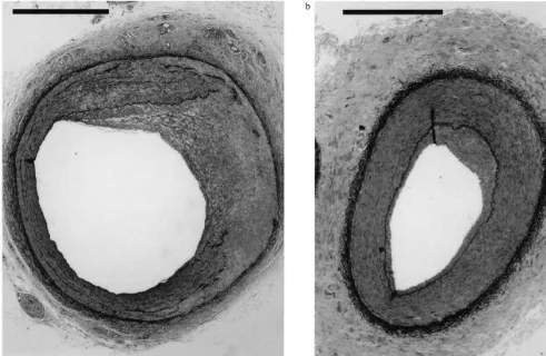
Fig. 3. Morphological features of coronary and iliac vessels 28 days after injury. Perfusion fixed, paraffin sections of balloon injured coronary (left) and iliac artery (right) stained with Masson’s Trichrome with Orcein (stains smooth muscle cells red, collagen green and elastin black). Scale bars represent 1 mm. Note the EEL is thicker in the iliac vessel. After balloon overstretch in the coronary vessel, extensive vessel wall injury with rupture of IEL has resulted in significant neointima formation which consists largely of smooth muscle cells and disordered collagen. In the iliac the IEL remains intact with only a minor amount of neointima formation but a with reduced lumen size mainly due to vessel shrinkage.
Although MMP activity has been reported to be re-duced in porcine and human coronary arteries after balloon injury these studies have been conducted very late in the course of restenosis and examined only the intima and media, not the adventitia where most colla-gen accumulates [21,22]. It is quite possible that early metalloproteinase activity may be important in inward remodelling after injury in coronary vessels.
There are some limitations of this study that should be considered. Firstly, the study was performed in normal porcine coronary and iliac vessels. The variable deformability of atherosclerotic plaque components (e.g. calcium vs lipid) may also significantly affect vessel compliance, the likelihood of tears of the elastic lami-nae, and response to balloon injury. Recent studies have found a similar importance for remodelling in lumen loss after balloon dilatation of artificially created atherosclerotic lesions in iliofemoral porcine vessels and naturally occurring atherosclerotic lesions in iliac ves-sels of cynomolgus monkey [23,24]. However there has not been a direct comparison between coronary and iliac vessels in these atherosclerotic models.
Secondly, in this study we were not able to reliably determine the absolute amount of remodelling (the change in vessel area from before balloon injury to follow-up). Two methods are commonly used to gener-ate such data: intravascular ultrasound (IVUS) and use of a histological reference segment. Accuracy of IVUS measurements relies on consistency of vascular tone during measurement. Despite the use of intraarterial nitrates the introduction of an IVUS catheter to the vessels of our animals resulted in significant reduction in angiographic lumen size (unpublished). Reference segments, which are widely used in both histological and IVUS-based studies to define remodelling, fre-quently undergo remodelling after nearby vessel injury [25]. We therefore chose to control for these problems by comparing coronary and iliac arteries which were of near identical size prior to injury to detect differences in remodelling responses.
examina-tion of many important issues, such as the influence of smooth muscle cell heterogeneity on intima formation, and these issues may have been resolved by assessment in greater numbers.
In summary we have shown that injury inflicted by balloon dilatation and the subsequent healing re-sponses are distinctly different between the coronary and iliac arteries justifying caution in extrapolation of data derived from injury of the porcine iliac vessels to human coronary restenosis. As the mechanisms of re-modelling in primary atherosclerotic and restenotic le-sions are only just beginning to be elucidated, it may be important to recognize that distinct mechanisms may be important in different vascular beds. Further investigation of the reasons underlying these differ-ences in remodelling may shed light on some of the basic causes of this important mechanism of lumen loss in both primary atherosclerotic and restenotic le-sions.
Acknowledgements
Dr Michael Ward is a recipient of an NH and MRC postgraduate medical research scholarship. These studies have in part been funded by the NH and MRC and the National Heart Foundation of Australia.
References
[1] Kimura T, Kaburagi S, Tamura T, Yokoi H, Nakagawa Y, Yokoi H, Hamasaki N, Nosaka H, Nobuyoshi M, Mintz GS, Popma JJ, Leon MB. Remodeling of human coronary arteries undergoing coronary angioplasty or atherectomy. Circulation 1997;96:475 – 83.
[2] Kakuta T, Currier JW, Haudenschild CC, Ryan TJ, Faxon DP. Differences in compensatory vessel enlargement, not inti-mal formation, account for restenosis after angioplasty in the hypercholesterolemic rabbit model. Circulation 1994;89:2809 – 15.
[3] Lafont A, Guzman LA, Whitlow PL, Goormastic M, Cornhill JF, Chisolm GM. Restenosis after experimental angioplasty. Intimal, medial, and adventitial changes associated with con-strictive remodelling. Circ Res 1995;76:996 – 1002.
[4] Flamme I, Frolich T, Risau W. Molecular mechanisms of vas-culogenesis and embryonic angiogenesis. J Cell Physiol 1997;173:206 – 10.
[5] Sims FH. Discontinuities in the internal elastic lamina: a com-parison of coronary and internal mammary arteries. Artery 1985;13:127 – 43.
[6] Fischer GM, Llaurado JG. Collagen and elastin content in canine arteries selected from functionally different vascular beds. Circ Res 1966;19:394 – 9.
[7] Groves PH, Banning AP, Penny WJ, Lewis MJ, Cheadle HA, Newby AC. Kinetics of smooth muscle cell proliferation and intimal thickening in a pig carotid model of balloon injury. Atherosclerosis 1995;117:83 – 96.
[8] Bonan R, Paiement P, Scortichini D, Cloutier MJ, Leung TK.
Coronary restenosis: evaluation of a restenosis injury index in a swine model. Am Heart J 1993;126:1334 – 40.
[9] Pasterkamp G, Schoneveld AH, van Wolferen W, Hillen B, Clarijs RJ, Haudenschild CC, Borst C. The impact of atherosclerotic arterial remodelling on percentage of luminal stenosis varies widely within the arterial system: a postmortem study. Arterioscler Thromb Vasc Biol 1997;17:3057 – 63. [10] Davenport HA. Staining Sections. In: Davenport HA, editor.
Histological and Histochemical Techniques. Philadelphia: W.B Saunders, 1960:217 – 62.
[11] Thieszen SL, Dalton M, Gadson PF, Patterson E, Rosenquist TH. Embryonic lineage of vascular smooth muscle cells deter-mines responses to collagen matrices and integrin receptor ex-pression. Exp Cell Res 1996;227:135 – 45.
[12] Badimon JJ, Ortiz AF, Meyer B, Mailhac A, Fallon JT, Falk E, Badimon L, Chesebro JH, Fuster V. Different response to balloon angioplasty of carotid and coronary arteries: effects on acute platelet deposition and intimal thickening. Atherosclerosis 1998;140:307 – 14.
[13] Andersen HR, Maeng M, Thorwest M, Falk E. Remodeling rather than neointimal formation explains luminal narrowing after deep vessel wall injury: insights from a porcine coronary (re)stenosis model. Circulation 1996;93:1716 – 24.
[14] Staab ME, Srivatsa SS, Lerman A, Sangiorgi G, Jeong MH, Edwards WD, Holmes DR Jr, Schwartz RS. Arterial remod-elling after experimental percutaneous injury is highly depen-dent on adventitial injury and histopathology. Int J Cardiol 1997;58:31 – 40.
[15] Shi Y O, Brien JE J, Fard A, Zalewski A. Transforming growth factor-beta 1 expression and myofibroblast formation during arterial repair. Arterioscler Thromb Vasc Biol 1996;16:1298 – 305.
[16] Coats WD, Whittaker P, Cheung DT, Currier JW, Han B, Faxon DP. Collagen content is significantly lower in restenotic versus nonrestenotic vessels after balloon angioplasty in the atherosclerotic rabbit model. Circulation 1997;95:1293 – 300. [17] Spears JR, Zhan H, Khurana S, Karvonen RL, Reiser KM.
Modulation by beta-aminopropionitrile of vessel luminal nar-rowing and structural abnormalities in arterial wall collagen in a rabbit model of conventional balloon angioplasty versus laser balloon angioplasty. J Clin Invest 1994;93:1543 – 53.
[18] Kristiansen SB, Schmidt MR, Danielsen CC, Falk HR, Syge-hus S. Inhibition of collagen cross-linking reduces (re)stenosis in a porcine model (abstract). Eur Heart J 1998;19:19. [19] Strauss BH, Robinson R, Batchelor WB, Chisholm RJ, Ravi
G, Natarajan MK, Logan RA, Mehta SR, Levy DE, Ezrin AM, Keeley FW. In vivo collagen turnover following experi-mental balloon angioplasty injury and the role of matrix metal-loproteinases. Circ Res 1996;79:541 – 50.
[20] de Smet BJGL, Robertus JLJMJR, van der Helm YJM, Borst C, Post MJ. Metalloproteinase inhibition reduces constrictive remodelling following balloon angioplasty: a study in the atherosclerotic yucatan micropig (abstract). J Am Coll Cardiol 1999;33:88.
[21] Tyagi SC, Meyer L, Schmaltz RA, Reddy HK, Voelker DJ. Proteinases and restenosis in the human coronary artery: extra-cellular matrix production exceeds the expression of proteolytic activity. Atherosclerosis 1995;116:43 – 57.
[22] Guarda E, Katwa LC, Campbell SE, Tanner MA, Webel RM, Laughlin H, Jenkins S, Myers PR. Extracellular matrix collagen synthesis and degradation following coronary balloon angio-plasty. J Mol Cell Cardiol 1996;28:699 – 706.
[24] Mondy JS, Williams JK, Adams MR, Dean RH, Geary RL. Structural determinants of lumen narrowing after angioplasty in atherosclerotic nonhuman primates. J Vasc Surg 1997;26:875 – 83.
[25] Kakuta T, Usui M, Coats WD Jr, Currier JW, Numano F, Faxon DP. Arterial remodelling at the reference site after angio-plasty in the atherosclerotic rabbit model. Arterioscler Thromb Vasc Biol 1998;18:47 – 51.