RESEARCH
ARTICLE
Curcumin alleviates oxidative stress, inflammation, and
renal fibrosis in remnant kidney through the Nrf2–keap1
pathway
Vivian Soetikno
1,2, Flori R. Sari
1, Arun P. Lakshmanan
1, Somasundaram Arumugam
1,
Meilei Harima
1, Kenji Suzuki
3, Hiroshi Kawachi
4and Kenichi Watanabe
11Department of Clinical Pharmacology, Faculty of Pharmaceutical Sciences, Niigata University of Pharmacy and Applied Life Sciences, Niigata City, Japan
2Department of Pharmacology, Faculty of Medicine, University of Indonesia, Jakarta, Indonesia
3Department of Gastroenterology and Hepatology, Niigata University Graduate School of Medical and Dental Sciences, Niigata, Japan
4Department of Cell Biology, Institute of Nephrology, Niigata University Graduate School of Medical and Dental Sciences, Niigata, Japan
Scope: We hypothesized that curcumin, by increasing the expression of nuclear factor-erythroid-2-related factor 2 (Nrf2), could reduce oxidative stress, inflammation, and renal fibro-sis in remnant kidney.
Methods and results: Sprague-Dawley rats were subjected to 5/6 nephrectomy and randomly assigned to untreated (Nx), curcumin-treated (75 mg/kg/day, orally), and telmisartan-treated groups (10 mg/kg/day, orally; as positive control). Sham-operated rats also served as controls. Five/sixth nephrectomy caused renal dysfunction, as evidenced by elevated proteinuria, blood urea nitrogen, and plasma creatinine, and decreased creatinine clearance that were ameliorated by curcumin or telmisartan treatment. The Nx rats demonstrated reduced Nrf2 protein expres-sion, whereas the Kelch-like ECH-associated protein 1 was upregulated and heme oxygenase-1 level was significantly diminished. Consequently, Nx animals had significantly higher kidney malondialdehyde concentration and lower glutathione peroxidase activity, which was associ-ated with the upregulation of nicotinamide adenine dinucleotide phosphatase oxidase subunit (p67phoxand p22phox), NF-kappaB p65, TNF-␣, TGF-1, cyclooxygenase-2, and fibronectin
accu-mulation in remnant kidney. Interestingly, all of these changes were ameliorated by curcumin or telmisartan.
Conclusion: These findings demonstrate that, by modulating Nrf2-Keap1 pathway, the cur-cumin effectively attenuates oxidative stress, inflammation, and renal fibrosis, which suggest that curcumin hold promising potential for safe treatment of chronic kidney disease.
Keywords:
Chronic kidney disease / Curcumin / Fibrosis / Inflammation / Oxidative stress
Received: August 13, 2012 Revised: September 18, 2012 Accepted: September 19, 2012
Correspondence: Professor Kenichi Watanabe, Department of Clinical Pharmacology, Faculty of Pharmaceutical Sciences, Ni-igata University of Pharmacy and Applied Life Sciences, 265–1 Higashijima Akiha-ku, Niigata City 956–8603, Japan
E-mail:[email protected] Fax:+81-250-25-5021
Abbreviations: ANG II, angiotensin II;ARB, AT1 receptor blockers; BP, blood pressure;BUN, blood urea nitrogen;CCr, creatinine clearance;CKD, chronic kidney disease;COX-2, cyclooxygenase-2; GPx, glutathione peroxidase; HO-1, heme oxygenase-1; Keap1, Kelch-like erythroid cell-derived protein with cap-n-collar homology-associated protein 1;MDA, malondialdehyde;
1
Introduction
The pathogenesis of chronic kidney disease (CKD) involves a complex interaction of oxidative stress, inflammation, and fibrotic processes that lead to glomerular and tubulointersti-tial scarring with subsequent progression toward end-stage renal disease [1, 2].
Oxidative stress may cause inflammation by direct toxic effects of reactive oxygen species (ROS) and by activation of redox-sensitive proinflammatory transcription factors and signal transduction pathways. Hence, oxidative stress and inflammation play a major role in the progression of renal in-jury [3]. The major ROS is superoxide, and in the kidney, this is mainly generated by NAD(P)H oxidase [4]. Redox systems including antioxidant and phase II detoxifying enzymes pro-vide protection against ROS-induced tissue injury. Nuclear factor-erythroid-2-related factor 2 (Nrf2) is an important cyto-protective transcription factor that induces the expression of several antioxidant and phase II detoxifying enzymes such as catalase, superoxide dismutase, uridine 5′-diphospho
(UDP)-glucuronosyltransferase, NAD(P)H:quinine oxidoreductase-1, heme oxygenase-1 (HO-1), glutamate cysteine ligase, glu-tathione S-transferase, glutathione peroxidase (GPx), and thioredoxin [5]. In resting cells, Nrf2 is sequestered in the cytoplasm as an inactive complex with the repressor Kelch-like ECH-associated protein 1 (Keap1). Under oxidative conditions, Nrf2 is released from Keap1 repression and translocates to the nucleus. After this translocation, Nrf2 forms heterodimers with other transcription factors, which in turn bind to the regulatory sequences encoding various antioxidants and phase II detoxifying enzymes [6]. Besides its role in regulating cellular detoxification and antioxidative defense, Nrf2 also has anti-inflammatory functions and thus represents a novel therapeutic approach for the treatment or prevention of inflammatory disorders [7].
Curcumin is a diferuloylmethane derived from the rhi-zomes of turmeric. Its chemopreventive effects have been extensively investigated and well defined [8]. Balogun et al. [9] reported that curcumin disrupted the Nrf2–Keap1 complex, leading to increases in the expression and activity of HO-1 in porcine renal epithelial proximal tubule cells. A very re-cent experimental study showed that short-term curcumin treatment in high-fat diet-fed mice ameliorated muscular ox-idative stress by activating Nrf2 function [10]. Moreover, cur-cumin treatment has been shown to decrease macrophage infiltration in the kidneys of chronic renal failure rats and to block transactivation of nuclear factor-kappa-light-chain-enhancer of activated B cells (NF-B), indicating that the anti-inflammatory property of curcumin may be responsible for alleviating disease in this animal model [11]. However, to the best of our knowledge, no attempt has been made to correlate the role of curcumin in Nrf2–Keap1 pathway and its downstream region in an experimental CKD model. The present study was designed to shed light on this issue.
2
Materials and methods
2.1 Study group
Male Sprague-Dawley rats with an average body weight of 150–200 g (age 6 wk of age) (Charles River, Japan Inc., Kana-gawa, Japan) were used in this study. The experimental
pro-tocol was approved by the Institutional Animal Care and Use Committee of the University of Pharmacy and Applied Life Sciences. Animals were housed in a climate-controlled vi-varium with a 12:12-h light–dark cycle and fed a standard laboratory diet and water ad libitum. The animals were ran-domly divided into four groups with six animals in each group. The control group underwent sham operation (sham), and the three other groups underwent 5/6 nephrectomy by surgical resection of the upper- and lower-thirds of the left kidney, followed by a right nephrectomy 7 days later. The procedures were carried out under general anesthesia (pen-tobarbital sodium, 50 mg/kg i.p.) using a sterile technique. In the second week after the second surgery, the animals were randomly divided into curcumin treatment (C75), telmisartan treatment (T10), and untreated (Nx) groups. The curcumin-treated group was subjected to gavage with 75 mg/kg cur-cumin daily [11]. The telmisartan group received 10 mg/kg oral telmisartan daily [12]. The untreated Nx animals received vehicle, which was 0.5% carboxymehtyl cellulose, daily. The animals were studied for 8 wk and in week 11 were killed, after which blood and kidney tissues were collected for fur-ther analysis. Twenty-four-hour urine samples were collected in metabolic cages at baseline, week 7, and week 10 for the determination of urinary levels of protein and creatinine.
2.2 Blood and urine chemistries
Blood samples were collected in EDTA vacutainer tubes by heart puncture. EDTA blood was centrifuge at 3000×gfor 15 min at 4⬚C for separation of plasma. The collected plasma was utilized for the subsequent determination of creatinine, blood urea nitrogen (BUN), plasma total cholesterol, triglyc-eride, LDL cholesterol, and HDL cholesterol (SRL Labora-tory, Japan). Urinary protein excretion was determined by the Bradford method [13]. Plasma and urinary creatinine levels were determined by Jaffe method [14]. BUN was determined by the diacetylmonoxamine method [15].
2.3 Measurement of malondialdehyde (MDA) content
Kidney tissues were rinsed, weighed, resuspended at 50 mg/mL in normal saline, and homogenized. After cen-trifugation at 5000 rpm for 10 min at 4⬚C, the supernatants were collected and analyzed with corresponding assay kits (OXItek, ZeptoMetrix Corporation, NY, USA) in accordance with the manufacturer’s instruction.
2.4 Measurement of GPx activity
spectrophotometer (Ultraspec 3100, Amersham Biosciences, Cambridge, UK). The oxidation of nicotinamide adenine din-ucleotide phosphate reduced (NADPH) to nicotinamide ade-nine dinucleotide phosphate was measured by the decrease in absorbance at 340 nm.
2.5 Longitudinal measurement of blood pressure by tail plethysmography
Blood pressure (BP) was noninvasively measured by a volume pressure-recording sensor and an occlusion tail-cuff (CODA System, Kent Scientific, Torrington, CT, USA). In brief, be-fore surgery, the conscious animal was placed in a restrainer and permitted to rest for 10 to 15 min. The cuff was then placed on the tail and was inflated and released several times to condition the animal to the procedure. After stabilization, BP was measured three times, and the average of the recorded values was used. In this study, we report the mean systolic BP (SBP) of each group. On the third day before surgery, BP was recorded (week 0). Two weeks after the second surgery, the animals were retrained and BP was recorded (week 3). Similar BP recording was carried out in weeks 6 and 10 postsurgery.
2.6 Kidney homogenates and nuclear extracts preparation
Briefly, 100 mg of kidney cortex was homogenized in 0.5 mL of buffer A containing 10 mM HEPES, pH 7.8, 10 mM KCl, 2 mM MgCl2, 1 mM DTT, 0.1 mM EDTA, 0.1 mM PMSF,
1M pepstatin, and 1 mMp-aminobenzamidine using a tis-sue homogenizer for 20 s. Homogenates were kept on ice for 15 min, and then 125L of a 10% Nonidet p40 solution were added and mixed for 15 s and the mixture was centrifuged for 2 min at 12 000 rpm. The supernatant containing cytoso-lic proteins was collected. The pelleted nuclei were washed once with 200L of buffer A plus 25L of 10% Nonidet p40, centrifuged, then suspended in 50L of buffer B containing 50 mM HEPES, pH 7.8, 50 mM KCl, 300 mM NaCl, 0.1 mM EDTA, 1 mM DTT, 0.1 mM PMSF, and 10% v/v glycerol, mixed for 20 min, and centrifuged for 5 min at 12 000 rpm. The supernatant containing nuclear proteins was stored at –80⬚C [16]. The protein concentrations in kidney ho-mogenates and nuclear extracts were determined by the bicin-choninic acid method.
2.7 Western blot analysis
Protein abundance of cycloxygenase-2 (COX-2), NADPH oxi-dase subunits (p22phoxand p67phox), transforming growth
fac-tor (TGF)-1, tumor necrosis factor (TNF)-␣, HO-1, Keap1, Nrf2 nuclear translocation, and p65 NF-B nuclear transloca-tion were measured by the Western blot technique. Antibod-ies against COX-2, p22phox, p67phox, TNF-␣, HO-1, Keap1, and
Nrf2 were purchased from Santa Cruz Biotechnology (Santa Cruz, CA, USA). Antibodies against p65 NF-B were
pur-chased from Cell Signaling Technology (Denver, CO, USA). Antibody against TGF-1 was purchased from Promega (Madison, WI, USA). Antibodies to -actin (Cell Signaling Technology) and Lamin A (Santa Cruz, CA, USA) were used for measurements of the housekeeping proteins for cytosolic and nuclear target proteins, respectively. Aliquots containing 70g of total protein (cytosol) and 50g of total protein (nu-clear extracts) were separated on a 7.5–12.5% SDS PAGE gel, and proteins were transferred to a nitrocellulose membrane. After being briefly washed in Tris-buffered saline (TBS; 10 mM Tris HCl, 0.85% NaCl, pH 7.2) containing 0.05% Tween-20 (TBS-T) and blocked for 1 h in 5% nonfat dry milk, membranes were incubated with appropriate antibodies in 5% nonfat dry milk overnight at 4⬚C. After being washed three times for 5 min in TBS-T, membranes were subse-quently incubated with secondary antibody appropriately di-luted in TBS-T for 1 h at room temperature. The membrane was washed four times and detection was performed using the enhanced chemiluminescence system (Amersham Bio-sciences, Buckinghamshire, UK) and exposed to X-ray film.
2.8 Immunohistochemistry of fibronectin
Formalin-fixed, paraffin-embedded kidney tissue sections were used for immunohistochemical staining. After deparaf-finization and hydration, the slides were washed in TBS. Endogenous peroxidase activity was quenched by incubat-ing the slides in methanol and 0.3% H2O2 in methanol.
After overnight incubation with the primary antibody, that is, fibronectin (diluted 1:50) (Santa Cruz, CA), at 4⬚C, the slides were washed in TBS and then horseradish peroxidase-conjugated secondary antibody was added and the slides were further incubated at room temperature for 1 h. The slides were washed in TBS and incubated with diaminoben-zidine tetrahydrochloride as the substrate and counterstained with hematoxylin. A negative control without primary anti-body was included in the experiment to verify the antianti-body specificity. For morphometry analysis, the percentage of pos-itive cells (stained brown) was determined with a computer-assisted color image analyzer (CIA-102; Olympus, Tokyo, Japan). Ten random fields per kidney were studied at×100 magnification for comparison of the different groups [17].
2.9 Histopathological analysis
Light microscopy was performed in the formalin-fixed sec-tions (4m) stained with Azan Mallory to stain collagen as an indicator of fibrosis and periodic acid-Schiff to analyze glomerulosclerosis and tubulointerstitial injury. Glomeru-losclerosis was graded from 0 to 4 (grade 0; normal, grade 1;
<25% involvement of the glomerular tuft, grade 2; 25–50%
1+)+(2×nglomeruli with 2+)+(3×nglomeruli with 3+)+ (4×nglomeruli with 4+)]×100/total number of glomeruli examined. Tubulointerstitial damage was graded according to the extension (%) of tubular damage (infiltra-tion, fibrosis, and tubular dilatation/atrophy). Interstitial fi-brosis was determined in ten random fields of the cortical area at×100 magnification with use of a computer-assisted color image analyzer (CIA-102; Olympus), with the observer blind to the study group. The results are presented as the ratio of the fibrotic area to the whole area of the kidney. The same instrument was used to determine glomerulosclerosis and tubulointerstitial injury [17].
2.10 Determination of TNF-␣expression by quantitative real-time RT-PCR
Kidney tissues were preserved by immersion in RNAlater (Ambion Inc., Austin, TX, USA) immediately after sam-pling. Total RNA was extracted after homogenization us-ing Ultra Turrax T8 (IKA Labortechinik, Staufen, Ger-many) in TRIzol reagent (Invitrogen Corp., Carlsbad, CA, USA) according to the standard protocol. cDNA was syn-thesized by reverse transcription using total RNA (2g) as a template (Super Script II; Invitrogen Corp). Gene expres-sion analysis was performed by real-time RT-PCR (Smart cycler; Cepheid, Sunnyvale, CA, USA) using cDNA syn-thesized from the samples specimen. Primer sequences were as follow: TNF-␣ (forward), CCCCAAAGGGATGA-GAAGTT, (reverse), CACTTGGTGGTTTGCTACGA; glycer-aldehyde 3-phosphate dehydrogenase (GAPDH) (forward) GCTCATTTCCTGGTATGACAACG, (reverse), AGGGGTC-TACATGGCAACTG. Real-time RT-PCR, monitoring with a TaqMan probe (TaqMan Gene expression assays; Applied Biosystems, Foster City, CA, USA) was performed according to the following protocol: 600 s at 95⬚C, followed by thermal cycles of 15 s at 95⬚C, and 60 s at 60⬚C for extension.
Rel-ative standard curves representing several tenfold dilutions of cDNA from kidney tissue samples were used for linear regression analysis of other samples. Results were normal-ized to GAPDH mRNA as an internal control and shown as relative mRNA levels.
2.11 Statistical analysis
Data are expressed as mean±SEM and were analyzed using one-way analysis of variance (ANOVA) followed by Tukey’s methods for post hoc analysis and two-tailed t-test when ap-propriate. A value ofp<0.05 was considered statistically
sig-nificant. For statistical analysis, GraphPad Prism 5 software (San Diego, CA, USA) was used.
3
Results
3.1 Effect of curcumin and telmisartan on general data
The final total number of animals used in this study was 24 (sham (6), Nx (6), T10 (6), C75 (6)). The survival rate after Nx surgery was 64.29% (18 of 28), this was not different between the three Nx groups (60% for Nx versus 66.67% for T10 versus 66.67% for C75,p>0.05) (data not shown).
Results obtained at 10 wk after 5/6 nephrectomy are sum-marized in Table 1. Compared with the sham group, the 5/6 nephrectomy groups had significantly decreased body weight, creatinine clearance (CCr), and HDL cholesterol, and increased left kidney weight, plasma creatinine, BUN, total cholesterol, LDL cholesterol, and triglyceride. Both curcumin and telmisartan treatments resulted in significant reductions of plasma creatinine and left kidney weight and an increase of CCr. The curcumin-treated and telmisartan-treated rats did not show any significant changes in body weight, total cholesterol, triglyceride, and HDL cholesterol in comparison
Table 1. General data obtained at 10 wk following surgerya)
Sham (n=6) Nx (n=6) T10 (n=6) C75 (n=6)
Body weight (g) 517.5±8.5 468.3±5.5b) 507.5±23.1 494.5±13.3
Left kidney weight (g) 1.45±0.06 1.96±0.07b) 1.52±0.05d) 1.54±0.04d)
Plasma creatinine (mg/dL) 0.65±0.3 2.27±0.18b) 0.82±0.34e) 1.04±0.29e)
BUN (mg/dL) 14±0.9 32.5±0.8b) 31.8±0.6b) 28.2±1.6b)
CCr (mL/min/kg) 5.3±1.4 1.3±0.08c) 2.8±0.4e) 3.2±0.6e)
Total cholesterol (mg/dL) 49.8±3.1 73.8±7.6c) 69.8±8.6 59±7.8
Triglyceride (mg/dL) 25±3.1 55.8±11.5c) 33.5±5.4 29.8±2.4
LDL cholesterol (mg/dL) 18±0.7 58.5±11.8c) 27.3±5e) 22.8±2.5e)
HDL cholesterol (mg/dL) 26.7±1.7 18±2.5c) 25.7±3.3 24.7±2.6
a) Values are given as mean±SEM.
b)p<0.01 versus sham based on two-tailed t-test. c)p<0.05 versus sham based on two-tailed t-test. d)p<0.01 versus Nx based on two-tailed t-test. e)p<0.05 versus Nx based on two-tailed t-test.
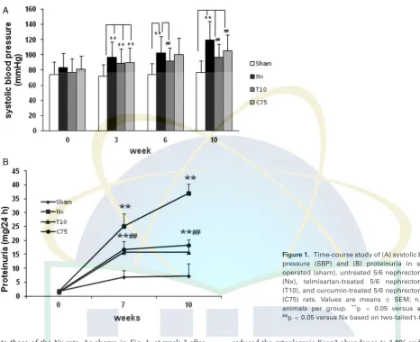
Figure 1. Time-course study of (A) systolic blood pressure (SBP) and (B) proteinuria in sham-operated (sham), untreated 5/6 nephrectomized (Nx), telmisartan-treated 5/6 nephrectomized (T10), and curcumin-treated 5/6 nephrectomized (C75) rats. Values are means ± SEM; n = 6 animals per group. **p
< 0.05 versus sham,
##p
<0.05 versus Nx based on two-tailedt-test.
to those of the Nx rats. As shown in Fig. 1, at week 3 after the operation, the SBP was markedly elevated in the 5/6 nephrectomy groups compared with that in the sham group (p<0.01). Telmisartan treatment significantly curtailed the
increase in SBP throughout the entire experimental period (p<0.01). In the curcumin group, the SBP was parallel to
that in the Nx group until week 10, but was subsequently reduced, although not to the extent in the telmisartan-treated animals (p< 0.01). The Nx group exhibited a significant
gradual increase in proteinuria. Curcumin was as effective as telmisartan in reducing proteinuria (p<0.01).
3.2 Effect of curcumin and telmisartan on Nrf2–Keap1 pathway
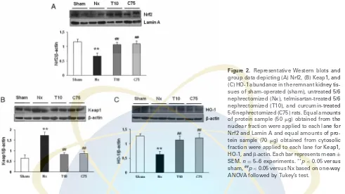
We next examined whether 8-wk treatment with curcumin or telmisartan affected the renal expression of Nrf2, Keap1, and HO-1. As shown in Fig. 2, 5/6 nephrectomy markedly reduced the nuclear expression of Nrf2 and increased the cytoplasmic Keap1 abundance. This was associated with sig-nificant reduction of renal tissue HO-1 protein abundance in the remnant kidney. Administration of curcumin or telmis-artan increased the nuclear expression of Nrf by 1.6%, and
reduced the cytoplasmic Keap1 abundance to 1.8% and 2%, respectively, and increased the HO-1 protein abundance in the remnant kidney by 1.9% and 1.8%, respectively. The effects of curcumin and telmisartan treatments on Nrf2, Keap1, and HO-1 protein expression were not significantly different.
3.3 Effect of curcumin and telmisartan on oxidative stress and inflammatory parameter
Compared with the Nx group, curcumin- and telmisartan-treated groups exhibited significantly attenuated p67phox
pro-tein expression, by 2.1-fold and twofold, respectively (Fig. 3A). Western blot analysis also demonstrated a significant increase of p22phoxprotein expression by twofold in the Nx group
com-pared with that in the sham group, which was significantly attenuated by treatment with curcumin by 1.2-fold. Telmis-artan treatment also reduced the expression of p22phox, but
Figure 2. Representative Western blots and group data depicting (A) Nrf2, (B) Keap1, and (C) HO-1 abundance in the remnant kidney tis-sues of sham-operated (sham), untreated 5/6 nephrectomized (Nx), telmisartan-treated 5/6 nephrectomized (T10), and curcumin-treated 5/6 nephrectomized (C75) rats. Equal amounts of protein sample (50g) obtained from the nuclear fraction were applied to each lane for Nrf2 and Lamin A and equal amounts of pro-tein sample (70g) obtained from cytosolic fraction were applied to each lane for Keap1, HO-1, and-actin. Each bar represents mean±
SEM.n=5–6 experiments.**p
<0.05 versus sham,##p
<0.05 versus Nx based on one-way ANOVA followed by Tukey’s test.
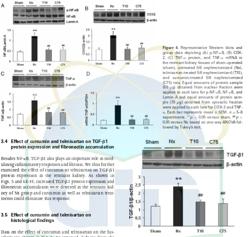
1.7-fold and 1.5-fold, respectively (Fig. 3C). GPx activity in the kidney was also significantly lower in the Nx group than in the sham group. Chronic treatment with curcumin or telmis-artan prevented this decrease in GPx activity (Fig. 3D). The increase of oxidative stress parameter was associated with a significant increase in NF-B activity, as indicated by the increased phosphorylation of NF-B in the nuclear fraction of the Nx group. Both curcumin and telmisartan treatments resulted in decreased activity of NF-B (Fig. 4A) and, as a
re-sult, decreases in the protein and mRNA expression of TNF-␣
(Fig. 4C and D). Similarly, COX-2 abundance was signifi-cantly increased in the remnant kidney of the Nx group com-pared with that of the sham group, which was significantly attenuated by treatment with curcumin as well as telmisar-tan (p<0.05) (Fig. 4B). The differences of NAD(P)H oxidase
subunit p67phox, MDA level, GPx activity, NF-B activity, and
COX-2 between curcumin- and telmisartan-treated groups were not significant.
Figure 3. Representative Western blots and group data depicting (A) p67phox and (B) p22phox abundance, (C) malondialdehyde (MDA) concentration, and (D) GPx activity in the remnant kidney tissues of sham-operated (sham), untreated 5/6 nephrectomized (Nx), telmisartan-treated 5/6 nephrectomized (T10), and curcumin-treated 5/6 nephrectomized (C75) rats. Equal amounts of protein sam-ple (70 g) were applied to each lane. Each bar represents mean±SEM.n=5–6 experiments.**p
Figure 4. Representative Western blots and group data depicting (A) p-NF-B, (B) COX-2, (C) TNF-␣ protein, and TNF-␣ mRNA in the remnant kidney tissues of sham-operated (sham), untreated 5/6 nephrectomized (Nx), telmisartan-treated 5/6 nephrectomized (T10), and curcumin-treated 5/6 nephrectomized (C75) rats. Equal amounts of protein sample (50g) obtained from nuclear fraction were applied to each lane for p-NF-B, NF-B, and Lamin A and equal amounts of protein sam-ple (70g) obtained from cytosolic fraction were applied to each lane for COX-2 and TNF-␣. Each bar represents mean±SEM.n=5–6 experiments.**p
<0.05 versus sham,##p< 0.05 versus Nx based on one-way ANOVA fol-lowed by Tukey’s test.
3.4 Effect of curcumin and telmisartan on TGF-1 protein expression and fibronectin accumulation
Besides NF-B, TGF-1 also plays an important role in mod-ulating inflammatory responses and fibrosis. We thus further examined the effect of curcumin or telmisartan on TGF-1 protein expression in the remnant kidney. As shown in Figs. 5 and 6E–H, increased TGF-1 protein expression and fibronectin accumulation were detected in the remnant kid-ney of Nx group and curcumin as well as telmisartan treat-ments could eliminate this response.
3.5 Effect of curcumin and telmisartan on histological findings
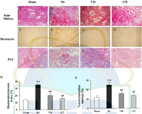
Data on the effect of curcumin and telmisartan on the his-tology are shown in Fig. 6. As expected, kidneys from the Nx group showed extensive fibrosis (Fig. 6B), significant glomerulosclerosis, tubular atrophy, and tubulointersitital in-jury (Fig. 6J). Treatment with telmisartan or with curcumin reduced fibrosis, glomerulosclerosis index, as well as tubu-lointerstitial injury in 5/6 nephrectomy rats (p<0.05).
4
Discussion
The removal of kidney, representing two-thirds of the to-tal functioning renal mass, by surgical procedures in this study was sufficient to trigger the progression of CKD, as evidenced by proteinuria, decreased CCr, increased BUN, and structural damage to the remnant kidney. This was
as-Figure 5. Representative Western blot and group data depict-ing TGF-1 in the remnant kidney tissues of sham operated (sham), untreated 5/6 nephrectomized (Nx), telmisartan-treated 5/6 nephrectomized (T10), and curcumin-treated 5/6 nephrec-tomized (C75) rats. Equal amounts of protein sample (70g) ob-tained from cytosolic fraction were applied to each lane. Each bar represents mean±SEM.n=5–6 experiments.**p
<0.05 versus sham,##p
<0.05 versus Nx based on one-way ANOVA followed by Tukey’s test.
Figure 6. Representative photomicrograph Azan Mallory staining (A–D) for fibrosis of the cross-sectional tissue slices of kidney. Fibrosis is indicated by the blue area (magnification×100), immunohistochemical staining of fibronectin is strong in epithelial tubular cells in the Nx group (E–H) (magnification×100), and PAS staining (I–L) (magnification×200) of the remnant kidney is shown in sham operated (sham), untreated 5/6 nephrectomized (Nx), telmisartan-treated 5/6 nephrectomized (T10), and curcumin-treated 5/6 nephrectomized (C75). Bar graphs depict the corresponding glomerulosclerosis index (M) and tubulointerstitial injury index (N).**p
<0.05 versus sham,##p<0.05 versus Nx based on one-way ANOVA followed by Tukey’s test.
clinical and experimental studies have revealed that activa-tion of ANG II type 1 receptor by ANG II can account for upregulation/activation of NAD(P)H oxidase and oxidative stress and that intervention by AT1 receptor blockers (ARBs) shows efficacy in slowing the onset of kidney disease and/or slowing progression of CKD [19, 20]. In fact, an experimental study of spontaneous focal glomerulosclerosis rats has indi-cated that ARB treatment prevented nephropathy, suppressed oxidative stress, inflammatory process, and endoplasmic reticulum stress-induced apoptosis as well as restored Nrf2 activation and expression of the antioxidant enzymes [21, 22]. A very recent study has revealed that telmisartan, an ARB, could suppress NAD(P)H oxidase and subsequently upreg-ulated Nrf2, leading to the amelioration of renal oxidative
stress and diabetic renal changes [23]. Moreover, it has been demonstrated that telmisartan attenuated the progression of CKD by blocking vascular dysfunction and inflammation in subtotal nephrectomy rat model [24]. To verify whether cur-cumin might have a beneficial role in CKD, we compared it with telmisartan. We showed here that curcumin gavage was as effective as telmisartan in alleviating the deterioration of renal function, as shown by biochemical and histologi-cal parameters, even though curcumin treatment could not significantly ameliorate the increased of SBP compared with telmisartan treatment.
regenerative ability of cells [25]. Pivotal to the antioxidant response is the transcription factor Nrf2. Under basal condi-tions, Nrf2 is mainly found sequestered in the cytoplasm as an inactive complex bound to a repressor molecule known as Keap1, which facilitates its ubiquitination. When challenged by oxidative stress derived from accumulation of ROS, Nrf2 can quickly translocate into the nucleus and induce genes en-coding antioxidant and phase II detoxifying enzymes, such as HO-1 [26]. Actually, disruption of Nrf2 in mice dimin-ishes or abrogates the induction of these genes [27]. More-over, Nrf2 gene ablation has been shown to cause a lupus-like autoimmune nephritis and exacerbate diabetes-induced inflammation, oxidative stress, and renal injury in experi-mental animals [28, 29]. It has been demonstrated that im-paired Nrf2 activation contributes to the severity of oxidative stress, inflammation, and the progression of tissue damage in the remnant kidney model [16]. Interestingly, curcumin has antioxidant and anti-inflammatory effects and in vitro studies have indicated its action in triggering Nrf2 signaling [9, 30]. Another study also reported that dimethoxycurcumin and curcumin induced HO-1 expression and Nrf2 nuclear translocation in RAW264.7 macrophages [31]. We therefore proposed the working hypothesis of this study that curcumin could alleviate oxidative stress and inflammation in the CKD model by its action via Nrf2 activation. We found that the remnant kidney in the CKD animals showed marked upreg-ulation of NAD(P)H oxidase isoforms (p67phox and p22phox),
significant elevation of lipid peroxidation product MDA, as well as depression of antioxidant activity GPx, denoting the presence of oxidation, while curcumin and telmisartan ad-ministration significantly reverses this deleterious effect. To further investigate the underlying mechanism of curcumin and telmisartan on regulating redox balance, we examined Nrf2 signaling in remnant kidney tissues. We found that CKD led to a reduced nuclear expression of Nrf2 and increased cy-toplasmic expression of Keap1, along with decreased levels of Nrf2 target gene products, HO-1 in remnant kidney. Kim and Vaziri [16] have recently reported that Nrf2 activity (nu-clear translocation) was mildly reduced at 6 wk and markedly reduced at 12 wk after 5/6 nephrectomy. This was accom-panied by reduced or unchanged of the Nrf2 target genes, HO-1, at 6 wk; and significantly diminished at 12 wk. In the present study, although curcumin and telmisartan adminis-tration slightly abrogated these changes; the differences reach a statistical significant. This perhaps related with the limited follow-up time in our model. Nevertheless, as far as we know, our study is the first evidence that treatment with curcumin and telmisartan for 8 wk starting 2 wk after 5/6 nephrectomy significantly reduced the degree of kidney structural damage and reversed oxidative stress in remnant kidney, and that these improvements were associated with increased nuclear expression of Nrf2. Importantly, this was achieved without in-creased mortality. We hence suggest that the activation of the major oxidative defense machinery Nrf2 in kidney is a novel mechanism behind curcumin’s alleviation of CKD induced-oxidative stress.
Furthermore, we here show that CKD animals exhibited significant activation of NF-B in the remnant kidney as evidenced by increased translocation of p65, the active subunit of this transcription factor. NF-B is the general transcription factor that plays a crucial role in mediating in-flammation in the kidney because it regulates the expres-sion of numerous genes involved in inflammation, includ-ing cytokines and adhesion molecules [32, 33]. In the CKD model, increased activity of the NF-B system was reported, and its decreased activity was associated with amelioration of proteinuria and renal structural damage [34, 35]. In fact, be-sides Nrf2’s role in regulating cellular antioxidative defense, it also has anti-inflammatory functions [7]. For instance, the in-creased susceptibility of Nrf2-deficient mice to dextran sulfate sodium-induced colitis and colorectal cancer was associated with decreased expression of antioxidant/phase II detoxify-ing enzymes in parallel with upregulation of proinflamma-tory cytokines, such as COX-2 and TNF-␣ [36]. It has also been reported that lipopolysachharide-induced NF-B activa-tion could be attenuated by diverse Nrf2 activators, such as phenethyl isothiocyanate and curcumin [37]. Furthermore, it was shown that Nrf2−/−mice exhibit prolonged
cholesterol. Curcumin and telmisartan treatment clearly ame-liorated the structural damage to remnant kidney and resulted in mild reduction of plasma triglyceride, total cholesterol, and LDL cholesterol, and an increase of HDL cholesterol; these improvements were most probably achieved via the activation of Nrf2 signaling.
In conclusion, long-term curcumin treatment attenuates oxidative stress, inflammation, and renal fibrosis in animals with CKD. All the above results suggest that the beneficial effect of curcumin occurs at least in part through modulation of Nrf2–Keap1 signaling pathway. Based on the above results and due to the safety profile as well as low cost of curcumin, we believe that these studies might facilitate future clinical trials with curcumin in the treatment of CKD.
This research was supported by Yujin Memorial Grant, Min-istry of Education, Culture, Sports and Technology of Japan and by a grant from the Promotion and Mutual Aid Corporation for Private Schools, Japan.
The authors have declared no conflict of interest.
5
References
[1] Fujimoto, S., Satoh, M., Horike, H., Hatta, H. et al., Olme-sartan ameliorates progressive glomerular injury in subto-tal nephrectomized rats through suppression of superoxide production.Hypertens. Res.2008,31, 305–313.
[2] Modlinger, P. S., Wilcox, C. S., Aslam, S., Nitric oxide, oxida-tive stress, and progression of chronic renal failure.Semin. Nephrol.2004,24, 354–365.
[3] Rodr´ıguez-Iturbe, B., Pons, H., Herrera-Acosta, J., Johnson, R. J., Role of immunocompetent cells in nonimmune renal diseases.Kidney Int. 2001,59, 1626–1640.
[4] Gill, P. S., Wilcox, C. S., NADPH oxidases in the kidney. An-tioxid. Redox. Signal.2006,8, 1597–1607.
[5] Li, W., Khor, T. O., Xu, C., Shen, G. et al., Activation of Nrf2-antioxidant signaling attenuates NFkappaB-inflammatory response and elicits apoptosis.Biochem. Pharmacol.2008, 76, 1485–1489.
[6] Surh, Y. J., Kundu, J. K., Na, H. K., Nrf2 as a master redox switch in turning on the cellular signaling involved in the induction of cytoprotective genes by some chemopreventive phytochemicals.Planta Med.2008,74, 1526–1539.
[7] Chen, X. L., Kunsch, C., Induction of cytoprotective genes through Nrf2/antioxidant response element pathway: a new therapeutic approach for the treatment of inflammatory dis-eases.Curr. Pharm. Des.2004,10, 879–891.
[8] Surh, Y. J., Chun, K. S., Cancer chemopreventive effects of curcumin.Adv. Exp. Med. Biol.2007,595, 149–172. [9] Balogun, E., Hoque, M., Gong, P., Killeen, E. et al., Curcumin
activates the haem oxygenase-1 gene via regulation of Nrf2 and the antioxidant-responsive element.Biochem. J.2003, 371, 887–895.
[10] He, H. J., Wang, G. Y., Gao, Y., Ling, W. H. et al., Curcumin attenuates Nrf2 signaling defect, oxidative stress in muscle and glucose intolerance in high fat diet-fed mice.World J. Diabetes2012,3, 94–104.
[11] Ghosh, S. S., Massey, H. D., Krieg, R., Fazelbhoy, Z. A. et al., Curcumin ameliorates renal failure in 5/6 nephrectomized rats: role of inflammation. Am. J. Physiol. Renal Physiol. 2009,296, F1146–F1157.
[12] Tsunenari, I., Ohmura, T., Seidler, R., Chachin, M. et al., Reno-protective effects of telmisartan in the 5/6 nephrectomised rats.J. Renin Angiotensin Aldosterone Syst.2007,8, 93–100. [13] Bradford, M. M., A rapid and sensitive method for the quan-titation of microgram quantities of protein utilizing the prin-ciple of protein-dye binding.Anal. Biochem.1976,72, 248– 254.
[14] Rapoport, A., Husdan, H., Endogenous creatinine clearance and serum creatinine in the clinical assessment of kidney function.Can. Med. Assoc. J.1968,99, 149–156.
[15] Foster, L. B., Hochholzer, J. M., A single-reagent manual method for directly determining urea nitrogen in serum. Clin. Chem.1971,17, 921–925.
[16] Kim, H. J., Vaziri, N. D., Contribution of impaired Nrf2-Keap1 pathway to oxidative stress and inflammation in chronic re-nal failure.Am. J. Physiol. Renal Physiol.2010,298, F662– F671.
[17] An, W. S., Kim, H. J., Cho, K. H., Vaziri, N. D., Omega-3 fatty acid supplementation attenuates oxidative stress, inflamma-tion, and tubulointerstitial fibrosis in the remnant kidney. Am. J. Physiol. Renal Physiol.2009,297, F895–F903. [18] Ram, C. V., Angiotensin receptor blockers: current status and
future prospects.Am. J. Med.2008,121, 656–663.
[19] Lee, Y. J., Cho, S., Kim, S. R., Jang, H. R. et al., Effect of losar-tan on proteinuria and urinary angiotensinogen excretion in non-diabetic patients with chronic kidney disease.Postgrad. Med. J.2011,87, 664–669.
[20] Vaziri, N. D., Bai, Y., Ni, Z., Quiroz, Y. et al., Intra-renal an-giotensin II/AT1 receptor, oxidative stress, inflammation, and progressive injury in renal mass reduction.J. Pharmacol. Exp. Ther.2007,323, 85–93.
[21] Kim, H. J., Sato, T., Rodr´ıguez-Iturbe, B., Vaziri, N. D., Role of intrarenal angiotensin system activation, oxidative stress, inflammation, and impaired nuclear factor-erythroid-2-related factor 2 activity in the progression of focal glomerulosclerosis.J. Pharmacol. Exp. Ther.2011,337, 583– 590.
[22] Aminzadeh, M. A., Sato, T., Vaziri, N. D., Participation of endo-plasmic reticulum stress in the pathogenesis of spontaneous glomerulosclerosis-Role of intra-renal angiotensin system. Transl. Res.2012,160, 309–318.
[23] Fujita, H., Fujishima, H., Morii, T., Sakamoto, T. et al., Modu-lation of renal superoxide dismutase by telmisartan therapy in C57BL/6-Ins2(Akita) diabetic mice.Hypertens. Res.2012, 35, 213–220.
[25] Small, D. M., Coombes, J. S., Bennett, N., Johnson, D. W. et al., Oxidative stress, anti-oxidant therapies and chronic kidney disease.Nephrology (Carlton)2012,17, 311–321. [26] Li, W., Kong, A. N., Molecular mechanisms of Nrf2-mediated
antioxidant response.Mol. Carcinog.2009,48, 91–104. [27] Chan, J. Y., Kwong, M., Impaired expression of glutathione
synthetic enzyme genes in mice with targeted deletion of the Nrf2 basic-leucine zipper protein.Biochim. Biophys. Acta 2000,1517, 19–26.
[28] Yoh, K., Hirayama, A., Ishizaki, K., Yamada, A. et al., Hyper-glycemia induces oxidative and nitrosative stress and in-creases renal functional impairment in Nrf2-deficient mice. Genes Cells2008,13, 1159–1170.
[29] Yoh, K., Itoh, K., Enomoto, A., Hirayama, A. et al., Nrf2-deficient female mice develop lupus-like autoimmune nephritis.Kidney Int. 2001,60, 1343–1353.
[30] Zhou, H., Beevers, C. S., Huang, S., The targets of curcumin. Curr. Drug Targets2011,12, 332–347.
[31] Jeong, S. O., Oh, G. S., Ha, H. Y., Soon Koo, B. et al., Dimethoxycurcumin, a Synthetic Curcumin Analogue, in-duces heme oxygenase-1 expression through Nrf2 activa-tion in RAW264.7 macrophages.J. Clin. Biochem. Nutr.2009, 44, 79–84.
[32] Donadelli, R., Zanchi, C., Morigi, M., Buelli, S. et al., Protein overload induces fractalkine upregulation in proximal tubu-lar cells through nuclear factor kappaB- and p38 mitogen-activated protein kinase-dependent pathways.J. Am. Soc. Nephrol.2003,14, 2436–2446.
[33] Ha, H., Yu, M. R., Choi, Y. J., Kitamura, M. et al., Role of high glucose-induced nuclear factor-kappaB activation in monocyte chemoattractant protein-1 expression by mesan-gial cells.J. Am. Soc. Nephrol.2002,13, 894–902.
[34] Gong, R., Rifai, A., Tolbert, E. M., Biswas, P. et al., Hepato-cyte growth factor ameliorates renal interstitial inflamma-tion in rat remnant kidney by modulating tubular expres-sion of macrophage chemoattractant protein-1 and RANTES. J. Am. Soc. Nephrol.2004,15, 2868–2881.
[35] Donadelli, R., Abbate, M., Zanchi, C., Corna, D. et al., Protein traffic activates NF-kB gene signaling and promotes
MCP-1-dependent interstitial inflammation.Am. J. Kidney Dis.2000, 36, 1226–1241.
[36] Khor, T. O., Huang, M. T., Kwon, K. H., Chan, J. Y. et al., Nrf2-deficient mice have an increased susceptibility to dextran sulfate sodium-induced colitis.Cancer Res. 2006,66, 11580– 11584.
[37] Jeong, W. S., Kim, I. W., Hu, R., Kong, A. N., Modulatory prop-erties of various natural chemopreventive agents on the ac-tivation of NF-kappaB signaling pathway.Pharm. Res.2004, 21, 661–670.
[38] Braun, S., Hanselmann, C., Gassmann, M. G., auf dem Keller, U. et al., Nrf2 transcription factor, a novel target of ker-atinocyte growth factor action which regulates gene expres-sion and inflammation in the healing skin wound.Mol. Cell Biol.2002,22, 5492–5505.
[39] Rangasamy, T., Cho, C. Y., Thimmulappa, R. K., Zhen, L. et al., Genetic ablation of Nrf2 enhances susceptibility to cigarette smoke-induced emphysema in mice.J. Clin. Invest.2004, 114, 1248–1259.
[40] Kim, H. J., Moradi, H., Yuan, J., Norris, K. et al., Re-nal mass reduction results in accumulation of lipids and dysregulation of lipid regulatory proteins in the remnant kidney. Am. J. Physiol. Renal Physiol. 2009, 296, F1297– F1306.
[41] Border, W. A., Noble, N. A., Transforming growth factor beta in tissue fibrosis.N. Engl. J. Med. 1994,331, 1286– 1292.
[42] Inoue, T., Okada, H., Kobayashi, T., Watanabe, Y. et al., Hepatocyte growth factor counteracts transforming growth factor-beta1, through attenuation of connective tissue growth factor induction, and prevents renal fibrogenesis in 5/6 nephrectomized mice.FASEB J.2003,17, 268–270. [43] Deelman, L., Sharma, K., Mechanisms of kidney fibrosis and
the role of antifibrotic therapies.Curr. Opin. Nephrol. Hyper-tens.2009,18, 85–90.