of Signal Transduction
in Prokaryotes
cell, the more abundant ones in several thousand cop-ies. Six different cytoplasmic components all interact with the cytoplasmic domains of the MCPs to form a supramolecular signaling complex (Figure 1). The signal-ing state of the MCPs regulates the autophosphorylation Rasika M. Harshey,1,* Ikuro Kawagishi,2
Janine Maddock,3and Linda J. Kenney4
1Section of Molecular Genetics
and Microbiology and
Institute of Cellular and Molecular Biology
activity of the histidine kinase CheA, whose association University of Texas, Austin
with the MCPs is stabilized by the coupling protein Austin, Texas 78712
CheW. In its active state, CheA phosphorylates two
re-2Division of Biological Science
sponse regulators—CheY and CheB. Phosphorylated Graduate School of Science
CheY (CheYⵑP) diffuses to the flagellar motor, where it Nagoya University
regulates swimming activity by interacting with the Chikusa-ku, Nagoya 464-8602
switch protein FliM. CheZ accelerates dephosphoryla-Japan
tion of CheYⵑP. Adaptation to the sensory stimulus
3Department of Molecular, Cellular
is mediated by methylation and demethylation of the and Developmental Biology
cytoplasmic domains of MCPs, catalyzed by the methyl-University of Michigan
transferase CheR and the phosphorylated form of the Ann Arbor, Michigan 48109
methylesterase CheB, respectively. CheA-CheY and
4Department of Molecular Microbiology
CheA-CheB are kinase response regulator members of and Immunology
a large family of two-component signaling proteins. L-220
In many species of bacteria, chemoreceptors are pre-Oregon Health & Science University
dominantly localized at one cell pole, forming an enor-3181 S.W. Sam Jackson Park Road
mous cluster (Figure 1; Gestwicki et al., 2000; Maddock Portland, Oregon 97239
and Shapiro, 1993). Although the basic chemoreceptor subunit is a symmetric dimer of identical subunits, the crystal structure of the soluble cytoplasmic domain re-Major areas covered at the Bacterial Locomotion and veals a trimeric assembly of three symmetric dimers Signal Transduction (BLAST) meeting included the (Kim et al., 1999). Signal propagation through higher-clustering of chemoreceptors and its significance to order complexes of these trimers of dimers could ac-signal amplification, organelle biogenesis, motility, de- count for the observed (but poorly understood) signal velopmental responses mediated by “chemotaxis” op- amplification in chemotaxis, where very small changes erons, and advances in two-component signaling in receptor occupancy produce a substantial chemotac-mechanisms. tic response (Duke et al., 2001; Segall et al., 1986; Sourjik and Berg, 2002). Measurements of CheA kinase activity Prokaryotes use their small size and metabolic diversity versus attractant concentration produce Hill coefficients to dominate every conceivable niche on earth. A large ranging from 1 to 3, consistent with this view of inter-part of this success comes from evolution of elaborate acting receptors (Bornhorst and Falke, 2001; Sourjik and sensory systems to monitor and respond to their envi- Berg, 2002). Experimental work, primarily with E. coli, ronment, to direct motility, and to program development. provides growing support for the notion that the recep-Research over the last decade has led to a fairly sophisti- tor arrays represent functional signaling units (Ames et cated understanding of many of these signaling mecha- al., 2002).
nisms in the model organismsEscherichia coliandSal- Role of Receptor Clustering
monella typhimurium. The influence of the genomics Interactions between neighboring MCP molecules may
era on these studies was clearly evident at the seventh significantly enhance receptor-stimulated kinase activ-ity by a mechanism not yet understood (Shimizu et al., biennial Bacterial Locomotion and Signal Transduction
2000). In membranes prepared from cells coexpressing (BLAST) meeting held in January 2003 in halcyon
sur-Tar and Tsr receptors, a synergistic increase in CheA roundings, in the mellow, sun-soaked, mountainous city
kinase activity, measured by accumulation of CheYⵑP, of Cuernavaca, Mexico (http://www.uic.edu/orgs/blast/).
was seen in vitro (Run-zhi Lai, Texas A&M University). This result suggests that mixed receptor populations Subcellular Organization of Supramolecular
enhance activation of CheA kinase and may explain how Signaling Complexes in Chemotaxis
low-abundance receptors signal in conjunction with Tar Bacterial chemotaxis is arguably the first complete
be-or Tsr. By suppbe-orting a high rate of CheYⵑP production, havioral system in biology to be understood at the
mo-mixed-receptor patches may also promote turnover of lecular level, yet the potential for complexity in chemore- CheY
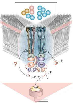
Figure 1. A Pie-Shaped Slice through anE. coliCell to Reveal Polar Clusters of Trans-membrane Chemoreceptors
Two trimers of receptor dimers are high-lighted. They can exist in pure or mixed ar-rangements (see inset). Higher-order config-urations of the trimer of dimers are thought to result from binding to CheW adaptors, linked, in turn, to CheA kinase in a giant sen-sory scaffold. CheA plays a central role not only in signal transduction, but also in tar-geting soluble components to the clusters, and possibly in the spread of conformational signals through the network. A His residue on the P1 domain of the dimeric CheA mediates phosphotransfer to CheY and CheB, which dock on CheA via the P2 domain. The direc-tion of phosphotransfer is indicated by red arrows. CheR and CheB add and remove CH3 groups (red circles). The pink stripes and tri-angle represent the inner membrane. Adapted from Stock and Levit (2000) and Falke (2002).
there is substantial variation in the levels of these pro- Targeting Soluble Signaling Components to Receptor Clusters
teins, but the ratio of the components with respect to one
another is relatively constant (Gerald Hazelbauer, Univer- The kinase activity of CheA is maximally expressed in ternary complexes with MCPs and CheW (Figure 1). With sity of Missouri). Images of higher-order complexes of the
cytoplasmic domain of Tsr complexed with CheW and GFP fusions, it has been shown that all the other proteins in the chemotaxis pathway colocalize with the MCP CheA indicate that the receptors may interact end to
end as well as side to side, implying that receptor signals clusters (Sourjik and Berg, 2000). Many of these interac-tions occur through the multiple domains of CheA (Fig-may be integrated via both axial and lateral contacts
(Peter Wolanin, Princeton University; Weis et al., 2003). ure 2). A CheB-GFP fusion is targeted via the N-terminal domain of CheB to the P2 domain of CheA (Satomi A simulation of chemotaxis in vivo was presented by
Thomas Shimizu (Keio University, Japan). Previous Banno, Nagoya University, Japan). Similarly, CheZ-GFP is targeted to the clusters by binding to the truncated models based on kinetic parameters can reproduce
many features of the attractant response but fail to ob- P1 domain of the short form of CheA (CheAs; Brian Cantwell, Texas A&M University). Immuoprecipitation tain the experimentally observed gain. The new
simula-tion incorporated a spatial interacsimula-tion between variously experiments were consistent with this conclusion (Christopher O’Connor, University of Illinois, Chicago). methylated receptors. The outcome was significantly
closer to recent in vivo results, suggesting that this Chemical modification of cysteine-substituted CheA proteins reveals that the CheW binding surface of the mechanism could account for the observed signal
am-plification, but discrepancies remain in the precise form P5 domain is occluded by the ATP binding domain in the CheA crystal structure, implying that the various of the dose-response relation between attractant
be differentially expressed under different growth condi-tions, the distinct clusters may respond to distinct exter-nal and interexter-nal environmental and metabolic cues.
Organelle Biogenesis and Motility Surface Motility
Bacteria move over surfaces by a variety of mecha-nisms. While flagella and type IV pili (TFP) mediate swarming and retractile motility, respectively, mecha-nism(s) underlying the gliding movement of many bacte-ria are not understood (McBride, 2001). Social gliding, or S motility, inMyxococcus xanthusis mediated by TFP and regulated by at least three che clusters. The frz
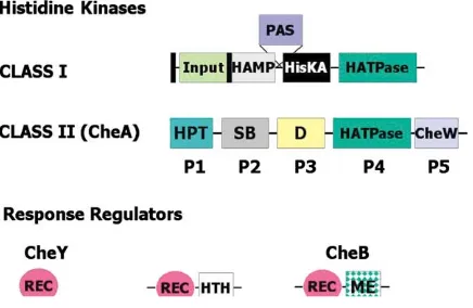
cluster controls cell reversal frequency, during which TFP alternate extension and retraction from opposite Figure 2. Modular Design of Histidine Kinases and Response
Regu-cell poles, while thedifcluster is responsible for produc-lators
tion of extracellular polysaccharide “fibrils” essential for The black rectangles indicate transmembrane domains. Most
two-S motility (two-Shimkets, 1999; Yang et al., 2000). Yinuo Li domain response regulators have a C-terminal DNA binding domain,
but a few have enzymatic activity (e.g., the methylesterase of CheB). (University of California, Los Angeles) reported that fi-The domains and their functions are as follows (domain, function): brils provide attachment sites for TFP and trigger pili input, ligand binding; HAMP, linker (named for presence in histidine retraction. Polysaccharides such as chitin (made of kinases, adenylyl cyclases, MCPs, and phosphatases); PAS, redox
N-acetylglucosamine) mimic this bioactivity, suggesting sensing, cofactor binding (named for presence in Per, Arnt, and
that similar residues in the fibrils serve as anchors for Sim); HisKA, dimerization (contains the phosphorylated histidine);
attachment of the tips of extended pili prior to retraction. HATPase, ATP binding; HPT, histidine phosphotransfer (contains
the phosphorylated histidine); SB, substrate binding (CheY/CheB); An attachment organelle is also important for gliding D, dimerization; CheW, regulatory coupling; REC, response regula- motility in mycoplasmas, parasitic bacteria that lack the tor receiver (contains the phosphorylated aspartate); HTH, helix- peptidoglycan layer, and do not have flagella, pili, or turn-helix DNA binding; ME, methylesterase.
a chemosensory system (Miyata et al., 2002). Several mycoplasma species have a distinct cell polarity and glide in the direction of the tapered end. Makoto Miyata shifts in solution, for example, during assembly of recep- (Osaka City University, Japan) reported thatM. mobile, tor signaling complexes (John Parkinson, University of which glides continuously on glass, exerts a force up
Utah). to 27 pN. Freeze-fracture EM has identified several 50
Comparative genomic analysis indicates that bacteria nm-long spikes that stick out from the cell membrane use a wide variety of domain organizations of these around the neck of this organism and grab the glass proteins (Igor Zhulin, Georgia Institute of Technology). surface. With a combination of nonbinding and nonglid-One of the unexpected findings was that many CheA ing mutants, as well as monoclonal antibodies that in-homologs lack the P2 domain and instead have a re- hibit gliding, two large protein targets (349 and 521 kDa) sponse regulator phosphorylation domain at their C ter- with distinct functions were localized to the neck. Gli349 mini. Further bioinformatics and experimental analyses is required for glass binding, while Gli521 is required for of such domain organization will provide important clues movement. A mechanical cycle of gliding was proposed, to the diversity of molecular interactions in chemotaxis. where Gli349 is involved in attachment, while Gli521 Cytoplasmic Clustering is responsible for the stroke or force generation that
Rhodobacter sphaeroideshas three Che operons with achieves gliding speeds of 2–4.5m/s.
multiple homologs of theE. colisensory proteins, most Organelle Biogenesis
(Aldridge and Hughes, 2002). The partially completed GGDEF, or DUF1, domain. GGDEF proteins are impli-structures serve as checkpoints in morphogenesis, sig- cated in the metabolism of the cofactor ci-diGMP (Tal naling sequential expression of the appropriate flagellar et al., 1998). Evidence presented by Urs Jenal (University operons arranged in a three-tiered transcriptional hierar- of Basel, Switzerland) suggested that ci-diGMP might chy. This regulatory strategy minimizes waste in case serve as a possible second messenger for regulating of unforeseen blocks in assembly. A flagellum-specific motility and development inC. cresentus.
type III secretion system (TTSS), which is homologous
to the secretion apparatus for virulence proteins in many Chemotaxis Operons as Mediators of Developmental pathogenic bacteria, allows the ordered exit of compo- Responses to External Stimuli
nents through the hollow core. The Hughes lab has char- Role in Development
acterized an anti-sigma factor that couples completion The first indication that chemotaxis genes may control of hook assembly to expression of sigma 28-dependent other behaviors came from studies on swarming inE. genes needed late in assembly; it is actively secreted coliandS. typhimurium, where these genes were shown via the completed hook basal body, freeing sigma 28 to be required for swarmer cell development and surface to transcribe late-assembly genes (Hughes et al., 1993). motility (Burkart et al., 1998). Chemotaxis, per se, is not Three factors influence secretion through the TTSS—the important for swarming in these organisms. Qingfeng N-terminal sequence of the exported protein, chaper- Wang (University of Texas, Austin) presented evidence ones that bind to their substrates and stabilize them for that, in the absence of other chemotaxis genes, a consti-export, and upstream elements of mRNA implicated in tutively active form of CheY is sufficient to confer swarm-coupling translation to assembly. Philip Aldridge (Uni- ing ability. Microarray analysis showed that approxi-versity of Washington, Seattle) described several spe- mately 100 genes were differentially regulated in acheY cific substrate-chaperone pairs. The chaperones have mutant, some of which belonged to flagellar operons. dual roles and also regulate transcription or translation However, CheY
ⵑP is unlikely to be directly involved in of specific substrates in other pairs needed at different regulating gene expression. Suppressors of a non-stages of assembly. This not only provides another swarmingcheYmutant mapped to the flagellar motor sensing mechanism to determine the stage of flagellar switch protein FliM, leading to possible models of how assembly, but also leads to a complex regulatory feed- CheY-FliM interaction affects swarming.
back, where expression of the three transcriptional tiers Control of motility is critical to the developmental cycle oscillates. Heather Bonifeld’s (University of Washington,
ofM. xanthus(Shimkets, 1999). Nine Che operons are now Seattle) results suggest that translation of the flagellar
discerned in this organism, which displays two kinds of filament gene may be targeted to the base of the flagel- motilities—adventurous (A) and social (S) motility. While lum itself, and Shin-Ichi Aizawa (CREST, JST, Japan)
Che1 (Frz system) controls both A and S motilities, Che4 presented EM evidence for the presence of flagellin at
was reported to exclusively modulate S motility (Hera these sites.
Vlamakis, University of California, Berkeley). The Che2, A Cautionary Tale of Two Locations
or Dif, operon has been known to control fibril biogene-InCaulobacter crescentus, cell differentiation and
asym-sis essential for bringing cells together during S motility metric cell division produce two cell types with distinct
(Yang et al., 2000). John Kirby (Georgia Institute of Tech-developmental programs: a stalked cell and a motile
nology) showed that Che3 plays a role in developmental and chemotactic swarmer cell. To replicate and divide,
gene expression. Mutations in several Che3 genes show motile swarmer cells have to differentiate into stalked
premature and inappropriate expression of develop-cells. During this process the chemotaxis machinery,
mentally regulated genes but do not affect motility. Inter-the flagellum and pili, are lost and are replaced by a
estingly, there is no CheY homolog in this operon, but stalk and adhesive holdfast structure at the same pole.
a divergently transcribed gene,crdA, is homologous to During the divison cycle, the cell assembles a new
flagel-NtrC-like response regulators. CheA3 and CrdA appar-lum and pili at the pole opposite the stalk (Poindexter,
ently interact, and CrdA mutants are delayed in develop-1964). Several two-component systems control polar
ment. Periplasmic stress was implicated as a possible organelle development and coordinate it with the cell
signal here and in parallel pathways regulating develop-cycle. Some of these, including the kinases PleC, DivJ,
ment (Heidi Kaplan, University of Texas Medical School, and CckA, localize to specific cell poles and undergo
Houston). Thus, Che operons control gene expression dynamic rearrangements as a function of the cell cycle
for both motility and development inM. xanthus. (Jenal and Stephens, 2002). Stephen Sciochetti
(Prince-A developmental role for a chemotaxis operon in
Rho-ton University) has identified the polar targeting signals
dospirillum centenumwas presented by James
Berle-of the stalked pole-specific kinase DivJ (Sciochetti et
man (Indiana University). There are three Che operons al., 2002) and, at this meeting, described the targeting
in this organism, all controlling some aspect of motility. signal of the swarmer cell pole-specific kinase PleC. A
Che3 plays an additional role in cyst formation, a starva-DivJ-PleC fusion protein containing the targeting
se-tion response. Mutase-tion of several Che3 genes, includ-quences of DivJ and the catalytic domain of PleC kinase
ingcheY, results in hypercyst formation in rich medium localized to the stalked pole yet was able to restore
but does not affect motility of vegetative cells. motility and stalk formation in a pleC mutant. These
Role in Biofilm Formation results suggest that the “correct” polar address of the
Pseudomonas aeruginosa is an opportunistic human
PleC kinase may not be critical for all its regulatory
pathogen with five Che operons and 26 MCP genes functions. Interestingly, both PleC and DivJ kinases
(Stover et al., 2000). One set of genes is involved in signal to the unorthodox response regulator PleD,
pilus-mediated motility. Carrie Harwood (University of Major questions in the field are, what are the structural consequences of phosphorylation in the receiver do-Iowa) reported that a third set ofchegenes (called
clus-ter II) has a minor involvement in chemotaxis and is main, and how is the information transmitted to the ef-fector domain? A significant advance was the determi-instead required for biofilm formation. Che II genes are
expressed in the stationary phase of cell growth. Mi- nation of the full-length structure of the response regulator DrrB fromThermotoga maritima(Victoria Rob-croarray studies showed that expression levels of a
common set of 116P. aeruginosagenes are altered in inson, University of Medicine and Dentistry, New Jer-sey). It is apparent that the interdomain interface
be-cheA2,cheB2, andcheY2 mutants. This result suggests
that a signal transduction complex comprised of cluster tween the receiver domain and the effector domain, across which information must be transmitted, is as II Che proteins may modulate gene expression, rather
than cell motility. Genes for anaerobic respiration were varied as the number of structures that have been solved to date (four thus far). Different surfaces are utilized in among those whose expression was most dramatically
affected. The interior of a biofilm is likely to be starved each interdomain interface. Thus, there is no conserved formula for activation that results from phosphorylation, for oxygen, and mutants that are unable to carry out
anaerobic denitrification may form biofilms with altered and the repertoire is astonishingly varied. Clearly, pat-terns will only become apparent after many more struc-structures. A challenging task will be the identification
of upstream signals for the multitudinous MCPs, as well tures have been determined. These results also provide a cautionary note about the use of chimeric proteins as mechanisms by which response regulators that lack
DNA binding domains alter developmental programs. that may disrupt the signaling interface.
Further advances came from the structural determina-Genomic Perspective
CheA is a member of the class II histidine kinase family tion of the individual N-terminal receiver domain of PhoP from Bacillus subtilis(Catherine Birck, CNRS, France) (HKII) of proteins (see Figure 2; Bilwes et al., 1999). Igor
Zhulin (Georgia Institute of Technology) reported that, and the central ATPase domain of NtrC from Aquifex aeolicus(David Wemmer, University of California, Berke-of 111 bacterial genomes surveyed, over half had at
least one HKII. Interestingly, all genomes possessing at ley). The interprotein surfaces of PhoP are asymmetrical, leaving a surface on each monomer of the dimer to drive least one CheA homolog also have at least one MCP,
implying that the two may function together. All bacteria oligomerization, creating a novel mode of association between receiver domains. A salt bridge between D60 containing at least one CheA are motile, but none of the
nonmotile bacteria have a CheA, suggesting that the on one surface and R113 on the other is critical for this interaction and for function (Chen et al., 2003). NtrC is ancestral chemotaxis system is involved in motility. Over
half of the motile bacteria possess more than one HKII. a response regulator that interacts with the54form of
RNA polymerase. Phosphorylation results in rearrange-Typically, these bacteria have more than one complete
chemotaxis system. It is apparent that chemotaxis ment of the dimer to form a different interaction surface and formation of a ring. While seven monomers were genes have also acquired a function typical of HKI
ki-nases, which control a wider variety of cellular pro- found in the ring in the crystals of the ATPase domain alone, there was considerable discussion of the possibil-cesses (Robinson et al., 2000).
Finally, the recently sequenced genome ofWolinella ity that, in vivo, with the other domains present, there may only be six (David Wemmer, University of California,
succinogenes, an obligate enterointestinal symbiont,
re-vealed that it has the highest percentage (5.5% of all Berkeley).
Identification of Targets of Essential Genes genes) of signal transduction genes of any bacterium
analyzed so far (Stephan Schuster, Max-Plank Institute, Only one of the 35 two-component systems inB. subtilis
(a gram-positive, soil-dwelling bacterium) is essential. Tubingen, Germany). Functional analyses of the
path-ways involved should illuminate mechanisms used by The YycF/G system is required for bacterial survival and cell division and is specific to “low G⫹C content” bacte-microorganisms not only for their survival and growth,
but also for transmission into larger host populations. ria, including many major pathogens, such as Staphylo-coccus aureus,Streptococcus pneumoniae, and Liste-ria monocytogenes. The genes that are regulated by this Advances in Two-Component Reaction Mechanism
system are largely unknown. For the identification of the Two-component regulatory systems represent the
ma-targets of YycF, a novel approach used a chimera in jor paradigm for signal transduction in prokaryotes and
which the phosphorylation (receiver) domain of PhoP lower eukaryotes. The simplest forms contain a sensor
was linked to the DNA binding domain of YycF. With kinase (HKI) that is phosphorylated on a conserved
histi-the known signal for PhoP (phosphate starvation condi-dine residue (see Figure 2). The phosphoryl group is
tions), an autolysin gene regulated by YycF was identi-transferred to a conserved aspartic acid residue on the
fied. DNase I footprinting with purified native protein receiver domain of the second component, the response
identified a 6 bp consensus repeat, and a search of the regulator. Most commonly, response regulators are
genome revealed additional potential members of the composed of two domains, with the effector, or output,
regulon, including a number of genes involved in cell domain binding to DNA.
wall synthesis and cell division (Tarek Msadek, Pasteur Interdomain Interfaces
Institute, France). Extending the genomic analysis toS.
In all species, protein modification is a common
mecha-aureus suggests that a number of potential virulence nism for altering activity. Many response regulators bind
factors may be regulated by this essential two-compo-to DNA with higher affinity when phosphorylated,
al-nent regulatory system. It therefore may represent a though some use phosphorylation to drive dimerization
Physical Sequestration of the Regulator that clustering of receptors can amplify signals down-stream of those receptors resonates within the eukary-InC. crescentus, an established mechanism for
control-ling signacontrol-ling is to sequester, or compartmentalize, the otic community. This type of amplification is likely to occur in T lymphocytes during assembly of the “immune components. A variation on this theme was reported,
in which the E. coli ROK transcriptional regulator (of synapse.” It may recapitulate some of the signal amplifi-cation that must occur within the postsynaptic densities
repressors,ORFs, andkinases) Mlc was prevented from
interacting with the transcriptional machinery by binding within neuronal cells that contribute to synaptic plastic-ity. This type of clustering may occur in all cells with to the transporter PtsG (Winfried Boos, University of
Konstanz, Germany). Under glucose-replete conditions, specialized membrane microdomains, for example, dur-ing maturation of lipid rafts into functional signaldur-ing PtsG is phosphorylated and Mlc represses transcription
of genes and operons encoding sugar-metabolizing en- units. This process is likely to involve a similar consolida-tion of membrane-associated signaling proteins. Fur-zymes and uptake systems. Under low-glucose
condi-tions, PtsG donates its phosphoryl group to glucose, thermore, the mechanisms of transactivation triggered by receptor clustering may provide insights into activa-enabling Mlc to bind and inactivating its repressor
func-tions. Thus, the location of the regulator is intimately tion of eukaryotic receptors, perhaps best exemplified by transactivation of heterodimers of the receptor tyro-tied to the active state of the transporter.
Signaling and Energy Coupling sine kinases.
The appreciation of methylation in eukaryotic signal-Many sensor kinases and most MCPs contain a
con-served structural element called the HAMP linker, lo- ing is in its infancy and represents one area that might well follow leads developed in bacterial systems. The cated between the transmembrane region and the
histi-dine kinase domain (see Figure 2). It was suggested that concept of checkpoints during organelle synthesis might have implications in the rapid regulation of neurite the HAMP linker is a negative regulator of output domain
activity. Deletion mutations in each of the amphipathic␣ outgrowth, although the need for rapid reversibility of organelle synthesis in bacteria may suggest that this helices in the HAMP linker (AS-1 and AS-2) had radically
different phenotypes. Deletions in AS-1 diminished type of regulation is unique to prokaryotes. The mecha-nisms that bacteria have developed for integrating mod-HAMP function, whereas AS-2 deletions resulted in a
reversed-response phenotype, suggesting that the heli- ular assembly and traffic of organelles with the signal transduction machinery will certainly find parallels in the ces play unique roles in signaling (J. Alex Appleman,
University of California, Davis). eukaryotic field of microtubular trafficking, where cargo proteins interact directly with signal transduction pro-Another regulatory element, the PAS domain, is found
in some histidine kinases and often binds a cofactor, teins. The studies in bacteria suggest that these interac-tions are necessary to provide the assembly machinery such as heme, FAD, or cinnaminic acid, depending on
whether signaling is in response to oxygen, redox poten- with a continuous reading of the environmental de-mands for the particular assembly. It will be very inter-tial, or light, respectively. The Aer sensor kinase contains
both HAMP and PAS domains. Second site suppressor esting to determine whether similar feedback mecha-nisms regulate cargo assembly and traffic in eukaryotes analysis of Aer suggests that the HAMP domain contacts
the PAS domain and stabilizes FAD binding. Interactions and whether parallels between the two worlds will also be found in mechanisms for targeting of mRNA transla-between HAMP domains across the dimer interface
(HAMP/HAMP⬘) as well as HAMP/PAS interactions are tion to sites of organellar assembly.
Lastly, phosphorylation strategies utilized by many crucial for FAD binding and normal Aer signaling (Kylie
Watts, Loma Linda University). two-component systems directly affect DNA binding. This situation mirrors the mechanism of eukaryotics
ig-InRhodobacter capsulatus, RegB senses oxygen by
monitoring redox and signaling to the response regula- naltransducers andactivators oftranscription (STAT) proteins. As additional prokaryotic strategies are unrav-tor RegA (Lee Swem, Indiana University). A reactive
cys-teine residue near the active site affects autophosphory- eled, they will certainly serve to stimulate the imagina-tion of scientists working in signal transducimagina-tion in all lation of RegB by ATP. The reduced form of RegB is the
active form, and metal binding promotes formation of organisms. the oxidized form. The reactivity of the cysteine is
proba-bly enhanced by two basic flanking residues. The kinase Acknowledgments is sensitive to many divalent metal ions, suggestive of
The following individuals coordinated the scientific and administra-a structuradministra-al role of the metadministra-al in promoting formadministra-ation of
tive aspects of the meeting: T. Bollinger, E. Calva, J. Falke, C. Har-the inactive form.
wood, R. Kadner, M. Manson, P. Matsumura, P. O’Neill, and J. Par-kinson. Financial support was provided by National Institutes of Links to the Eukaryotic World Health (R13 GM066736). We thank Kristina Schlegel for Figure 1 The reliance on two-component signal transduction dis- and Igor Zhulin for providing a template for Figure 2. L.J.K. is grateful to Philip Stork (Vollum Institute, Portland, OR) for helpful discussions tinguishes the prokaryotes from higher eukaryotes.
and input on eukaryotic links. However, the strategies utilized by bacteria to
coordi-nate environmental signals are so diverse that fruitful
comparisons to eukaryotic cells can be found. References
Perhaps the most compelling strategy is the utilization
Aldridge, P., and Hughes, K.T. (2002). Regulation of flagellar assem-of receptor arrays for signal amplification. The details
bly. Curr. Opin. Microbiol.5, 160–165. elucidated in bacteria have implications for the
Collaborative signaling by mixed chemoreceptor teams inEsche- Hickey, M.J., Brinkman, F.S., Hufnagle, W.O., Kowalik, D.J., Lagrou, M., et al. (2000). Complete genome sequence ofPseudomonas aeru-richia coli. Proc. Natl. Acad. Sci. USA99, 7060–7065.
ginosaPA01, an opportunistic pathogen. Nature406, 959–964. Bilwes, A.M., Alex, L.A., Crane, B.R., and Simon, M.I. (1999).
Struc-Tal, R., Wong, H.C., Calhoon, R., Gelfand, D., Fear, A.L., Volman, ture of CheA, a signal-transducing histidine kinase. Cell96, 131–141.
G., Mayer, R., Ross, P., Amikam, D., Weinhouse, H., et al. (1998). Bornhorst, J.A., and Falke, J.J. (2001). Evidence that both ligand
Three cdg operons control cellular turnover of cyclic di-GMP in binding and covalent adaptation drive a two-state equilibrium in the
Acetobacter xylinum: genetic organization and occurrence of con-aspartate receptor signaling complex. J. Gen. Physiol.118, 693–710.
served domains in isoenzymes. J. Bacteriol.180, 4416–4425. Burkart, M., Toguchi, A., and Harshey, R.M. (1998). The chemotaxis
Wadhams, G.H., Martin, A.C., Porter, S.L., Maddock, J.R., Mantotta, system, but not chemotaxis, is essential for swarming motility in
J.C., King, H.M., and Armitage, J.P. (2002). TlpC, a novel chemotaxis
Escherichia coli. Proc. Natl. Acad. Sci. USA95, 2568–2573.
protein inRhodobacter sphaeroides, localizes to a discrete region Chen, Y., Birck, C., Samama, J.P., and Hulett, F.M. (2003). Residue
in the cytoplasm. Mol. Microbiol.46, 1211–1221. R113 is essential for PhoP dimerization and function: a residue
Weis, R.M., Hirail, T., Chalah, A., Kessell, M., Pers, P., and Subraman-buried in the asymmetric PhoP dimer interface determined in the
iam, S. (2003). Electron microscopic analysis of membrane assem-PhoPN three-dimensional crystal structure. J. Bacteriol. 185,
blies formed by the bacterial chemotaxis receptor Tsr. J. Bacteriol., 262–273.
in press. Duke, T.A., Novere, N.L., and Bray, D. (2001). Conformational spread
Yang, Z., Ma, X., Tong, L., Kaplan, H.B., Shimkets, L.J., and Shi, W. in a ring of proteins: a stochastic approach to allostery. J. Mol. Biol.
(2000).Myxococcus xanthusdif genes are required for biogenesis
308, 541–553.
of cell surface fibrils essential for social gliding motility. J. Bacteriol. Falke, J.J. (2002). Cooperativity between bacterial chemotaxis
re-182, 5793–5798. ceptors. Proc. Natl. Acad. Sci. USA99, 6530–6532.
Gestwicki, J.E., Lamanna, A.C., Harshey, R.M., McCarter, L.L., Kies-sling, L.L., and Adler, J. (2000). Evolutionary conservation of methyl-accepting chemotaxis protein location in Bacteria and Archaea. J. Bacteriol.182, 6499–6502.
Hughes, K.T., Gillen, K.L., Semon, M.J., and Karlinsey, J.E. (1993). Sensing structural intermediates in bacterial flagellar assembly by export of a negative regulator. Science262, 1277–1280.
Jenal, U., and Stephens, C. (2002). The Caulobacter cell cycle: tim-ing, spatial organization and checkpoints. Curr. Opin. Microbiol.5, 558–563.
Kim, K.K., Yokota, H., and Kim, S.H. (1999). Four-helical-bundle structure of the cytoplasmic domain of a serine chemotaxis recep-tor. Nature400, 787–792.
Maddock, J.R., and Shapiro, L. (1993). Polar location of the chemore-ceptor complex in theEscherichia colicell. Science259, 1717–1723.
McBride, M.J. (2001). Bacterial gliding motility: multiple mechanisms for cell movement over surfaces. Annu. Rev. Microbiol.55, 49–75. Miyata, M., Ryu, W.S., and Berg, H.C. (2002). Force and velocity of mycoplasma mobile gliding. J. Bacteriol.184, 1827–1831. Poindexter, J.S. (1964). Biological properties and classification of the Caulobacter group. Bacteriol. Rev.28, 231–295.
Porter, S.L., Warren, A.V., Martin, A.C., and Armitage, J.P. (2002). The third chemotaxis locus ofRhodobacter sphaeroidesis essential for chemotaxis. Mol. Microbiol.46, 1081–1094.
Robinson, V.L., Buckler, D.R., and Stock, A.M. (2000). A tale of two components: a novel kinase and a regulatory switch. Nat. Struct. Biol.7, 626–633.
Sciochetti, S.A., Lane, T., Ohta, N., and Newton, A. (2002). Protein sequences and cellular factors required for polar localization of a histidine kinase inCaulobacter crescentus. J. Bacteriol.184, 6037– 6049.
Segall, J.E., Block, S.M., and Berg, H.C. (1986). Temporal compari-sons in bacterial chemotaxis. Proc. Natl. Acad. Sci. USA83, 8987– 8991.
Shimizu, T.S., Le Novere, N., Levin, M.D., Beavil, A.J., Sutton, B.J., and Bray, D. (2000). Molecular model of a lattice of signalling proteins involved in bacterial chemotaxis. Nat. Cell Biol.2, 792–796. Shimkets, L.J. (1999). Intercellular signaling during fruiting-body de-velopment of Myxococcus xanthus. Annu. Rev. Microbiol. 53, 525–549.
Sourjik, V., and Berg, H.C. (2000). Localization of components of the chemotaxis machinery ofEscherichia coliusing fluorescent protein fusions. Mol. Microbiol.37, 740–751.
Sourjik, V., and Berg, H.C. (2002). Receptor sensitivity in bacterial chemotaxis. Proc. Natl. Acad. Sci. USA99, 123–127.
Stock, J., and Levit, M. (2000). Signal transduction: hair brains in bacterial chemotaxis. Curr. Biol.10, R11–14.