PENGANTAR ANATOMI
DAN FISIOLOGI
AMI RACHMI 15 JULI 2011
PERATURAN
1. TOLERANSI WAKTU 10 MENIT 2. HP VIBRASI
3. TIDAK MAKAN DAN MINUM
4. PAKAIAN RAPIH, SOPAN, TIDAK MEMAKAI SANDAL 5. BILA TIDAK HADIR MEMBERITAHU LANGSUNG
DOSEN, SURAT
ANATOMI
BERASAL DARI BAHASA LATIN YAITU, * ANA : BAGIAN, MEMISAHKAN
* TOMI (TOMIE) : IRIS/ POTONG
ANATOMI ADALAH ILMU YANG
MEMPELAJARI BENTUK DAN SUSUNAN
TUBUH BAIK SECARA KESELURUHAN
MAUPUN BAGIAN-BAGIAN SERTA
HUBUNGAN ALAT TUBUH YANG SATU DENGAN YANG LAIN
ILMU URAI YANG MEMPELAJARI SUSUNAN
TUBUH DAN HUBUNGAN BAGIAN
-BAGIANNYA SATU SAMA LAIN
FISIOLOGI
BERASAL DARI BAHASA LATIN YAITU : * FISI (PHYSIS) : ALAM/ CARA KERJA * LOGOS (LOGI) : ILMU PENGETAHUAN
FISIOLOGI ADALAH ILMU YANG
MEMPELAJARI FAAL ATAU PEKERJAAN DARI TIAP-TIAP JARINGAN TUBUH ATAU
BAGIAN DARI ALAT-ALAT TUBUH DAN
SEBAGAINYA
FISIOLOGI MEMPELAJARI FUNGSI ATAU KERJA TUBUH MANUSIA DALAM KEADAAN NORMAL
ANATOMI-FISIOLOGI
ADALAH ILMU PENGETAHUAN YANG
MEMPELAJARI TENTANG SUSUNAN ATAU POTONGAN TUBUH DAN
BAGAIMANA ALAT TUBUH TERSEBUT BEKERJA
ISTILAH YANG DIPAKAI UNTUK MENUNJUKAN ILMU YANG DIPAKAI
• SITOLOGI
(ILMU PENGETAHUAN TENTANG STRUKTUR DAN FUNGSI SEL)
• HISTOLOGI
(ILMU PENGETAHUAN TENTANG SEL DAN JARINGAN SECARA MIKROSKOPIK)
• OSTEOLOGI
(ILMU PENGETAHUAN TENTANG TULANG)
• ARTHROLOGI
(ILMU PENGETAHUAN TENTANG SENDI)
• MIOLOGI
(ILMU PENGETAHUAN TENTANG OTOT)
• NEUROLOGI
(ILMU PENGETAHUAN TENTANG SARAF & STRUKTUR SARAF)
ISTILAH LOKASI ANATOMI
Bidang anatomi adalah bidang yang melalui tubuh dalam posisi anatomi:
• Bidang median: bidang yang membagi tepat tubuh menjadi bagian kanan dan kiri.
• Bidang sagital: bidang yang membagi tubuh menjadi dua bagian dari titik tertentu (tidak membagi tepat dua bagian). Bidang ini sejajar dengan bidang median.
• Bidang horizontal: bidang yang terletak melintang melalui tubuh (bidang X-Y). Bidang ini membagi tubuh menjadi bagian atas (superior) dan bawah (inferior).
• Bidang koronal: bidang vertikal yang melalui tubuh, letaknya tegak lurus terhadap bidang median atau
sagital. membagi tubuh menjadi bagian depan (frontal) dan belakang (dorsal).
ARAH DAN BIDANG ANATOMI
• Superior (=atas) atau kranial: lebih dekat pada kepala. Contoh: Mulut terletak superior terhadap dagu.
• Inferior (=bawah) atau kaudal: lebih dekat pada kaki. Contoh: Pusar terletak inferior terhadap payudara.
• Anterior (=depan): lebih dekat ke depan.
Contoh: Lambung terletak anterior terhadap limpa.
• Posterior (=belakang): lebih dekat ke belakang.
Contoh: Jantung terletak posterior terhadap tulang rusuk.
• Superfisial: lebih dekat ke/di permukaan.
Contoh: Otot kaki terletak superfisial dari tulangnya.
• Profunda: lebih jauh dari permukaan.
Contoh: Tulang hasta dan pengumpil terletak lebih profunda dari otot lengan bawah.
• Medial (=dalam): lebih dekat ke bidang median.
Contoh: pangkal lengan terletak medial terhadap tubuh.
• Lateral (=luar): menjauhi bidang median.
Contoh: Telinga terletak lateral terhadap mata.
• Proksimal (=dekat): lebih dekat dengan batang tubuh atau pangkal. Contoh: Siku terletak proksimal terhadap telapak tangan.
• Distal (=jauh): lebih jauh dari batang tubuh atau pangkal.
ISTILAH GERAKAN ANATOMI
• Fleksi dan ekstensi
Fleksi adalah gerak menekuk atau membengkokkan. Ekstensi adalah gerakan untuk meluruskan. Contoh: gerakan ayunan lutut pada kegiatan gerak jalan. Gerakan ayunan ke depan merupakan (ante)fleksi dan ayunan ke belakang disebut (retro)fleksi/ekstensi. Ayunan ke belakang lebih lanjut disebut hiperekstensi.
• Adduksi dan abduksi
Adduksi adalah gerakan mendekati tubuh. Abduksi adalah gerakan menjauhi tubuh. Contoh: gerakan membuka tungkai kaki pada posisi istirahat di tempat merupakan gerakan abduksi (menjauhi tubuh). Bila kaki digerakkan kembali ke posisi siap merupakan gerakan adduksi (mendekati tubuh).
• Elevasi dan depresi
Elevasi merupakan gerakan mengangkat, depresi adalah gerakan menurunkan. Contohnya: Gerakan membuka mulut (elevasi) dan menutupnya (depresi)juga gerakan pundak keatas (elevasi) dan kebawah (depresi)
• Inversi dan eversi
Inversi adalah gerak memiringkan telapak kaki ke dalam tubuh. Eversi adalah gerakan memiringkan telapak kaki ke luar. Juga perlu diketahui untuk istilah inversi dan
eversi hanya untuk wilayah di pergelangan kaki.
• Supinasi dan pronasi
Supinasi adalah gerakan menengadahkan tangan. Pronasi adalah gerakan menelungkupkan. Juga perlu diketahui istilah supinasi dan pronasi hanya digunakan untuk wilayah pergelangan tangan saja
• Endorotasi dan eksorotasi
Endorotasi adalah gerakan ke dalam pada sekililing sumbu panjang tulang yang bersendi (rotasi).
Sedangkan eksorotasi adalah gerakan rotasi ke luar.
GERAKAN ANATOMI
• FLEXION / ENTENSION
GERAKAN ANATOMI
• ABDUCTION / ADDUCTION
GERAKAN ANATOMI
• ROTATION MEDIAL & LATERAL
GERAKAN ANATOMI
• ELEVATION / DEPRESSION
GERAKAN ANATOMI
• PRONATION / SUPINATION
GERAKAN ANATOMI
• INVERSION / EVERSION
STRUKTUR TUBUH MANUSIA
SEL
(UNSUR DASAR JARINGAN TUBUH YANG TERDIRI ATAS INTI SEL/ NUCLEUS DAN PROTOPLASMA)
↓
JARINGAN
(KUMPULAN SEL KHUSUS DENGAN BENTUK & FUNGSI YANG SAMA)
↓
ORGAN
(BAGIAN TUBUH/ ALAT MANUSIA DGN FUNGSI KHUSUS)
↓
SISTEM
(SUSUNAN ALAT DENGAN FUNGSI TERTENTU)
JARINGAN
• Ada empat tipe jaringan dasar yang membentuk tubuh semua
hewan, termasuk tubuh manusia dan organisme multiseluler tingkat rendah seperti serangga.
* Jaringan epitel.
Jaringan yang disusun oleh lapisan sel yang melapisi permukaan organ seperti permukaan kulit. Jaringan ini berfungsi untuk
melindungi organ yang dilapisinya, sebagai organ sekresi dan penyerapan.
* Jaringan pengikat.
Sesuai namanya, jaringan pengikat berfungsi untuk mengikat
jaringan dan alat tubuh. Contoh jaringan ini adalah jaringan darah.
* Jaringan otot.
Jaringan otot terbagi atas tiga kategori yang berbeda yaitu otot licin yang dapat ditemukan di organ tubuh bagian dalam, otot lurik yang dapat ditemukan pada rangka tubuh, dan otot jantung yang dapat ditemukan di jantung.
* Jaringan saraf.
Adalah jaringan yang berfungsi untuk mengatur aktivitas otot dan organ serta menerima dan meneruskan rangsangan.
ORGAN / SISTEM
• Sistem kardiovaskular: memompa darah ke seluruh tubuh
• Sistem pencernaan: pemrosesan makanan dengan mulut, perut, dan usus
• Sistem endokrin: komunikasi dalam tubuh dengan hormon
• Sistem kekebalan: mempertahankan tubuh dari serangan benda yang menyebabkan penyakit
• Sistem integumen: kulit, rambut
• Sistem limfatik: struktur yang terlibat dalam transfer limfa antara jaringan dan aliran darah
• Sistem otot: menggerakkan tubuh
• Sistem saraf: mengumpulkan, mengirim, dan memproses informasi dalam otak dan saraf (SS. PUSAT, SS. PERIFER, SS. OTONOM)
• Sistem reproduksi: organ seks
• Sistem pernafasan: organ yang digunakan bernafas, paru-paru
• Sistem rangka: sokongan dan perlindungan struktural dengan tulang
• Sistem urin: ginjal dan struktur yang dihubungkan dalam produksi dan ekskresi urin
SECARA FUNGSI ADA 4 SISTEM DALAM
SPEECH PATHOLOGY :
1. SISTEM RESPIRASI/PERNAFASAN
2. SISTEM FONASI TDD SISTEM RESPIRASI DAN SISTEM DIGESTIVE YG BERHUBUNGAN DGN PRODUKSI SUARA (LARING)
3. SISTEM ARTIKULASI/RESONANSI (STRUKTUR WAJAH, MULUT DAN HIDUNG)
4. SISTEM SARAF ( SISTEM SARAF YANG MENGONTROL PROSES BICARA)
• Saluran nafas yang dilalui udara adalah
hidung, faring, laring, trakea, bronkus,
bronkiolus dan alveoli. Di dalamnya
terdapat suatu sistem yang sedemikian
rupa dapat menghangatkan udara
sebelum sampai ke alveoli. Terdapat juga
suatu sistem pertahanan yang
memungkinkan kotoran atau benda asing
yang masuk dapat dikeluarkan baik
• adalah pipa berotot yang berjalan dari dasar tengkorak sampai persambungan-nya dengan oesopagus pada ketinggian tulang rawan krikoid. Maka letaknya di belakang larinx (larinx-faringeal). Orofaring adalah bagian dari faring merupakan
gabungan sistem respirasi dan
• Otot-otot kecil yang melekat pada cartilago arytenoidea, cricoidea, dan thyroidea, yang dengan kontraksi dan relaksasi
dapat mendekatkan dan memisahkan
• Percabangan saluran nafas dimulai dari trakea yang bercabang menjadi bronkus kanan dan kiri. Masing-masing bronkus terus bercabang sampai dengan 20-25 kali
sebelum sampai ke alveoli. Sampai
• Bagian terakhir dari perjalanan udara adalah di alveoli. Di sini terjadi pertukaran
oksigen dan karbondioksida dari
• Sistem pernafasan pada dasarnya dibentuk oleh jalan atau saluran nafas dan
paru-paru beserta pembungkusnya
(pleura) dan rongga dada yang
• Paru-paru terdapat dalam rongga thoraks pada bagian kiri dan kanan. Paru-paru memilki :
1. Apeks, Apeks paru meluas kedalam
leher sekitar 2,5 cm diatas calvicula 2. permukaan costo vertebra, menempel
pada bagian dalam dinding dada
3. permukaan mediastinal, menempel
pada perikardium dan jantung.
• Rongga dada diperkuat oleh tulang-tulang yang membentuk rangka dada. Rangka dada ini terdiri dari costae (iga-iga),
sternum (tulang dada) tempat sebagian
iga-iga menempel di depan, dan vertebra
torakal (tulang belakang) tempat
• Terdapat otot-otot yang menempel pada rangka dada yang berfungsi penting sebagai otot pernafasan. Otot-otot yang berfungsi dalam bernafas adalah sebagai berikut :
- interkostalis eksterrnus (antar iga luar) yang
mengangkat masing-masing iga.
- sternokleidomastoid yang mengangkat sternum
(tulang dada).
- skalenus yang mengangkat 2 iga teratas.
- interkostalis internus (antar iga dalam) yang
menurunkan iga-iga.
- otot perut yang menarik iga ke bawah sekaligus membuat isi perut mendorong diafragma ke atas. - otot dalam diafragma yang dapat menurunkan
Proses fisiologis respirasi di mana oksigen dipindahkan dari udara ke dalam jaringan-jaringan, dan karbon dioksida dikeluarkan ke udara ekspirasi dapat dibagi menjadi tiga stadium.
1. Stadium pertama adalah ventilasi, yaitu masuknya
campuran gas-gas ke dalam dan ke luar paru-paru.
2. Stadium ke dua, transportasi, yang terdiri dari beberapa aspek :
(a) difusi gas-gas antara alveolus dan kapiler paru-paru
(respirasi eksterna) dan antara darah sistemik dan selsel jaringan;
(b) distribusi darah dalam sirkulasi pulmoner dan
penyesuaiannya dengan distribusi udara dalam alveolus-alveolus; dan
(c) reaksi kimia dan fisik dari oksigen dan karbon dioksida dengan darah.
• Oksigen dapat ditranspor dari paru-paru ke jaringan melalui dua jalan :
1. secara fisik larut dalam plasma atau 2. secara kimia berikatan dengan
hemoglobin sebagai oksihemoglobin (HbO2).
• Transport CO2 dari jaringan keparu-paru melalui tiga cara sebagai berikut:
1. Secara fisk larut dalam plasma (10 %) 2. Berikatan dengan gugus amino pada
Hb dalam sel darah merah (20%)
3. ditransport sebagai bikarbonat plasma (70%) Karbon dioksida berikatan dengan air dengan reaksi seperti dibawah ini:
+HCO3-1. Medulla Oblongata 2. Pons
Secara garis besar bahwa Paru-paru memiliki fungsi sebagai berikut:
1. Terdapat permukaan gas-gas yaitu
mengalirkan Oksigen dari udara atmosfer kedarah vena dan mengeluarkan gas
carbondioksida dari alveoli keudara atmosfer. 2. Menyaring bahan beracun dari sirkulasi
3. Reservoir darah
FISIOLOGI
PENDENGARAN DAN
KESEIMBANGAN
Anatomi telinga
• Telinga luar (auris eksterna) : daun telinga, liang telinga
• Telinga tengah ( auris media) : membran timpani, kavum timpani, tuba eustakius, prosesus mastoideus
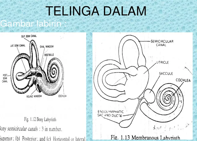
Anatomi Fisiologi Telinga Dalam
• Telinga dalam terletak di dalam pars petrosus os temporale
TELINGA DALAM
TRANSMISI BUNYI
TELINGA LUAR
• Gelombang bunyi ditangkap oleh daun telinga dan ditransmisikan ke dalam
MEMBRANA TYMPANI
• Gelombang bunyi vibrasi membrane timpani
• Sifat membrane elastic mudah bergetar bila tekanan pada kedua sisinya bersifat atmosferik
• Membrana timpani tidak akan bergetar dengan baik bila tuba tersumbat dan tekanan kedua sisi tidak sama.
• Amplitude getaran membrane proporsional dengan intensitas bunyi
OSIKEL
• Getaran membrane timpani ditangkap oleh malleus, yang melekat pada permukaan
dalamnya dan ditransmisikan melalui incus ke stapes.
• membrane timpani 15 – 20 kali lebih besar dari pada fenestrum ovalem gaya vibrasi pada fenestrum lebih besar dari pada gaya pada membrane timpani
• Muskulus stapedius dan tensor timpani
berkontraksi secara reflektorik sebagai respons terhadap bunyi yang keras berkontraksi
KOKLEA
• Vibrasi fenestrum ovale menyebabkan
gelombang tekanan dalam perilimf telinga dalam
• Ketika gelombang mencapai fenestrum rotundum pada bagian dasar, membrane menutup fenestrum tersebut
ORGAN CORTI
• Gerakan membrane basalis, dihasilkan oleh gelombang yang berjalan naik turun didalam koklea, menggerakkan sel-sel rambut dan mengeksitasinya
• gelombang yang dihasilkan oleh bunyi
berfrekuensi tinggi hanya berjalan sedikit di dalam koklea sebelum teredam, dan
• Amplitudo kerasnya bunyi
• Pembedaan oleh telinga antara suara dengan berfrekuensi yang berbeda
agaknya diakibatkan oleh pola getaran
• Nada / frekwensi tinggi resonansinya terjadi di dekat basis koklea dan nada / frekwensi rendah merangsang apeks koklea.
HUBUNGAN SENTRAL
• Nerves auditorius pars koklearis
menstranmisikan sensasi pada otak.
• Tempat sensasi tersebut diinterpretasikan di dalam pars auditorius Globus
temporalis.
FISIOLOGI PENDENGARAN
• Bunyi ditangkap daun telinga membran
timpani tulang pendengaran fenestra ovale
menggerakkan perilimfe pada skala vestibuli
melalui membran reissner mendorong
endolimfe menimbulkan gerak relatif membran basilaris dan membran tektoria defleksi
stereosilia sel rambut kanal ion terbuka
terjadi pertukaran ion depolarisasi sel rambut
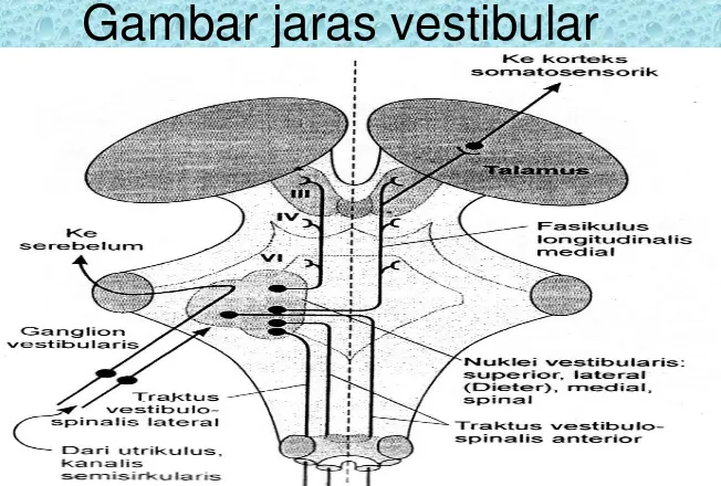
KESEIMBANGAN
• Kanalis semisirkularis, sakulus dan utrikulus
• Kanalis semisirkularis berperan pada gerakan kepala berputar gerakan
• Otolit sakulus dan utrikulus; bergerak oleh perubahan posisi kepala
•
Rangsangan ditransmisikan sepanjang serat saraf
nervus kranialis kedelapan ( auditorius) pars vestibularis ke otak tengah , medulla oblongata, serebelum , dan
medulla spinalis.
• Rangsangan ini memulai perubahan refleks pada otot-otot leher , mata, badan, dan ekstremitas untuk
FISIOLOGI
• Informasi keseimbangan tubuh akan
ditangkap oleh reseptor vestibuler, visual dan propioseptik.
• Dari ketiga jenis reseptor tersebut,
reseptor vestibuler yang punya kontribusi paling besar ( >50% ) disusul kemudian reseptor visual dan yang paling kecil
• bila ada gerakan atau perubahan dari
kepala atau tubuh perpindahan cairan endolimfe di labirin hair cells menekuk
• Tekukan hair sel menyebabkan permeabilitas membran sel berubah
• Influx Ca menyebabkan depolarisasi dan juga merangsang pelepasan NT
eksitator (glutamat) saraf aferen (vestibularis) pusat-pusat
• Pusat Integrasi alat keseimbangan tubuh
pertama di inti vestibularis (menerima impuls aferen dari propioseptik, visual dan vestibuler)
• Serebellum merupakan pusat integrasi kedua juga pusat komparasi informasi yang sedang berlangsung dengan informasi gerakan yang sudah lewat
• LARING MERUPAKAN PENGHUBUNG ANTARA FARING DAN TRAKEA, DIDESAIN UNTUK
MEMPRODUKSI SUARA (FONASI).
• LARING INI TERDIRI DARI 9 KARTILAGO, 3
KARTILAGO YANG BERPASANGAN, DAN 3 YANG TIDAK BERPASANGAN. ORGAN INI TERLETAK
PADA MIDLINE DI DEPAN CERVIKAL VERTEBRA KE 3 SAMPAI 6.
• ORGAN INI DIBAGI KE DALAM 3 REGIO: 1. VESTIBULE
2. VENTRICLE 3. INFRAGLOTIC
• VOCAL FOLD (TRUE CORD) DAN VESTIBULAR FOLD
(FALSE CORD) TERLETAK PADA REGIO VENTRICLE
AMI RACHMI
14 NOPEMBER 2011
ANATOMI FONASI
Cavity of the Larynx
Cavity of the Larynx
• Vestibule – boundaries:
– Anterior: posterior surface of epiglottis
– Posterior: interval between arytenoid cartilages
– Lateral: inner surface of aryepiglottic folds and upper surfaces of the false cord
Cavity of the Larynx
– Glottis (rima glottidis) • Abduction: Respiration,
wide and triangular • Adduction: Phonation, slit-like appearance
• Ventricle
• Saccule – conical pouch at anterior part
Cavity of the Larynx
False Cords (ventricular bands)
Anteriorly: angle of the thyroid cartilage
Posteriorly: bodies of the arytenoid cartilage
True cords
Voice production
Protection of lower respiratory tract
Anteriorly,: angle of thyroid cartilage
Posteriorly: vocal processes of the arytenoid cartilages
Enclose vocal ligament and a major part of the vocalis muscle
Laryngeal Cartilages
• Paired
– Arytenoid cartilage
– Corniculate cartilage
– Cuneiform cartilage
• Unpaired:
– Thyroid cartilage
– Cricoid cartilage
– Epiglottis
Thyroid Cartilage
• Hyaline cartilage
• Largest
• Encloses the larynx anteriorly and laterally
• Two alae
• Ossification
Cricoid Cartilage
Hyaline cartilage
Directly below the thyroid cartilage
Stongest
Shape: Signet ring
Lamina – flat portion
Only complete annular support of the larynx
Articulates w/ Inferior cornu of the thyroid cartilage
Epiglottis
• Fibroelastic cartilage
• Leaf-shaped structure
• Petiole – small narrow portion of the glottis
Arytenoid Cartilage
• Mostly hyaline cartilage
• Smaller in size
• Responsible for opening and closing of the larynx
• Shape: pyramidal
Arytenoid Cartilage
• Anterior
– Vocal process -receives the
attachement of the mobile end of each VC
• Lateral
– Muscular process
• Articulation
– Cricoarytenoid joint
Corniculate Cartilages
• Fibroelastic
• Cartilages of Santorini
• Small cartilages above the arytenoid and in the aryepiglottic folds
Cuneiform Cartilages
• Firboelastic cartilages
• Cartilages of Wrisberg
• Elongated pieces of small yellow elastic cartilage in the
aryepiglottic folds
Triticeous Cartilage
• Cartilago triticea
• Small elastic cartilage in the
lateral thyrohyoid ligament
Laryngeal Ligaments
• Extrinsic
– Thyrohyoid membrane and ligaments
– Cricothyroid membrane and ligaments
– Cricotracheal ligament
– Epiglottis
• Intrinsic
– Elastic membrane
– Quadrangular membrane
– Conus elasticus
(cricovocal membrane)
– Median cricothyroid ligament
– Vocal Ligament
– Thyroepiglottic ligament
Extrinsic Ligaments
Thyrohyoid membrane
pierced on each side by:
1. Superior laryngeal vessels 2. Internal branch of superior laryngeal nerve
Median thyrohyoid ligament
– thickened median portion
Lateral thyrohyoid ligament
– thickened posterior border - where cartilago triticea is often found
Extrinsic Ligaments
• Cricothyroid
membrane and ligaments
– May be pierced for emergency tracheotomy (cricothyrotomy)
Extrinsic Ligaments
• Cricotracheal Ligament
– Attaches the cricoid cartilage to the first attached ring
• Epiglottis
– suspended in position by membranous connections to the hyoid bone, thyroid cartilage and base of the tongue
Intrinsic Ligaments
• Elastic membrane
– Divided into upper and lower parts by the ventricle of the larynx
• Quadrangular membrane
– Upper part of the elastic membrane
– Boundaries
• Epiglottis , arytenoid, corniculate cartilage, false cord
– Forms part of wall between upper pyriform sinus and laryngeal vestibule
Intrinsic Ligaments
• Conus elasticus (cricovocal membrane)
– Lower part of elastic membrane
– Composed mainly of yellow elastic tissue
– Boundaries
• Inferior: superior border of cricoid cartilage
• Superoanterior: deep surface of angle thyroid cartilage
• Superoposterior: vocal process of arytenoid cartilage
– Median cricothyroid ligament – thickened anteior part
– Vocal Ligament – free upper edge
• Thyroepiglottic ligament
Laryngeal Joints
• Cricothyroid Joint
– Between inferior cornu of the thyroid cartilage and facet on the cricoid
cartilage at the
junction of the arch and lamina
– Two movements:
– Rotation
– Gliding
• Cricoarytenoid Joint
– bet. base of the arytenoid cartilage and the facet on the upper border of the lamina of the cricoid cartilage
– Two movements:
– Rotation
– Gliding
• BENTUK LARING MENYERUPAI LIMAS SEGITIGA TERPANCUNG, DENGAN BAGIAN ATAS LEBIH BESAR DARIPADA BAGIAN BAWAH. GERAKAN
LARING DILAKSANAKAN OLEH KELOMPOK OTOT-OTOT EKSTRINSIK DAN OTOT-OTOT-OTOT-OTOT INSTRINSIK
• OTOT-OTOT EKSTRINSIK TERUTAMA BEKERJA
PADA LARING SECARA KESELURUHAN, SEDANGKAN OTOT-OTOT INSTRINSIK
MENYEBABKAN GERAK BAGIAN-BAGIAN LARING TERTENTU YANG BERHUBUNGAN DENGAN
GERAKAN PITA SUARA
OTOT EKSTRINSIK :
1. DIATAS TULANG HIOID (SUPRAHIOID) BERFUNGSI MENARIK LARING KE BAWAH
2. DIBAWAH TULANG HYOID (INFRAHIOID) BERFUNGSI MENARIK LARING KE ATAS.
OTOT-OTOT EKSTRINSIK YANG SUPRAHIOID IALAH
M.DIGASTRIKUS, M.GENIOHIOID, M.STILOHIOID,DAN M.MILOHIOID
OTOT EKTRINSIK YANG INFRAHIOID IALAH
M.STERNOHIOID, M.OMOHIOID, DAN M.TIROHIOID.
OTOT INSTRINSIK
1. TERLETAK DIBAGIAN LATERAL LARING : M.KRIKOARITENOID LATERAL, M.TIROEPIGOTIKA, M.VOKALIS,M.TIROARITENOID,M.ARIEPIGLOTIKA DAN M.KRIKOTIROID
2. TERLETAK DIBAGIAN POSTERIOR : M.ARITENOID TRANSVERSUM, M.ARITENOID OBLIK DAN M.KRIKOARITENOD POSTERIOR
• SEBAGIAN BESAR OTOT-OTOT INSTRINSIK ADALAH OTOT ADDUKTOR (KONTRAKSINYA AKAN MENDEKATKAN KEDUA PITA SUARA KE TENGAH) KECUALI M.KRIKOARITENOID POSTERIOR YANG MERUPAKAN OTOT ABDUCTOR
( KONTRAKSINYA AKAN MENJAUHKAN KEDUA PITA SUARA KE LATERAL.
• Pergerakan pita suara (abduksi, adduksi dan tension) dipengaruhi oleh otot-otot yang terdapat disekitar laring, dimana fungsi otot-otot tersebut adalah:
1. M. Cricothyroideus : menegangkan pita suara
2. M. Tyroarytenoideus (vocalis) : relaksasi pita suara
Laryngeal Muscles
• Intrinsic Muscles
– Interarytenoid muscle • Transvers
• Oblique
– Post. Cricoarytenoid m.
– Lateral cricoarytenoid m.
– Thyroarytenoid m.
– Cricothyroid m.
Extrinsic Muscles
Depressor group
Elevator group
Constrictor muscles
Pharyngeal muscles
Depressor muscles
Origin Insertion Action
Sternohyoid (C2, C3)
manubrium of sternum and medial end of clavicle
oblique line of the thyroid cartilage
depresses/stabilize s the hyoid bone
Thyrohyoid (C1) oblique line of the thyroid cartilage
lower border of the hyoid bone
elevates the larynx; depresses/stabilizes the hyoid bone
Omohyoid (C2, C3)
superior border of scapula near the
suprascapular notch
inferior border of hyoid bone
depresses, retracts and
steadies the hyoid during swallowing and speaking
Elevator muscles
Origin Insertion Action
Geniohyoid (C1)
inferior mental spine of
mandible
body of hyoid bone
pulls the hyoid bone
anterosuperiorly, and shortens the floor of the mouth and
widens the pharynx
Digastrics (Ant. CN V; Post. N. VII)
anterior belly -digastric fossa of mandible,
posterior belly -mastoid notch of temporal bone
intermediate tendon to body and greater horn of hyoid bone
depresses the mandible and raises the hyoid bone. Also, it steadies the hyoid bone during swallowing and speaking
Mylohyoid (V)
mylohyoid line of mandible
raphe and body of hyoid bone
elevates the hyoid bone, floor of the mouth and the tongue
during swallowing and speaking
Stylohyoid (VII)
styloid process of the temporal
bone
body of hyoid bone
elevates and retracts the hyoid bone, thereby elongating the floor of the mouth
Elevator muscles
Origin Insertion Action
Stylopharyngeus (CN IX) styloid process of temporal bone posterior and
superior borders of thyroid cartilage with palatopharyngeus muscle
elevates the pharynx and larynx and expands the sides of the pharynx
Salpingopharyngeu s (pharyngeal
plexus)
cartilaginous part of the auditory tube
blends with
palatopharyngeus muscle
elevates the pharynx and larynx and opens the
orifice of the auditory tube during swallowing
Palatopharyngeus hard palate and palatine aponeurosis
lateral wall of pharynx
tenses the soft palate and pulls the walls of the
pharynx superiorly, anteriorly and medially during swallowing
Muscles Controlling the
Laryngeal Inlet
Intrinsic Muscles
Origin Insertion Action
Interarytenoid m., oblique (RLN)
muscular process of the arytenoid cartilage
posterior surface of the contralateral arytenoid cartilage, near its apex
draws arytenoid cartilages together, adducting the vocal folds (closure of glottis)
Thyroepiglottic (ELN)
inner surface of the thyroid cartilage near the laryngeal prominence
lateral surface of the epiglottic cartilage
draws the epiglottic cartilage downward
Muscles Controlling Movements of the Vocal Cords
Intrinsic Muscles
Origin Insertion Action
Cricothyroid (ELN)
arch of the cricoid cartilage
inferior border of the thyroid cartilage
draws the thyroid cartilage forward, lengthening the vocal ligaments, tenses vocal cords
Thyroarytenoid (vocalis, ILN)
inner surface of the thyroid
cartilage
lateral border of the arytenoid cartilage
relaxes and adducts the vocal folds
Lateral
cricoarytenoid (ILN)
arch of the cricoid cartilage
muscular process of the arytenoid
cartilage
Adducts the vocal cords by rotating the arytenoid cartilage
Muscles Controlling Movements of the
Vocal Cords
Intrinsic Muscles
Origin Insertion Action
Posterior
cricoarytenoid (ILN)
posterior surface of the lamina of the cricoid
cartilage
muscular process of the arytenoid
cartilage
Adducts the vocal cords by rotating the arytenoid cartilage
Interarytenoid m., transverse (ILN)
posterior surface of the arytenoid cartilage
posterior surface of the contralateral arytenoid cartilage
Closes posterior part of rima glottidis by approximating
arytenoid cartilages
Mucous Membrane
• Stratified squamous epith.: over vocal
cords and upper part of vestibule of larynx
• Ciliated columnar epith.: remainder of the cavity
• Mucous glands:
– Ventricles and sacculi
– Posterior surface of epiglottis
– Margins of aryepiglottic folds
• Reinke’s layer of connective tissue: No
Nerve Supply
• Supplied by Vagus nerve:
– Superior laryngeal n.
• Internal branch (sensory) – areas above the glottis
• External branch (motor and sensory) Motor – Cricothyroid muscle
Sensory – Anterior infraglottic larynx at level of cricothyroid membrane
– Inferior (recurrent) laryngeal n.
• Motor – all intrinsic laryngeal muscles of SAME side (except cricothyroid) and interarytenoid
muscle of BOTH sides
• Sensory – areas below the glottis
Blood Supply
• Upper Larynx
– External carotid artery
– Superior thyroid artery
– Superior laryngeal artery
• Lower Larynx
– Subclavian artery
– Thyrocervical artery
– Inferior thyroid artery
– Inferior laryngeal artery
ANATOMY ARTICULATION
ARTICULATION
• Articulation is the joining of two elements together.
MAJOR ARTICULATORS
Mobile • Tongue • Mandible • Lips • Velum • Cheeks* • Larynx* • HyoidBone* • Fauces*• Pharynx (resonance)*
• *Assistive
Immobile
• Alveolar Ridge
• Maxillae
• Palate (hard)
SOURCE FILTER THEORY OF
SPEECH PRODUCTION
• A widely accepted theory
• The vocal signal coming from the lower vocal tract (produced through both
respiration and phonation) is shaped through articulation
CAVITIES OF THE VOCAL TRACT
• Oral Cavity
• PharyngealCavity
• Oropharynx
• Laryngopharynx
• Nasopharynx
• Velopharyngeal Port
• Nares (nostrils)
• Nasal Choanae (portal connecting the
• Hard Palate
• Rugae(laterally running ridges)
• Median Raphe
• Velum
• Uvula
• Anterior Faucial Pillars
• Posterior Faucial Pillars
• Buccal Cavity (Space
between cheeks and teeth)
BONES OF THE
ARTICULATORY SUBSYSTEM
• Just like all our speech subsystems thus far, our discussion will begin with the articulatory subsystem's support.
• Bones of the face
– Mandible
– Maxillae
– Nasal Bones
– Palatine bone and nasal conchae
– Zygomatic Bone
– Vomer
– Lacrimal Bone
THE MANDIBLE
• Commonly named lower jaw.
• When viewed from above, it appears to
have a “u” shape. The portion making up
this arch is called the body, and the point where the two halves are joined is called the mental symphysis. At birth, these halved are connected via connective tissue which ossifies in the 1styear of life.
• Note:
Mental protuberance Mental tubercles
Mental Foramen (mental nerve) Angle of the mandible
Ramus•Condylar and Conoid Processes
separated by the mandibular notch Mandibular foramen
THE MAXILLAE
• Bones making up the upper jaw. These bones make up most of the hard palate.
• Note:
Frontal Process Orbital Process
Infraorbital foramen (nerve) Zygomatic Process
Anterior Nasal Spine Nasal Crest
THE MAXILLAE
• Note:
PalatineProcess (again) Intermaxillary suture
Incisive Foramen (nerve)
NASAL BONES
• Two small oblong plates of bones,
PALATINE BONE
• Located at the back of the nasal cavity and they
contribute to the formation of the floor and lateral wall of the nasal cavity and roof of mouth. The horizontal
plate’s inferior surface
makes up the posterior ¼ of the palate‐articulating
NASAL CONCHAE
• Small bones locatedon the lateral surfaces of the nasal cavity.
Inferior‐middle and
superior‐Covered with mucosal linings which allow air passing
through the nasal cavity to be warmed and
VOMER
• Makes up the
inferiorand posterior nasal septum. The
complete nasal septum is formed by the
ZYGOMATIC BONE
• Make up the cheek bones
• Articulateswith the
LACRIMAL BONES
• Form part of the
medical walls of the
HYOID BONE
• The hyoid bone is an anchoring structure for the root of the tongue. Besides the role it plays in phonation, it serves as a support structure for the tongue and other muscles of articulation (such as submental
BONES OF THE CRANIUM
Those bones which make up the cranial cavity (where the brain is held)
– Temporal bone (2)
– Occipital bone (1)
– Frontal bone (1)
– Parietal bone (2)
– Sphenoid bone (1)
DENTITION
• Human dentition
MUSCLES INVOLVED IN
ARTICULATION
• Muscles of
articulationare
generally described as being located:
•In the face
•In the tongue
•In the velum
FACIAL MUSCLES
• Interactive Facial Muscle
• Orbicularis oris
• Risorius
• Buccinator
• Levator labii superioris
• Zygomatic minor
• Levator labii superioris alaequa nasi
• Levator anguli oris
• Zygomatic major
• Depressor labii inferioris
• Depressor anguli oris
• Mentalis
Copyright 2010, John Wiley & Sons, Inc.
TONGUE ANATOMY
TONGUE MUSCLES
• Intrinsic
INTRINSIC TONGUE
MUSCLES
• Superior Longitudinal‐ Function: elevates,
assists in retraction, or deviates tongue tip
INTRINSIC TONGUE
MUSCLES
• Transverse Muscle‐ Function: Provides a mechanism for
narrowing the tongue
• Vertical Muscle‐
EXTRINSIC TONGUE
MUSCLES
• Genioglossus‐
Function: Anterior fibers retract tongue; posterior fibers
protrude tongue;
together both anterior and posterior fibers depress the tongue
• Hyoglossus‐
EXTRINSIC TONGUE
MUSCLES
• Styloglossus‐
Function: Draws the tongue back and up
• Chondoglossus‐(not pictured)
Function: Depresses the tongue
• Palatoglossus‐
MUSCLES OF THE VELUM
• Levator veli palatini
• Musculus uvulae
• Tensor veli palatini
• Palatoglossus
LevatorVeli Palatini‐
Function: Elevates and retracts the posterior velum
MusculusUvula‐(not labeled) Function: Shortens the velum
Tensor veli palatini‐
Function: Dilates eustaciantube and stiffens and lowers palatine
aponeurosis
Palatoglossus‐
Function: Elevates tongue or depresses soft palate
Palatopharyngeus‐
Superior pharyngealconstrictor‐
Function: pulls pharyngeal wall forward and constricts pharyngeal diameter
Middle pharyngeal constrictor‐
Function: Narrows diameter of pharynx
Inferiorpharyngeal constrictor (thyropharyngeus)‐
Function: Reduces diameter of lower pharynx
Inferior pharyngeal constrictor (cricopharyngeus)‐
• Salpingopharyngeus‐ Function: Elevates
lateral pharyngeal wall (shortening the
pharynx)
• Stylopharyngeus‐
ANATOMI LARYNX
AMI RACHMIMekanisme bicara = mechanical system
-power supply - tekanan - respirasi -getar elemen - fonasi
-katup dan filter - artikulasi
getar-elemen
Laryngeal Cartilages
1. Berpasangan
– Arytenoid cartilage
– Corniculate cartilage
– Cuneiform cartilage
2. Tidak berpasangan
– Thyroid cartilage
– Cricoid cartilage
Thyroid Cartilage
• Hyaline cartilage
• Largest
• Encloses the larynx anteriorly and laterally
• Two alae
Cricoid Cartilage
Hyaline cartilage
Tepat di bawah kartilago tiroid
Stongest
Bentuk: cincin Pagoda Lamina - bagian datar
Hanya dapat mendukung annular yang lengkap
dari laring
Epiglottis
• fibroelastik tulang rawan
• Struktur berbentuk daun
Arytenoid Cartilage
• Sebagian kartilago hialin
• Ukurannya lebih kecil
• Bertanggung jawab untuk membuka dan menutup laring

