Corresponding author: [email protected]
Optimization of mice model of painful
diabetic neuropathy (PDN)
Fajrin FA1,2*, Nurrochmad A3, Nugroho AE3, Susilowati R4
1Postgraduate Programme, Faculty of Pharmacy, Universitas Gadjah Mada, Yogyakarta
2Departement of Clinical and Community Pharmacy, Faculty of Pharmacy, Jember
University, Jember, 3Department of Pharmacology and Clinical Pharmacy, Faculty of
Pharmacy, Universitas Gadjah Mada, Yogyakarta, Indonesia, 4Departement of Histology
and Cell Biology, Faculty of Medicine, Universitas Gadjah Mada, Yogyakarta.
DOI: http://dx.doi.org/10.19106/JMedSci004903201701
ABSTRACT
Painful diabetic neuropathy (PDN) is a complication of long-term diabetes mellitus (DM) characterized by hyperalgesia and allodynia. In streptozotocin (STZ)-induced diabetic mice, higher dose of STZ and lengthened hyperglycemic condition results in better model
of PDN. However, higher dose of STZ tend to induce mortality. The aim of the study was to evaluate the doses of STZ that caused PDN with less mortality rate and the timing of pain behavior development in mice model of PDN. BALB/c mice were divided into non-diabetic and STZ-induced diabetic group. The doses of STZ were started from 180 mg/kg BW i.p. Serum glucose levels were measured 7 days (one week) after induction. Mice with glucose levels ≥ 200 mg/dL were considered as diabetic. Pain behaviour was determined by four method i.e. hot plate, tail lick test, von Frey ilament and randall selitto, measured on week-0 (baseline), 1, 2, 3, 4 and 5. Data were presented as mean ± SEM. The mean differences between weeks were evaluated by One-Way ANOVA and
the mean differences between two groups by independent t-test. STZ doses 180, 150 and 120 mg/kg BW caused 100% death and STZ 90 mg/kg BW failed to induce diabetic condition. STZ 110 mg/kg BW resulted in 0% mortality while it induced diabetes in 100% of the mice. Latency time toward thermal stimulus decreased to 5.8 ± 0.4 sec at 1st week after the mice become diabetes (p<0.05) and it was continued to decrease
until 4th week. The same result was also showed in tail lick test and Randal Selitto. The
pain sensitivity determined by von Frey ilament decreased to 1.37 ± 0.12 g at 1st week
(p<0.05) and continued decrease until 5th week. In conclusion, optimum dose of STZ to
induce PDN was 110 mg/kg BW. Pain behaviour of diabetic group was observed at 1st
week after diabetes and continued until 4th week.
ABSTRAK
menggunakan 4 metode yaitu metode hot plate, tail lick, von Frey ilament dan randall
selitto yang diukur pada minggu ke 0 (data awal), 1, 2, 3, 4 dan 5. Data disajikan sebagai rerata ± SEM. Perbedaan rerata antara perilaku nyeri setiap minggu dievaluasi dengan ANOVA satu jalan dan perbedaan antara 2 kelompok dievaluasi dengan uji t. Dosis STZ 180, 150 dan 120 mg/kg BB menyebabkan 100% kematian dan dosis STZ 90 mg/kg BW gagal menginduksi diabetes. Dosis STZ 110 mg/kgBB menghasil 0% kematian dan menginduksi diabetes 100% mencit. Waktu latensi terhadap stimulus panas menurun 5,8 ± 0,4 detik pada minggu pertama setelah mencit diabetes (p<0,05) dan berlanjut menurun hingga minggu ke 4. Hasil yang sama juga ditunjukkan pada uji dengan tail ick
dan Randal Selitto. Sensitivitas nyeri yang diukur dengan von Frey ilament menurun
menjadi 1,37 ± 0,12 g pada minggu pertama (p<0,05) dan berlanjut menurun hingga minggu ke 5. Dosis optimum STZ untuk menginduksi NND adalah 110 mg/kg BB. Perilaku nyeri kelompok diabetes teramati pada minggu pertama setelah diabetes dan berlanjut hingga minggu ke 4.
Keywords: NND, hot plate, tail lick test, von Frey ilament, randall selitto
INTRODUCTION
Diabetes mellitus (DM) is the leading cause of degenerative diseases with the highest mortality rate.1 Diabetes mellitus is caused by disturbance in insulin secretion, insulin action, or both that characterized by hyperglycemia.2 Hyperglycemia and reduce cellular source of energy lead to many complication of diabetes. Painful Diabetic Neuropathy (PDN) is the most common complication of diabetes, occurs in 25-50% diabetic patients, as a result of abnormal pain signal system due to peripheral nerves damage.3 Patient with PDN usually complains chronic pain with hyperalgesia and allodynia, which interfere their daily activities.4 Therefore, the prevention of PDN development in diabetic patients are crucial in maintaining the patient’s quality of life.3,4
For the purpose of drug development, animal model of PDN is needed. One of the most common animal model is Streptozotocin (STZ)-induced diabetic mice. From the literature, the doses of STZ used for diabetic induction in mice are varied.5,6,7,8 Higher dose may induce lethal hyperglycemia that may prevent complete measurement and analysis. An appropriate dose of STZ is important,
balancing the need to induce hyperglycemia and PDN, while at the same time reducing animal mortality rate.
PDN state occurs as early as 2-4 weeks after establishment of hyperglicemia. Prolonged hyperglicemia at more than six weeks caused hypoalgesia, related to loss of
nerves ibers.9 Several studies reported more than four weeks of hyperglicemia is needed to develop PDN in mice.7,10,11 However, prolonged hyperglycemia increases diabetic mice mortality rate. With many animals died,
the purpose of investigation won’t be fulilled.
Therefore, it is important to evaluate the doses of STZ that induce PDN with less mortality rate in mice. The timing of pain behavior development in mice model of PDN also needs to be investigated.
MATERIALS AND METHODS
Materials
from Sigma (Singapore). The pain behavior was determined using hot plate (UgoBasile,
Italy), tail lick test (Columbus Instrument, USA), von Frey ilament (Aesthesio, USA)
and randall selitto (UgoBasile, Italy).
Preparation of STZ
Streptozotocin 1.1% was dissolved in freshly prepared ice cold citrate buffer (pH 4.5) before used.
Preparation of experimental animals
Fourty-three of male BALB/c strain mice (25-35 g, 8-10 w) were obtained from Faculty of Pharmacy, Gadjah Mada University, Yogyakarta, Indonesia. Mice were placed in clean cages and maintained at temperature
25±1 °C with 12 hours light/dark cycle.
Mice had free access on food and water ad libitum. Ethical clearance was obtained from the Medical and Health Research Ethic
Committee, Faculty of Medicine, Universitas Gadjah Mada-Dr. Sardjito General Hospital, Yogyakarta number Ref. KE/FK/559/
EC/2016.
Experimental protocol
Mice were fasted overnight for 12 hours, before the experiment. Each group of mice were given a single-high-dose of STZ (180, 150, 120, 110 or 90 mg/kg BW) i.p. The fasting-blood glucose levels were checked 7 days after STZ induction using GOD-POD kit. Mice with fasting-blood glucose levels ≥200 mg/dL were considered diabetes. Measurement of blood glucose levels were conducted once every week until week 5.
Pain behavioral assays
Pain behavior assays were measured before STZ induction and at week 1, 2, 3, 4 and 5 after diabetes. All experiments were
taken in three replications.
Hot plate test
Each mouse was placed individually on a
hot plate that adjusted at 50±0.5°C. The latency
time toward thermal stimulus was evaluated when the mice showed pain responses such as
licking, jumping, tapping, rearing, lattering and frizzing (whatever which came irst).
The cut off time was 30 sec in order to avoid damage of the nerves.12,13
Tail lick test
The tail of each mouse was exposed to nichrome-radiant heat. The intensity of the radiant heat was adjusted to give a basal latency of 6-8 sec in both diabetic and
non-diabetic groups. Tail lick latency was time interval that taken by mice to lick its tail after
the exposure of radiant heat. The maximum cut-off time was 15 sec to prevent tissue damage.14,15
von Frey ilament test
The sensitivity of mechanical stimulus
was measured by von Frey ilament. Mice
were placed in an individual glass-caged that completed by wire-mesh-bottom. A von Frey
ilament was applied 10 times for 5 sec to the
foot pads of the hind paw and the number of positive response (such as jumping, scratching or scraping) was recorded. The increasing of sensitivity was interpreted as more than 5 positive responses from 10 times test.12,13
Randall selitto test
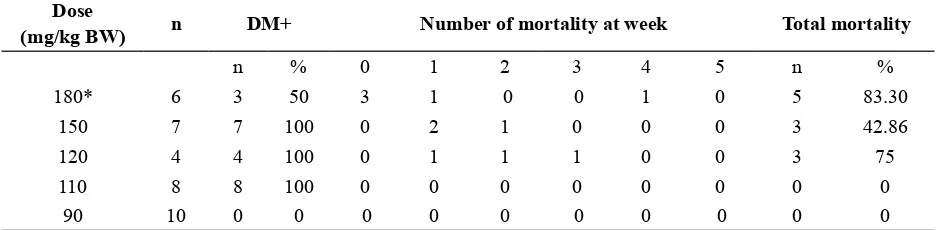
TABLE 1. The percentage of diabetic mice and mortality in various doses of STZ induction until 5 weeks
Dose
(mg/kg BW) n DM+ Number of mortality at week Total mortality
n % 0 1 2 3 4 5 n %
180* 6 3 50 3 1 0 0 1 0 5 83.30
150 7 7 100 0 2 1 0 0 0 3 42.86
120 4 4 100 0 1 1 1 0 0 3 75
110 8 8 100 0 0 0 0 0 0 0 0
90 10 0 0 0 0 0 0 0 0 0 0
* 3 mice died before the irst examination of blood glucose level
Statistical analysis
Data was statistically analyzed using SPSS version 20. All values were described as mean ± SEM. The mean differences of pain behaviour measurement between weeks were compared using One-Way ANOVA and between two groups (diabetic and non-diabetic) using independent t-test. The
signiicance level were set at 95%.
RESULTS
The result of the diabetic induction and subsequent survival of the mice after administration different dose of STZ was presented in TABLE 1. At day 7 after STZ
administration, doses 180, 150, 120 and 110 mg/kg BW resulted in 100% diabetic induction. Lower STZ dose at 90 mg/kg BW failed to induce diabetic condition in any mice. Although STZ dose 180, 150 and 120 mg/kg BW were successful in inducing diabetes, the mice survival was a problem. Most of the mice were died before week 3 and all of the mice from 180 and 120 mg/kg BW groups were died at week 7. The mean blood glucose level of the died mice at week 1 were 378.77±74.45 mg/dL, higher than the mean of blood glucose level of the surviving mice (281.64±66.84 mg/dL). However, it was not
signiicantly different (p >0.05).
STZ dose 110 mg/kg BW induced diabetes at 100% rate, but the dose did not cause any mortality. All of the mice in this group survived until week 7. Since, the STZ dose 110 mg/kg BW gave the best result, this group was used in subsequent analysis. In this group, the blood glucose level reached
# indicated signiicant differences compared to non-diabetic group
FIGURE 1. Blood glucose levels between diabetic and non-diabetic groups at weeks-0 (baseline), 1, 4 and 5.
FIGURE 2. The body weight of diabetic and non-di-abetic groups at weeks-0 (baseline), 1, 2, 3, 4 and 5
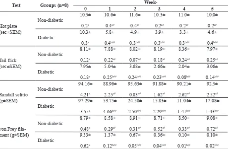
TABLE 2. Pain behaviour assessment in non-diabetic and diabetic groups after STZ induction dose 110 mg/kg BW.
Test Groups (n=8)
Week-0 1 2 3 4 5
Data analysis between groups using independent t test (p<0.05). Different letter indicated signiicant differences of pain be -haviour between diabetic and non-diabetic groups of each day. # indicated no signiicant differences compared to baseline
Pain behaviour in mice were evaluated using four different methods. Hyperalgesia
was determined by hot plate, tail lick test
and randall selitto. Allodynia was determined
by von Frey ilament. The pain behaviour of
diabetic mice and control mice were described in TABLE 2. Diabetic mice shortened latency time toward thermal stimulus using hot plate
from 10.3 sec at baseline to 5.8 sec in the irst
week after STZ induction and lasted up fourth week with consecutive latency time 4.9, 3.9,
and 3.3 sec. This result was signiicantly
different with non-diabetic group, which maintained the latency time arround 10 sec (p<0.05). At week-5, latency time toward thermal stimulus become 4.6 sec, longer than
previous week but still signiicantly lower
(p<0.05) than non-diabetic group and not returned yet to baseline.
The pain behavior evaluation using other
tests such as tail lick and Randall Selitto
gave similar results. Tail latency time using
tail lick test shortened from 7.95 s to 5.04 at
week-1 after STZ induction. This condition lasted up 4th week. Decreasing of withdrawal load threshold using randall selitto test was observed from 97.29 g at baseline to 53.75 g at week-1 after STZ induction and had been reducing until week-4. At the end of the study (week-5), the pain responses were less severe than the previous week.
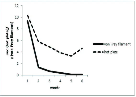
Measurement of pain behaviour using
von Frey ilament showed slightly different
outcome. The tactile sensitivity towards mechanical stimulus was continuously decreased until 5th week from 9.33
±0.62 g to 1.37 ± 0.12 g, 0.67 ± 0.05 g, 0.36 ± 0.04 g, 0.10 ± 0.01 g and 0.10 ± 0.02 g and signiicantly different with non-diabetic group (p<0.05).
DISCUSSION
In this study, the dose of STZ 110 mg/ kg BW was found to be the most effective in inducing diabetic state while preventing mortality of the mice. The STZ dose was lower than previous report i.e. 150 mg/kg, 180 mg/kg and 200 mg/kg.5,6,8 Administration of higher dose of STZ induced higher level of blood glucose more than 450 mg/dl. In our study, mouse with blood glucose levels up to 450 mg/dl was more vulnerable to die. When we used STZ dose 110 mg/kg BW, the blood glucose level was found in the range of 231.89 until 413.96 mg/dL (mean 281.64±66.84 mg/ dL). Our result was slightly similar to the report by Hayashi et al. (2006).16 Their study showed that dose 100 and 125 mg/kg BW of STZ induced non-insulin dependent diabetes in BALB/c mice model and showed a slowly progressive diabetic state until 12 weeks.16
However, our result showed signiicantly
increasing of high glucose level in the beginning of the study. The glucose levels continued to increase until week 5, more than the treshold (200 mg/dL).
Animal gender and strain may inluence
the response after STZ induction. Male pancreatic islet ß-cells are more prone than female cells to STZ-induced toxicity, that is why male rodent is more popular for diabetic study. Different strain of animals also display different sensitivity to STZ. Based
on Cardinal et al (1998),17 the action of STZ in ß-cells varies in different strain due to differences in the uptake or metabolism of the drug, the rate of DNA repair, the activity of Poly (ADP-ribose) polymerase (PARP) or damage to Nicotinamide adenine dinucleotide (NAD+) generating mechanism.17 CD-1 and
C57BL/6 mice were reliably sensitive to STZ,
STZ-induced diabetes mellitus.18 In this study, we showed that BALB/c mice could be used as STZ-induce diabetic animal model.
At week-0 (baseline), there were no pain behavior found in all groups. One week after STZ induction, the latency time toward
thermal stimulus using hot plate and tail lick
test declined in the hyperglycemia group compared to the baseline and the control group, supporting previous studies.13,14,19
According to Saikh and Somani (2010),9
mechanical hyperalgesia and tactile allodynia occur within 1-8 weeks after STZ induction.9 In this study, both of mechanical hyperalgesia tested by Randall Selitto and tactile allodynia
tested by von Frey ilament were developed
one week after STZ induction. This result was faster compared to other report that showed the occurance of mechanical hyperalgesia and tactile allodynia in mice model of PDN varies within 1-8 weeks after STZ induction.15,19,23-26 The report from Sugimoto et al (2013)15 showed that mechanical hyperalgesia was
started to be observed at 6th week after diabetic state and this condition continued until 10-11 weeks.15
Based on our result, hyperalgesia and allodynia in diabetic condition occured at the same time (Figure 2). Thermal stimulus-induced hyperalgesia started in the early phases of diabetic condition until week 4. There was a trend of reducing hyperalgesia in week 5, suggested failure to report the stimulus to the central nervous system due
to severe nerve ibers damage. Mechanical
stimulus-induced allodynia also occured at the early phases of diabetic condition and continues until week 5. Hyperalgesia usually
implicates the damage of C and A∂ nerve iber, but in allodynia, Aß nerve ibers are also
involved.21 The smaller diameter and
non-myelinated nature of C-ibers require higher
energy consumption, hence more prone to the diabetic condition.21 Furthermore, C and A∂
nerve ibers contain a lot of TRPV1 molecules
that are responsible to early hyperalgesia and advanced hypoalgesia.22
CONCLUSION
Optimum dose of STZ to induce PDN is 110 mg/kg BW. Pain behaviour of diabetic group is reached at 1st week after diabetic and continued until 5th week.
ACKNOWLEDGEMENT
The authors wished to thank Dr. D. Pakaya for excellent technical assistance.
REFERENCES
1. World Health Organization (WHO).
Noncommunicable diseases country proiles.
Geneva: WHO Publishing, 2011.
2. International Diabetes Federation. Atlas Diabetes, 6th Edition. Brussels: International
Diabetes Federation, 2013.
3. Alleman CJ, Westerhout KY, Hensen M, Chambers C, Stoker M, Long S, et al.
Humanistic and economic burden of painful diabetic peripheral neuropathy in europe: a
review of the literature. Diabetes Res Clin
Pract 2015; 109(2):215-25. http://dx.doi. org/10.1016/j.diabres.2015.04.031
4. Haanpaa M, Hietaharju A. Halting the march of painful diabetic neuropathy. IASP: Pain
Clinical Update 2015; 23(2):1-8.
5. Lennertz RC, Medler KA, Bain JL, Wright DE, Stucky CL. Impaired sensory nerve
function and axon morphology in mice with
diabetic neuropathy. J Neurophysiol 2011;
106(2):905-14. http://dx.doi.org/10.1152/ jn.01123.2010
6. Micov A, Tomic M, Pecikoza U, Ugresic N, Stepanovic-Petrovic R. Levetiracetam synergises with common analgesics in producing antinociception in a mouse model of painful diabetic neuropathy. Pharmacol Res 2015; 97:131-42. http://dx.doi.org/10.1016/j. phrs.2015.04.014
7. Reda HM, Zaitone SA, Moustafa YM. Effect of levetiracetam versus gabapentin on peripheral neuropathy and sciatic degeneration in streptozotocin-diabetic mice:
inluence on spinal microglia and astrocytes. Eur J Pharmacol 2016; 771:162-72. http://
dx.doi.org/10.1016/j.ejphar.2015.12.035 8. Tanaka K, Nakanishi Y, Sekino S, Ikegami
M, Ikeda H, Kamei J. Fentanyl produces
an anti-hyperalgesic effect through the suppression of sodium channels in mice with
painful diabetic neuropathy. Eur J Pharmacol
2014; 733:68-74. http://dx.doi.org/10.1016/j. ejphar.2014.03.042
9. Shaikh AS, Somani RS. Animal models and biomarkers of neuropathy in diabetic rodents.
Indian J Pharmacol 2010; 42(3):129-34.
http://dx.doi.org/10.4103/0253-7613.66833 10. Zangiabadi N, Mohtashami H, Shabani
M, Jafari M. Neuroprotective effect of
cerebrolysin on diabetic neuropathy: a study
on male rats. J Diabetes Metab 2014; 5(4):1-
7.http://dx.doi.org/10.4172/2155-6156. 1000355
11. Raposo D, Morgado C, Pereira-Terra P,
Tavares I. Nociceptive spinal cord neurons of laminae I-III exhibit oxidative stress damage during diabetic neuropathy which is prevented by early antioxidant treatment
with epigallocatechin-gallate (EGCG). Brain
Res Bull 2015; 110:68-75. http://dx.doi. org/10.1016/j.brainresbull.2014.12.004
12. Fong SW, Lin HC, Wu MF, Chen CC, Huang YS. CPEB3 Deiciency Elevates TRPV1
Expression in Dorsal Root Ganglia Neurons to Potentiate Thermosensation. PLOS ONE 2016; 11:e0148491. http:/dx.doi.org/10.1371/ journal.pone.0148491
13. Parkar N, Addepalli V. Effect of nobiletin on diabetic neuropathy in experimental rats.
Austin J Pharmacol Ther 2014; 2(5):1-5.
Inhibitory effect of Thymus caramanicus
Jalas on hyperglycemia-induced apoptosis
in in vitro and in vivo models of diabetic
neuropathic pain. J Ethnopharmacol 2014;
153(3):596-603. http://dx.doi.org/10.1016/j. jep.2014.02.049
15. Sugimoto K, Baba M, Suzuki S, Yagihashi S. The Impact of Low-Dose Insulin on Peripheral Nerve Insulin Receptor Signaling in Streptozotocin-Induced Diabetic Rats. PLoS ONE 2013; 8:e74247. http:/dx.doi. org/10.1371/journal.pone.0074247
16. Hayashi K, Kojima R, Ito M. Strain differences in the diabetogenic activity of streptozotocin in mice. Biol. Pharm. Bull 2006; 29(6):1110– 1119. http:/dx.doi.org/10.1248/bpb.29.1110
17. Cardinal JW, Allan DJ, Cameron DP.
Differential metabolite accumulation may be the cause of strain differences in
sensitivity to streptozotocin-induced β cell
death in inbred mice. Endocrinology 1998; 139(6):2885–2891. http:/dx.doi.org/10.1210/ endo.139.6.6048.
18. Furman BL, 2015. Streptozotocin-Induced Diabetic Models in Mice and Rats: Streptozotocin-Induced Diabetic Models, in:
Enna SJ, Williams M, Frechette R, Kenakin T, McGonigle P, Ruggeri B (Eds.). Current Protocols in Pharmacology. John Wiley & Sons, Inc., Hoboken, NJ, USA, pp. 5.47.1–
5.47.20.
19. Tian R, Yang W, Xue Q, Gao L, Huo J, Ren D, Chen X. Rutin ameliorates diabetic
neuropathy by lowering plasma glucose and decreasing oxidative stress via Nrf2
signaling pathway in rats. Eur J Pharmacol
2016; 771:84-92. http:// dx.doi.org/10.1016/j. ejphar.2015.12.021
20. Yadav SK, Nagori BP, Desai PK.
Pharmacological characterization of different fractions of Calotropis procera
(Aslepiadaceae) in streptozotocin induced
experimental model of diabetic neuropathy. J
Ethnopharmacol 2014; 152(2):349-57. http:// dx.doi.org/10.1016/j. jep.2014.01.020
21. Xu ZZ, Kim YH, Bang S, Zhang Y, Berta
T, Wang F, Oh SB, Ji RR. Inhibition of
mechanical allodynia in neuropathic pain
by TLR5-mediated A-iber blockade. Nat.
Med 2015; 21(11):1326–1331. http:/dx.doi. org/10.1038/nm.3978
22. Hong S, Wiley JW. Early Painful Diabetic
Neuropathy Is Associated with Differential
Changes in the Expression and Function of Vanilloid Receptor 1. J. Biol. Chem 2005;
280(1):618–627. http:/dx.doi.org/10.1074/ jbc.M408500200
23. Dyck PJ, Dyck PJ, Larson TS, O’Brien PC, Velosa JA. Patterns of quantitative sensation
testing hypoesthesia and hyperalgesia are predictive on diabetic polyneuropathy: a study of three cohorts. Nerve growth factor
study group. Diabetes Care 2000; 23(4):510-
7. http://dx.doi.org/10.2337/diacare.23.4.510
24. Calcutt NA, Jorge MC, Yaksh TL, Chaplan
SR. Tactile allodynia and formalin hyperalgesia in streptozotocin-diabetic rats: effects of insulin, aldose reductase inhibition and lidocaine. Pain 1996; 68(2):293-9. http:// dx.doi.org/10.1016/S0304-3959(96)03201-0
25. Courteix C, Eschalier A, Lavarenne J. Streptozocin-induced diabetic rats:
behavioural evidence for a model of chronic pain. Pain 1993; 53(1):81-8. http://dx.doi. org/10.1016/0304-3959(93)90059-X
26. Fox A, Eastwood C, Gentry C, Manning D, Urban L. Critical evaluation of the