Preservation of Natural Colorant Extract of Jalawe Fruit Peel (Terminalia bellirica)
in Water-Based Solution
Edia Rahayuningsih
*, Ayub Wijayanto, and Putri Nurfitasari
Chemical Engineering Department, Faculty of Engineering, Universitas Gadjah MadaJl Grafika 2, Kampus UGM, Yogyakarta, 55281, Indonesia
Received May 2, 2016; Accepted July 10, 2016
ABSTRACT
The general objective of this study is to preserve natural colorant extract of jalawe fruit peel in water-based solution during storing. The specific objectives in this research are finding suitable type and amount of antimicrobial compound to prevent biological degradation of the natural colorant jalawe extract, as well as determining the colorant half-life by evaluating its degradation rate. The colorant extract solution was added to a reactor followed by addition of an antimicrobial compound. Samples were taken at certain periods of time and they were then centrifuged to separate the flock from the mixture. The amounts of colorant compound in the solution and microbes in the flock were analyzed using a gravimetric method. The results showed that solutions of formaldehyde in water and chitosan in acetic acid could inhibit the degradation of jalawe extract. The most effective concentrations of formaldehyde and chitosan in the jalawe extract were 0.015 and 0.125%, respectively, with respect to total volume of the extract. The half-life of jalawe extract in a water based solution with the addition of formaldehyde and chitosan was 140 and 180 days respectively, while that without any addition of the antimicrobial compounds was 25 days.
Keywords:natural colorant; jalawe extract; antimicrobial compound; biodegradation rate
ABSTRAK
Tujuan umum kajian ini adalah untuk mengawetkan ekstrak pewarna alami dari kulit buah jalawe dengan pelarut air selama penyimpanan. Adapun tujuan khusus penelitian ini adalah untuk mendapatkan jenis dan jumlah senyawa antimikrobia yang dapat menghambat reaksi biodegradasi ekstrak pewarna alami dari kulit buah jalawe dengan pelarut air, juga menentukan waktu paruh degradasi ekstrak pewarna alami tersebut dengan mengevaluasi kecepatan biodegradasinya. Ekstrak pewarna alami dan senyawa antimikrobia dimasukkan ke dalam reaktor yang dilindungi dari sinar ultra violet. Setiap selang waktu tertentu cuplikan diambil dan disentrifugasi untuk memisahkan padatan biomassa yang terbentuk dan cairan ekstrak. Konsentrasi pewarna alami dalam cairan ekstrak dan jumlah padatan biomassa dianalisa secara gravimetri. Berdasarkan hasil penelitian ini dapat dinyatakan bahwa larutan formalin dalam air dan larutan kitosan dalam larutan asam asetat dapat menghambat reaksi biodegradasi pewarna alami dalam ekstrak kulit buah jalawe dengan pelarut air. Konsentrasi formalin dan kitosan paling efektif masing-masing 0,015 dan 0,125% terhadap volume total ekstrak. Waktu paruh berkurangnya konsentrasi pewarna alami dalam ekstrak dengan penambahan antimikrobia formalin dan kitosan masing-masing 140 dan 180 hari, sedangkan waktu paruh berkurangnya konsentrasi pewarna alami dalam ekstrak tanpa penambahan senyawa antimikroba adalah 25 hari.
Kata Kunci:pewarna alami; ekstrak jalawe; senyawa antimikroba; reaksi biodegradasi
INTRODUCTION
There is a growing interest in the use of natural colorants for textile applications due to the strict environmental standards imposed by many countries in response to the toxic and allergic reactions associated with the use of synthetic colorants [1]. In general, natural colorants are highly compatible with environment due to their lower toxicity, antibacterial, anti allergic, deodorizing, and anti-cancer properties, as well as their novelty in the harmonization of natural shades [2].
market, easy to use, competitive in price, and high in quality.
Natural colorants for textile applications that have been known and used in Indonesia are extracted from tingi bark (Ceriops candolleanaarn), tegeran wood (Cudraina javanensis), jalawe fruit peel (Terminalia bellirica), merbau wood (Intsia bijuga), coconut coir (Cocos nucifera), as well as fromIndigofera tinctoria[3]. Polar natural colorants can be easily extracted using water (water-based recovery) and directly used to color textiles. Water-based natural colorant solutions are usually cheap and easy to handle during extraction and during its applications. However, the organic natural compounds in water are prone to degradation caused by microbes. To prevent microbial growth and degradation of the natural colorants, antimicrobial compounds can be added to the solutions.
Some researchers have studied the effects of antimicrobial compounds in preventing degradation of natural colorants. Among them, Shahid et al. [5] studied the use of antimicrobial compounds for textile, which are classified into inorganic, organic, and natural antimicrobial compounds. Inorganic antimicrobial compounds mainly refer to some metallic compounds including nano-sized metals and metal oxides like silver, copper and mercury. These compounds inhibit the activity of enzymes that are responsible for microbial metabolism. Organic antimicrobial compounds include citric acid, benzoic acid, and aldehyde compounds e.g. formaldehyde, while natural antimicrobial compounds are usually bioactive compounds found in plants and animals. Chitosan as an antimicrobial compound was used by Ghoranneviss et al. [6] and Kumar [7]. Ghoranneviss et al. [5] also used poly biguanides, n-halamine, and triclosan, while Kumar [7] used tulsi leaf extract as antimicrobial compounds. Simoncic and Tomsic [8] reported that natural bioactive compounds having antimicrobial properties become increasingly important because they are safe in use and environment-friendly. Natural antimicrobial compounds extracted from plants contain phenolics and polyphenols such as simple phenols, phenolic acids, quinines, flavonoids, flavones, flavonols, tannins and coumarins, terpenoids, essential oils, alkaloids, lectins, polypeptides and polyacetylenes. These compounds, besides having antimicrobial properties, sometimes pose coloring properties. Lee et al. [9] investigated the fabrics that were colored with five kinds of natural colorants, namely peony, pomegranate, clove, coptis chinensis, and gallnut extracts. The authors stated that the application of natural colorants to the fabrics reduced the activity of Staphylococcus aureus up to 96.8%. These results demonstrate the antimicrobial properties of the natural colorants.
Rahayuningsih et al. [10] observed five different natural colorant extracts of Merbau wood (Intsia bijuga), Tegeran wood (Cudraina javanensis), Tingi bark (Ceriops candolleanaarn), jalawe fruit peel (Terminalia bellirica), and coconut coir (Cocos nucifera). It was observed that there was no microbial growth in the extract solutions of Merbau wood, Tegeran wood, and Tingi bark during storage for 5 (five) months. This observation is in a good agreement with the results published by Jansen et al. [11], which reported that tannin from wood is classified into phenolic compounds that possess antimicrobial properties. Furthermore, Fengel [12] stated that tannin from merbau wood consists of polar polyphenol macromolecules. In the contrary, it was found that the extract solutions of jalawe fruit peel and coconut coir were prone to degradation during the storage, in which microbial growth was observed after storage for 1 (one) week.
The jalawe fruit peel contains glucoside, gallo-tannic acid, coloring matter, resins and a greenish yellow oil, ellagic acid, gallic acid, lignans (termilignan and taninlignan), 7-hydroxy 3’4’ flavone and anolignan molecules [13], while the coconut coir contains lignocellulose materials. Its constituents include cellulose, hemicelluloses, and extractives such as pectin and tannin [14]. Furthermore, both jalawe fruit peel and coconut coir contains glucose, fructose, and pectin that are suitable for the growth medium of microbes. It is therefore necessary to add suitable antimicrobial compounds to the extract solutions of jalawe and coconut coir to prolong the shelf-life of the natural colorant extract solutions.
In this study, solutions of formaldehyde in water, cinnamon extract in water, betel extract in water, and chitosan in acetic acid, were added to the jalawe fruit peel extract. Their antimicrobial activities were then evaluated. According to Oliveira [15] tannins contained in jalawe are condensed tannins. These condensed tannins cannot be hydrolyzed except in acidic conditions. Therefore the degradation of the natural colorant of jalawe is only caused by a biodegradation process.
Darkuni [16] reported that microbial growth is influenced by many factors in the environment. Physical parameters that influence microbial growth include temperature, pH, and osmotic pressure, while chemical parameters include water, carbon source, nitrogen, oxygen, minerals, and other growth factors. Aerobic microbial growth is an autocatalytic reaction, which in general can be expressed as follows [17]:
culture media
2 2
cells + carbon source + oxygen source + phosphor source more cells + products +H O + CO
(1)
Equation (1) can be simplified as:
The reaction rates of substrate decrease and cell growth are expressed in the Monod equations [18] as follow:
o s c
Cc= Cell concentration, (g/L)
Cs= Substrate concentration, (g/L)
Ks= Monod constant, (g/L)
kd= Specific death rate constant, (s-1)
rc= Rate of cell growth, (g/L/s)
rs= Rate of mass substrates used, (g/L/s)
Yi= Mass ratio of cell formed and substrates used μ0= Specific growth rate, (s-1)
In the batch system, the decrease of the colorant (substrate) and the increase of microbial (cell) concentrations can be expressed as follow:
S
Cc0= Cell concentration at initial time, (g/L)
Cs0= Substrate concentration at initial time, (g/L) For certain values of μ0, Ks, kd and Yi, the
concentration of the substrate and the cells can be determined by Equations (3), (4), (5), (6), and (7), simultaneously solved using Runge-Kutta method [19]. The values of μ0, Ks, kd and Yi, were determined using
curve fitting method between the experimental and the calculated concentrations of substrate, as well as between the experimental and the calculated concentrations of cells.
Once the μ0, Ks, kd and Yi values have been
determined, the half-life of the substrate concentration can be calculated. Half-life is the amount of time
required for the substrate concentration to fall to half of its initial value.
EXPERIMENTAL SECTION
Materials and Equipments
The materials used in this study are jalawe fruit peel extract (3 g colorant in 100 mL water}, formaldehyde (technical grade, C.V. Bratachem), chitosan (technical grade, C.V. Chemix), acetic acid (technical grade, C.V. Bratachem), cinnamons bark extract (7.6 g antimicrobial compound in 100 mL tap water), betel leaf extract (12 g antimicrobial compound in 100 mL tap water).
The experimental set-up is depicted in Fig 1.
Procedure
The experiments were divided into two steps; First, selection of type and concentration of antimicrobial compounds, second, evaluation of the degradation rate and half-life of colorant compound.
Selection of type and concentration of antimicrobial compounds
Forty milliliters (40 mL) extract solution of jalawe fruit peel was added to a batch reactor, followed by addition of a certain amount of an antimicrobial compound. Various types and volumes of antimicrobial compounds added to the extract are shown in Table 1.
Fig 1.The experimental set-up
Table 1.Type, composition, and volume of antimicrobial compounds added to the jalawe extract.
No. Antimicrobial compound Volume added to the
40 mL jalawe extract, mL
Type Composition
1 Formaldehyde 1 mL formaldehyde in 100 mL water
0.2; 0.4; 0.6; 0.8; and 1.0
2 Cinnamons bark extract
7.6 g antimicrobial compound from cinnamons bark in 100 mL water
1; 2; 5; 10; and 15
3 Betel leaf extract 12 g antimicrobial compound from betel leaf in 100 mL water
1; 2; 5; 10; and 15
4 Chitosan 2.5 g chitosan in 10 mL of 2% volume of acetic acid solution in water
The reactor was covered with aluminum foil to avoid UV exposure to the solution. Their physical change was visually observed by observing the flock formation in the solution. At the end of the observation, the extract solution was used to color textile in order to test its performance. The test was conducted by comparing the color quality of the textile that was colored with the aged extract solution and that with the new extract.
From the data shown in Table 1, it was possible to calculate the final concentration of the antimicrobial compounds in the extract solution. Suitable types of antimicrobial compounds were chosen based on visual observation (flock formation and coloring quality). Then, suitable concentrations were determined based on the degradation rate of colorant upon addition of various volumes of the chosen antimicrobial compounds.
Evaluation of the degradation rates and half-life of colorant compounds
Once the most suitable types and concentrations of antimicrobial compounds for jalawe extract have been determined as described above, the degradation rate and half-life of the colorant compounds were evaluated. Three hundred mL (300 mL) jalawe extract solution was centrifuged to separate its solid from liquid. The liquid was then used in the experiment to determine the degradation rates and half-life time of colorant
compounds. The experiments were conducted in a batch reactor. To evaluate the initial colorant concentration, 5 mL sample was taken and analyzed using a gravimetric method. Subsequently, 10 mL samples were taken at a certain period of time and centrifuged at 4000 rpm for 15 min to separate its flock from the mixture. Then, the amount of colorant in the solution and microbes as the flock was analyzed using a gravimetric method. Gravimetric method was done as follow, the sample was weighed before and after drying; the difference between the two masses could be determined the content of water or solids in the sample.
RESULT AND DISCUSSION
Selection of Type and Amount of Antimicrobial Compounds
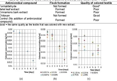
The qualitative observation of the physical changes and the coloring quality of the extract solution of jalawe fruit peel, with addition of various antimicrobial compounds, after 7 days are summarized in Table 2.
From Table 2, it can be seen that the addition of formaldehyde and chitosan can inhibit the microbial growth. This fact was observed visually, i.e. no flock
Table 2.Qualitative observations of jalawe extract preservation after 7 days
Antimicrobial compound Flock formation Quality of colored textile
Formaldehyde Not formed Good*
Betel leaf extract Formed Poor
Cinnamons bark extract Formed Poor
Chitosan Not formed Good
Control (No addition of antimicrobial
compound) Formed Poor
* Good = the same quality as the textile that was colored with new extract.
formation in the colorant extract, and good quality of colored textile after being colored with the aged colorant. Fig. 2(a) shows the colorant concentration in the extract solution of jalawe fruit peel without addition of antimicrobial compound. It shows that the colorant concentration steeply decreases after two days of aging. The decreasing rate of the colorant due to the microbial activity after two days is approximately 0.003 g colorant/(mL extract solution)(day) Fig. 2(b) shows the colorant concentration as a function of time upon addition of formaldehyde of various concentrations ((mL of formaldehyde/100 mL extract solution) x 100%). It is observed that after 2 days, the addition of formaldehyde of 0.015% can inhibit the growth of microbial better than the other concentrations. Fig. 2(c) plots the colorant concentration as a function of time upon addition of chitosan of various concentrations ((gram of
chitosan/100 mL extract solution) x 100%). It is observed that addition of 0.125% chitosan in acetic acid results in the lowest rate of colorant decrease.
Evaluation of the Degradation Rate and Half-Life Time of Colorant Matter
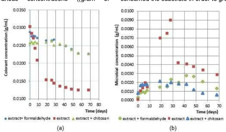
The experimental results of both the colorant and the microbial concentrations in the jalawe extract solution as a function of time are presented in Fig. 3(a) and 3(b), respectively.
Fig. 3(a) and 3(b) show that in the extract without antimicrobial compounds, the steep decrease of colorant concentration during 0-30 days corresponds to a steep increase of microbial concentration. This observed phenomenon indicates that the microbes consumed the substrate in order to grow. The microbial
Fig 3.(a) The colorant concentration and (b) The microbial concentration, as a function of time
Fig 5.Colorant and microbial concentration as a function of time in jalawe fruit peel extract with addition of 0.015% formaldehyde
Fig 6.Colorant and microbial concentration as a function of time in jalawe fruit peel extract with addition of 0.125% chitosan
concentration then decreases slowly after 30 days, which indicates substrate depletion. Addition of antimicrobial compounds, either formaldehyde or chitosan, to the extract significantly slows down the decrease of colorant and the increase of microbial concentrations. Thus, the slight decreases of colorant concentration correspond to slight increases of microbial concentration. This phenomenon indicates that there is an activity of the antimicrobial compounds that inhibits the microbial growth, thus preventing the colorant from being consumed.
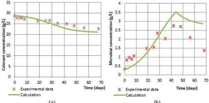
In Fig. 4, 5, and 6 show the results of curve-fitting between the experimental and calculated data of colorant and microbial concentrations as a function of time. It is seen in Fig. 4(a), 5(a), and 6(a) that the calculated data of colorant concentration satisfactorily fit the experimental ones. However, Fig. 4(b), 5(b), and 6(b) demonstrate that the calculated data of microbial concentration do not satisfactorily fit the experimental
data. The discrepancy may be caused by the four parameters, μ0, Ks, kd and Yi, that must be
simultaneously evaluated by curve-fitting between the experimental data and calculated of colorant concentration; and between the experimental data and the calculated of microbial concentration. Another explanation could be that there were some other settled substances in addition to the microbes during centrifugation, which results in slight errors when determining the experimental microbial concentration.
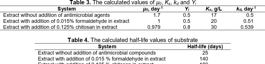
The values of the parameters μ0, Ks, kdand Yiare
chosen if the sum square error between the experimental data and the calculated of colorant concentrations; and the experimental data and the calculated of microbial concentrations are minimum. The calculated results of the parameters are shown in Table 3.
Using the calculated values of μ0, Ks, kd and Yi,
Table 3.The calculated values ofµ0,Ks,kdandYi
System µ0, day-1 Y
i Ks, g/L kd, day-1
Extract without addition of antimicrobial agents 1.7 0.5 17 0.5 Extract with addition of 0.015% formaldehyde in extract 1 0.5 20 0.51 Extract with addition of 0.125% chitosan in extract 0.979 0.8 30 0.539
Table 4.The calculated half-life values of substrate
System Half-life (days)
Extract without addition of antimicrobial compounds 25 Extract with addition of 0.015 % formaldehyde in extract 140 Extract with addition of 0.125 % chitosan in extract 180
calculated. The calculated half-life values are shown in Table 4.
Data shown in Table 4 demonstrate that the addition of 0.015% formaldehyde and 0.125% chitosan significantly increase the half-life of the natural colorant in jalawe extract up to 5.6 and 7.2 times, respectively. This means that the antimicrobial compounds could inhibit the microbial growth-rate in the extract and preserve the colorant from degradation.
CONCLUSION
Formaldehyde in water and chitosan in acetic acid can be utilized for the preservation of the natural colorant in extract solution of jalawe fruit peel. The effective concentrations of formaldehyde and chitosan in jalawe extract to inhibit the microbial growth are 0.015 and 0.125%, respectively. The result shows that rate of colorant degradation and the rate of microbial growth are represented with Monod model. The half-life values of the extracted colorant with addition of antimicrobial compounds chitosan and formaldehyde are 180 and 140 days, respectively. While the half-life of the extracted colorant without addition of any antimicrobial compounds is 25 days.
REFERENCES
1. Erkurt, E.A., Ünyayar, A., and Kumbur, H., 2007, Process Biochem., 42 (10), 1429–1435.
2. Saxena, S., and Raja, A.S.M., 2014, “Natural Dyes: Sources, Chemistry, Application and Sustainability Issues” in Roadmap to Sustainable Textiles and Clothing, Textile Science and Clothing Technology, S.S. Muthu, (Eds.), Springer Science-Business Media, Singapore,: 37–80.
3. Badan Pusat Statistik (BPS), 2015, available: http://www.bps.go.id./data import of synthetic dyes for textiles, accessed March 2015.
4. Rini, S., Sugiharti, and Riswati, M.K., 2011,Pesona Warna Alam Indonesia, available: http://www.kehati.or.id/images/publikasi/Buku/01_Pe sona Warna Alam Indonesia_final.pdf
5. Shahid, M., ul-Islam, S., and Mohammad, F., 2013, J. Cleaner Prod., 53, 310–331.
6. Ghoranneviss, M., Shahidi, S., Anvari, A., Motaghi, Z., Wiener, J., and Šlamborová, I., 2011, Prog. Org. Coat., 70 (4), 388–393.
7. Kumar, M.S.Y, Raghu, T.S., Varghese, F.V., Shekar, R.I., Kotresh, T.M., Rajendran, R., Babu, K.M., and Padaki, V.C., 2016, J. Ind. Text., 45, 1115–1127.
8. Simoncic, B., and Tomsic, B., 2009, Materials, 2, 10–21.
9. Lee, Y.H., Hwang, E.K., and Kim, H.D., 2009, Materials, 2 (1), 10–21.
10. Rahayuningsih, E., Wijayanto, A., and Fitasari, P.N., 2014, Laporan Penelitian, JurusanTeknik Kimia, FakultasTeknik, UGM.
11. Jansen, P.C.M., and Cardon, D., 2005, Prota 3: Dyes and Tannins ed., Wageningen.
12. Fengel, D., and Wegener, G., 1995, Kayu: Kimia Ultra Struktur Reaksi, Gadjah Mada University Press, Yogyakarta.
13. Motamarri, S., Karthikeyan, M., Kannan, M., and Rajasekar, S., 2012, Int. J. Res. Pharm. Biomed. Sci., 3, 1, 96–99
14. Israel, A.U., Ogali, R.E., Akaranta, O., and Obot, I.B., 2011, Songklanakarin J. Sci. Technol., 33 (6), 717–724.
15. Oliveira, L.M., Bevilaqua, C.M., Costa, C.T., Macedo, I.T., Barros, R.S., Rodrigues, A.C., Camurça-Vasconcelos, A.L., Morais, S.M., Lima, Y.C., Vieira, L.S., and Navarro, A.M., 2008, Vet. Parasitol., 159 (1), 55–59.
16. Darkuni, M.N., 2001, Mikrobiologi (Bakteriologi, Virologi, dan Mikologi),Universitas Negeri Malang, JICA.
17. Shuler, M.L., and Kargi, F., 2002, Bioprocess Engineering Basic Concepts, 2nded., Prentice-Hall,
Inc. New Jersey, 161.
18. Rahayuningsih, E., 2009, Analisis Kuantitatif Perilaku Pestisida di Tanah, UGM Press, 123-155. 19. Sediawan, W.B., and Prasetya, A., 1997,