Summary We investigated the synthesis and accumulation of vegetative storage proteins (VSPs) in poplar plantlets and the homology between poplar seed storage proteins (SSPs) and VSPs. One-dimensional SDS polyacrylamide gel electropho-resis confirmed that both seed and vegetative storage proteins contained two predominant polypeptides of MW 32 and 36 kDa, but the subunit composition of the polypeptides differed. The 32- and 36-kDa polypeptides were highly abundant in basal leaves, stems, and roots of poplar plantlets. The 36-kDa subunit was synthesized in all plantlet tissues examined, but the 32-kDa subunit was not, suggesting that the 36-kDa polypeptide is a precursor of the 32-kDa polypeptide. The 36-and 32-kDa polypeptides of both SSPs 36-and VSPs were glyco-sylated and both were found to be albumins. In addition, both polypeptides cross-reacted with a VSP antibody. Protein fin-gerprint patterns generated with two different proteolytic en-zymes were identical for the 36-kDa polypeptide isolated from seeds or from stem tissue. Our study provides evidence that poplar SSPs and VSPs exhibit homology, and that expression is neither tissue-specific nor regulated solely by photoperiod.
Keywords: Alnus crispa, electrophoresis, Larix × eurolepis, poplar, protein fingerprint, Salix microstycha.
Introduction
Plants contain proteins for the storage of nitrogen and carbon (Bewley and Black 1994). These proteins are called vegetative storage proteins (VSPs) when located in vegetative tissues, and seed storage proteins (SSPs) when located in seeds. Seed and vegetative storage proteins have generally been studied in separate experimental systems. In soybean, it is thought that VSPs and SSPs are distinct from one another, although they share similar characteristics (Staswick 1990). Research on SSPs has focused on their nutritional value for human and animal consumption, whereas VSPs have been targeted for plant improvement.
Seed storage proteins are synthesized in the developing seed and hydrolyzed during imbibition and germination, providing a source of reduced nitrogen and carbon for early seedling
growth. Seed storage protein synthesis is specific to embryo-genesis, is environmentally regulated by nitrogen and sulfur availability, and is specific to the seed (reviewed by Bewley and Black 1994). Vegetative storage proteins accumulate in leaves, stems and roots during plant development and growth and provide a source of reduced nitrogen and carbon (O’Ken-nedy and Titus 1979, Staswick 1989a, 1989b, Langheinrich and Tishner 1991). Their synthesis is regulated by seasonal changes in source--sink relationships (Clausen and Apel 1991, Coleman et al. 1991, Coleman et al. 1992, Sauter and van Cleve 1992), nitrogen availability (Staswick et al. 1991), drought (Mason and Mullet 1990) and wounding (Mason and Mullet 1990). Vegetative storage protein synthesis is not tissue specific (Staswick 1990); however, VSP accumulation may be cell specific.
Two VSPs with molecular weights of 32 and 36 kDa have been identified in various poplar species (Wetzel et al. 1989, Coleman et al. 1991) and Salix microstachya Turz. (Wetzel and Greenwood 1991). In poplar, these VSPs are located in bark, wood and roots (Coleman et al. 1991, Stepien and Martin 1992, Langheinrich 1993), where they make up more than 25% of total soluble proteins (Langheinrich 1993). Accumulation of VSPs correlates positively with rapid growth, winter hardiness and the onset of dormancy (O’Kennedy and Titus 1979, Wetzel et al. 1989, Clausen and Apel 1991, Coleman et al. 1991). Shortened photoperiod is thought to trigger VSP accumula-tion, as supported by reports of increases in VSP mRNA synthesis in the fall (Clausen and Apel 1991, Coleman et al. 1991, Coleman et al. 1992). Reports show that VSP synthesis can also be induced by high nitrogen availability under long-day laboratory conditions (van Cleve and Apel 1993, Coleman et al. 1993).
We characterized poplar SSPs to determine if they exhibit homology with poplar VSPs. Such homology would make poplar an ideal system for studies on storage protein gene regulation in trees. We also attempted to induce VSP synthesis in various tissues of poplar plantlets. The ability to induce VSP synthesis would allow the natural cycle of active growth
fol-Characterization of seed storage proteins in
Populus
and their
homology with
Populus
vegetative storage proteins
TANNIS BEARDMORE,
1SUZANNE WETZEL,
2DARWIN BURGESS
3and
PIERRE J. CHAREST
31
Canadian Forest Service, P.O. Box 4000, Regent St. South, Fredricton, NB, E3B 5P7, Canada
2 Canadian Forest Service, P.O. Box 490, 1219 Queen St. E., Sault Ste. Marie, ON, P6A 5M7, Canada
3
Canadian Forest Service, P.O. Box 2000, Chalk River, ON, K0J 1J0, Canada
Received March 16, 1995
lowed by dormancy to be shortened significantly and would thus be helpful in future VSP research.
Materials and methods
Plant material
Poplar plantlets were obtained by direct organogenesis of Populus alba L. according to Son and Hall (1990) and were placed in modified Phytacon vessels (116 mm diameter, Sigma Co., St. Louis, MO). In addition, two 0.2 µm CR-PTFF filters (Gelman Sciences, Ann Arbor, MI) were placed on top of the vessels. Organogenesis was stimulated with 100 ml basal me-dium, pH 5.7 at 25 °C, containing 0.44% MS salts (0.355 g nitrogen per plantlet) (Murashige and Skoog 1962), 0.1% B5 vitamins (Sigma Co. St. Louis, MO), 3% sucrose and 0.9% agar, a 16-h photoperiod and a photon flux of 80 µmol m−2 s−1.
Stem, root and leaf sections for protein extraction were se-lected from plantlets approximately 6 cm in height. There were 16 to 20 leaves on each plantlet, with approximately 11--15 of the leaves fully expanded. Two types of leaf tissue samples were taken: (1) apical (the first four fully opened leaves), and (2) basal (middle four leaves).
Seeds of Populus grandidentata Michx., P. balsamifera L., P. tremuloides Michx., P. deltoides Bartr. ex Marsh, Alnus crispa (Ait.) Pursh, Larix × eurolepis A. Henry, and Salix microstachya were obtained from the National Tree Seed Cen-tre (Chalk River, Ontario). Populus grandidentata seeds were used for the characterization of polypeptides in poplar seeds because P. alba seeds were unavailable. The P. grandidentata seeds were imbibed with 5 ml distilled water in petri dishes (0.1 g seeds per 60 × 15 mm petri dish) containing two layers of Whatman No. 1 filter paper in a growth chamber (25 °C, 16-h photoperiod, 80 µmol m−2 s−1). Samples were removed
for protein extraction 24, 48, and 72 h after imbibition.
Protein extraction
Seeds and plantlet tissues were homogenized in an extraction buffer [1.0 M sodium chloride (NaCl), 2% sodium dodecyl sulfate (SDS), 10% glycerol, 1 mM phenylmethylsulfonyl fluoride (PMSF, Sigma Co. St. Louis, MO), 62.5 mM Tris-HCl, pH 6.8] at 4 oC, with a liquid nitrogen-cooled mortar and pestle. After centrifugation at 16,000 ×g for 5 min, the super-natant was collected and heated at 100 oC for 5 min. For the preparation of reduced protein, 50 mM dithiothreitol (DTT) was added to the extraction buffer; protein samples intended for non-denaturing polyacrylamide gel electrophoresis were homogenized in extraction buffer without SDS or DTT. Pro-tein was quantified with the DC proPro-tein assay (Bio-Rad Inc., Rockford, IL) and a bovine serum albumin standard.
To determine solubility characteristics, protein extractions from P. grandidentata were carried out sequentially in double distilled water, 0.5 M NaCl, 70% propanol, 60% glacial acetic acid, 0.1 M sodium hydroxide, and finally, in a pH 10 solution of 0.1 M sodium borate, 1% SDS, and 50 mM DTT (Hu and Esen 1981). Protein content in each sequential extraction was determined by the DC protein assay, and the percentage of total protein in each sequential extraction was calculated.
Radioisotope labeling and in vivo protein synthesis
To characterize the kinetics of protein synthesis, P. alba plant-let tissue (0.1 g of stem, root, apical and basal leaves) and P. grandidentata dry seeds (0.1 g), were incubated with 4.0 MBq [35S]-methionine (ICN Biomedical, Irvine, CA) in distilled water for 2 h at 24 °C. Samples were rinsed with distilled water and the proteins were isolated in extraction buffer containing 50 mM DTT. Incorporation of radiolabeled methionine was determined by cold acetone precipitation (Gif-ford and Bewley 1984).
Polyacrylamide gel electrophoresis
One-dimensional SDS-PAGE under reducing (50 mM DTT) and dissociating (without DTT) conditions was preformed through 14% polyacrylamide gels (Laemmli 1970). Either 20 µg protein or 5 × 10⁵ cpm of [35S]-methionine were loaded per well. The proteins were visualized by staining the gels for 1 h with 0.1% Coomassie Brilliant Blue R-250 in metha-nol/water/acetic acid (5/4/1) and destaining with water/etha-nol/acetic acid (67/25/8). The gels were stored in water/acetic acid/methanol (88/7/5). Alternatively, proteins were visualized by immunoblotting, according to fluorography procedures de-scribed by Gifford and Bewley (1984).
Protein glycosylation was determined by soaking SDS-PAGE gels in 1% periodic acid and 3% acetic acid for 50 min, washing six times for 10 min each in double distilled water, staining with fuchsin-sulfite (1% basic fuchsin in 0.15 M HCl) in the dark for 50 min, rinsing in 0.5% metabisulfite three times for 10 min each, and washing in distilled water. Ovalbumin was run alongside the samples as a control. The gels were stored in 5% acetic acid (Zacharius et al. 1968).
To determine the abundance of disulfide bonds, two-dimen-sional gel electrophoresis was performed under dissociating conditions in the first dimension, and under reducing condi-tions in the second dimension (Krochko and Bewley 1988). For the second dimension, a dissociating gel slice was incu-bated for 30 min in extraction buffer with 50 mM DTT. It was then placed on top of the second dimension gel and run for 45 min at 175 V.
The MW of the native proteins was calculated by Ferguson plots (Weber and Osborn 1969).
Isoelectric focusing (IEF) was performed with a Mini Isoelectric Focusing Unit (Bio-Rad Inc., Rockford, IL) accord-ing to a modification of the procedure described by O’Farrell (1975). Gels were prefocused for 30 min at 200 V. After focusing, gels were extruded and incubated in a sample buffer (2% SDS, 10% glycerol, 80 mM Tris-HCl, pH 6.8) for 20 min. The pH gradient across the gel was determined using Bio-Rad IEF standards (pI range 4.6--9.6). For the second dimension, gels were overlaid on an SDS-PAGE gel and run as previously described.
Immunoblotting
Proteins separated by SDS-PAGE under reducing conditions were transferred to nitrocellulose membranes (Bio-Rad Inc., Rockford, IL) with a mini trans-blot electrophoretic transfer cell (Bio-Rad Inc., Rockford, IL), according to the manufac-turer’s instructions. The nitrocellulose membranes were probed with immunoglobin-G (Ig-G) against the 32-kDa vege-tative storage protein from Salix microstaycha (Wetzel and Greenwood 1991), according to the procedure of Towbin et al. (1979). Binding of the Ig-G to the membrane was visualized using goat--anti-rabbit Ig-G-conjugated alkaline phosphatase (Jackson Immuno Research Labs., West Grove, PA) and the Bio-Rad immuno-blot assay kit for goat--anti-rabbit alkaline phosphatase conjugates.
Protein fingerprinting
The SDS-PAGE gels were run under reducing conditions as previously described using protein isolated from P. alba plant-let stems and P. grandidentata seeds. The 36- and 32-kDa polypeptides were cut out of stained gels. The excised gel pieces were incubated for 30 min in 25 mM Tris base, 0.192 M glycine, 2% SDS, pH 8.3 buffer, and were then placed in the wells of an SDS-PAGE gel (4--5% and 17% acrylamide in the stacking and separating gel, respectively). Twenty percent (w/v) endoproteinase Glu-C (25 units of specific activity) (Promega, Madison, WI) in 10 mM ammonium bicarbonate, pH 7.8, and 20% (w/v) alkaline protease (25 units of specific activity) (Promega, Madison, WI) in 50 mM ammonium ace-tate, pH 4.0 were underloaded into the wells. Endoproteinase Glu-C cleaves the carboxylic side of lysine, and alkaline pro-tease cleaves aromatic residues. The initial applied voltage was 100 V. Once the protein had migrated to the interface of the stacking and separating gels, the power was turned off for 30 min and then reapplied at 175 V until the protein reached the end of the gel. The proteolytic products were visualized by silver staining, according to the procedure of Wray et al. (1981).
Results
Characterization of seed storage proteins
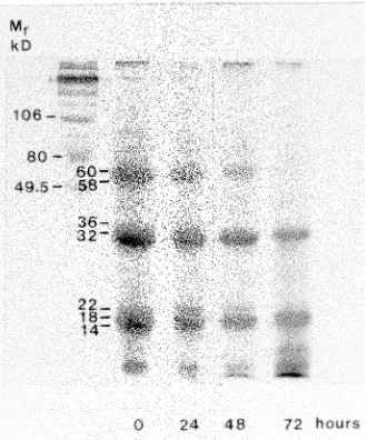
Three groups of polypeptides were classified as seed storage proteins because they were present in large quantities in the
P. grandidentata seed and were almost completely mobilized 72 h after imbibition (seeds germinated by 72 h) (Figure 1). The three groups of SSPs had subunit molecular weights of approximately 60 and 58 kDa, 36 and 32 kDa, and 22, 18 and 14 kDa (Figure 1). All three groups were abundant in the embryo and present in low amounts in the testa (results not shown).
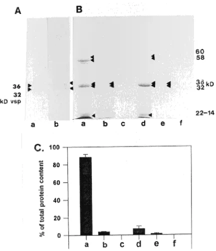
The poplar SSP was characterized concurrently with the poplar VSP. In its native form, the poplar VSP had a molecular weight of 500 kDa (see Figure 4). The identity of the 500 kDa protein was verified by two-dimensional electrophoresis. No difference was observed between the poplar stem VSP and the willow bark VSP (Wetzel and Greenwood 1991). The poplar seed 36- and 32-kDa polypeptides were glycosylated (Figure 2A) and water soluble (Figures 2B and 2C). The 60- and 58-kDa polypeptides, and 22- to 14-kDa polypeptides were also water soluble (Figures 2B and 2C).
A large protein mass of approximately 100--120 kDa was identified when seed proteins were electrophoretically sepa-rated under non-denaturing conditions (Figure 3Ai). When dissociated and reduced, this protein mass yielded two similar groups of three polypeptides, based on molecular weight (Fig-ure 3Aii). It was difficult to determine differences in the polypeptide constituents of the proteins because of diffusion in both dimensions (Figure 3Aii), a problem inherent to this technique (Krochko and Bewley 1988). Dissociation of the native proteins without reduction yielded 60- and 58-kDa subunits (Figures 3Bi and 4Aii). With reduction, these subunits yielded 36-, 32-, 22-, 18-, and 14-kDa polypeptides (Figures 3Bii and 4Aiii). The 60- and 58-kDa subunits were still present in varying amounts (Figures 3C, 4Aiii, 7A and 8), suggesting that subunits in these samples were not fully reduced. Com-plete reduction of the 60- and 58-kDa polypeptides could be
Figure 1. One-dimensional gel electrophoresis of proteins extracted from Populus grandidentata seeds during imbibition. Twenty µg of
achieved by using 0.5 M DTT, but this resulted in extensive distortion of the polypeptides (results not shown). A summary of the composition of the 36- and 32-kDa VSP and SSP polypeptides is presented in Figure 4.
The 32- and 36-kDa polypeptides and 60- and 58-kDa subunits exhibited charge heterogeneity (Figure 3C). There were approximately 8--12 isoforms of the 36- and 32-kDa polypeptides (pI 7.0--8.2) and four isoforms of the 60- and 58-kDa subunits (pI 7.0--7.2). The 18- and 14-kDa polypep-tides did not exist as isoforms and both had a pI of 7.0 (Figure 3C). The 22-kDa polypeptide was not present in the IEF separation of the proteins, suggesting that its pI was less than 5.0 or greater than 8.5.
The homology between the 36-kDa VSP and the 36-kDa SSP was examined using protein fingerprints generated by the proteolytic digestion of the SSP and VSP 36-kDa polypeptides (Figures 5A and 5B). The proteolytic enzymes, alkaline pro-tease and endoproteinase Glu-C, were not detectable on the stained gels (results not shown), therefore all visible bands corresponded to the 36-kDa polypeptides and their resultant
Figure 2. Glycosylation and solubility of seed storage proteins from
Populus grandidentata seeds. (A) PAS stain of reduced (a) seed pro-teins and (b) stem propro-teins, separated by one-dimensional SDS-PAGE. Molecular weight markers are in kilodaltons (kD). (B) One-dimen-sional SDS-PAGE separation of reduced proteins sequentially ex-tracted from poplar seeds: (a) distilled water extract; (b) sodium chloride extract; (c) 70% propanol extract; (d) acetic acid extract; (e) sodium hydroxide extract; and (f) sodium borate extract. Twenty µg of protein was loaded in each lane. (C) Percent of total protein in each seqential protein extraction from poplar seeds: (a) distilled water extract; (b) sodium chloride extract; (c) 70% propanol; (d) acetic acid extract; (e) sodium hydroxide extract; (f) sodium borate extract. Values are the mean of protein content of three replicates (one replicate represents one sequential extraction) ± SE.
proteolytic digestion fragments (Figures 5A and 5B). The digestion of the SSP and VSP 36-kDa polypeptides with endo-proteinase Glu-C and with alkaline protease was incomplete,
as the 36-kDa polypeptide was evident in these samples (Fig-ures 5A and 5B). The endoproteinase Glu-C digestions of the 36-kDa VSP and SSP each yielded seven peptide fragments, which had the same molecular weights in SSP and VSP (Figure 5A). Six different peptide fragments were generated from the alkaline protease digestion of either the SSP or VSP 36-kDa polypeptides and the MW of each of these six peptide frag-ments was the same in the SSP and VSP samples (Figure 5B).
Immuno-relationship of the Salix vegetative storage protein to proteins in the seeds of Populus and other genera
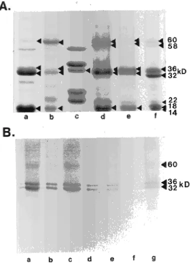
The presence of SSP 36- and 32-kDa polypeptides was inves-tigated in Populus grandidentata, P. balsamifera and P. del-toides, as well as in Alnus crispa, Larix × eurolepis, and Salix microstachya (Figure 6). A 36-kDa and a 32-kDa polypeptide were present in all seeds(Figure 6A). The antibody specific to the 32-kDa VSP in Salix cross-reacted with the 36- and 32-kDa polypeptides in all samples except Larix (Figure 6B). There was a faint cross-reactivity of the antibody with the 60-kDa
Figure 4. Summary of the composition of Populus seed storage pro-teins and vegetative storage propro-teins. (A) Seed storage protein; (B) vegetative storage protein isolated from P. alba stems. One-dimen-sional electrophoresis of (i) non-dissociated protein (ND), (ii) dissoci-ated protein (SDS-PAGE), and (iii) dissocidissoci-ated and reduced protein (SDS-PAGE + DTT).
Figure 5. One-dimensional SDS-PAGE analysis of the proteolytic digestion products of the 36-kDa polypeptide isolated from Populus alba stem and Populus grandidentata seeds. A total of 25 units of specific activity was used for each digestion. (A) Endoproteinase Glu-C (EGC) proteolysis. Lane 1, SSP 36-kDa polypeptide digested with EGC; Lane 2, VSP 36-kDa polypeptide digested with EGC. (B) Alkaline protease (AP) proteolysis. Lane 1, VSP 36-kDa polypeptide digested with AP; Lane 2, 36-kDa polypeptide digested with AP.
Figure 6.Identification of the seed storage proteins in Populus gran-didentata, P. balsamifera, P. deltoides, Alnus crispa, Salix micro-stachya and Larix × eurolepis seeds. (A) SDS-PAGE separation of reduced proteins extracted from seeds of (a) poplar (P. grandidentata), (b) poplar (P. balsamifera), (c) poplar (Populus deltoides), (d) alder (Alnus crispa), (e) willow (Salix microstachya) and (f) larch (Larix × eurolepsis). (B) Western blot of an SDS-PAGE separation of reduced proteins extracted from (a) P. grandidentata, (b) P. balsamifera, (c) P. deltoides, (d) A. crispa, (e) S. microsyachya, (f) L. × eurolepsis seeds and (g) S. microstachya bark. Twenty µg of protein was loaded in each
polypeptide in P. grandidentata, P. balsamifera, P. deltoides, S. microstachya, and A. crispa seeds and with low molecular weight polypeptides in P. grandidentata, and P. deltoides. The low molecular weight polypeptides corresponded to the 22-, 18- and 14-kDa polypeptides of the SSP. The cross-reactivity to the low molecular weight polypeptides was also evident in the protein isolated from S. microstachya bark. The preim-mune antibody did not cross-react with any polypeptide in the seeds (results not shown).
Accumulation and synthesis of the VSP in poplar plantlets and seeds
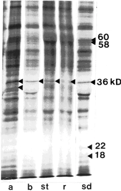
Vegetative tissue from poplar plantlets was examined to deter-mine whether VSPs were present in plantlets grown in vitro. The 36- and 32-kDa polypeptides were abundant in the stem, root and basal leaves of Populus alba plantlets, as identified by SDS-PAGE (Figure 7A). Only the 36-kDa polypeptide was present in apical leaves, and only in small quantities (Figures 7A and 7B). Western blotting confirmed that the polypeptides were immunologically related to the Salix 32-kDa VSP (Figure 7B). The preimmune antibody did not react to any of the polypeptides present in the plantlet tissues examined (results not shown).
Incorporation studies showed that the 36-kDa polypeptide was synthesized in basal and apical leaves, stem, root and seeds (Figure 8). The 60-, 58- 22- and 18-kDa polypeptides were synthesized in the seed (Figure 8), and the 32-kDa polypeptide was not synthesized in the plantlet tissues or in the seed.
Discussion
Although both VSPs and SSPs are localized in vacuoles and provide a source of nitrogen and carbon for growing or
devel-oping plant tissue, they are considered to be distinct forms of plant storage proteins (Staswick 1990). Previous studies com-paring VSPs with SSPs in soybean showed that they differ in polypeptide composition and in native protein and subunit composition (Gayler and Sykes 1981, Spielmann et al. 1982, Wittenbach 1983). Also, the soybean VSP and SSP were not immunologically related to one another (Wittenbach 1983, Staswick 1989a), and the VSP did not exhibit DNA sequence homology to the SSP (Staswick 1989b).
Contrary to the results of studies on soybean, we have found homology between the poplar 36-kDa SSP and VSP. This suggests that storage protein genes expressed during seed development could be related to those expressed seasonally during vegetative growth. To our knowledge, this is the first finding of homology between seed and vegetative storage proteins in poplar. We obtained three types of evidence sup-porting homology between VSPs and SSPs: (1) similar pro-teolytic digestion patterns; (2) antibody cross-reactivity; and (3) similar chemical characteristics. The SSP and VSP 36-kDa polypeptides yielded peptide fragments of identical molecular weight when digested with either alkaline protease or endopro-teinase Glu-C. This provides strong evidence that the SSP and
Figure 7. Identification of vegetative and seed storage proteins in
Populus plantlets and seeds. (A) SDS-PAGE separation of reduced proteins; (B) Western blot of proteins. Twenty µg of protein was loaded in each lane. The western blot was probed with an antibody to the Salix VSP; (a) apical leaves; (b) basal leaves; (st) stem; (r) root; (sd) seed. Molecular weight markers in kilodaltons (kD).
Figure 8. Fluorographs of a one-dimensional gel electrophoresis sepa-ration of proteins synthesized in vivo in Populus alba plantlets and
VSP 36-kDa polypeptides have similar amino acid sequences. The SSP and VSP 32- and 36-kDa polypeptides also cross-re-acted to the same VSP antibody, further indicating homology in amino acid sequence. The polypeptides were both water soluble (albumin storage proteins) and glycoslylated. In addi-tion, the isoelectric point of the SSP 32- and 36-kDa polypep-tides corresponded to those published for the willow VSP (pI 7.2--7.8, Wetzel and Greenwood 1991).
Although the 36- and 32-kDa VSP and SSP polypeptides shared certain characteristics, they differed with respect to their native proteins and subunit composition. Specifically, the SSP 32- and 36-kDa polypeptides were disulfide bonded, whereas the VSP 32- and 36-kDa polypeptides were hydrogen-bonded. Because disulfide bond formation is a post-transla-tional process (Alberts et al. 1989), the SSP 32- and 36-kDa polypeptides must be transcribed as one larger polypeptide. Two scenarios are possible: (1) the SSPs and the VSPs are encoded by different genes that are likely derived from the same ancestral gene, or (2) they are encoded by the same gene but are processed in different ways in seeds and in vegetative tissues.
Previously, VSPs have been characterized in poplar (Langheinrich and Tischner 1991, Stepien and Martin 1992) and willow (Wetzel and Greenwood 1991) stem and bark tissue. Using two-dimensional PAGE, Stepien and Martin (1992) determined that the native VSP in Populus eurameri-cana stems has a MW of 116--230 kDa and contains 32-, 36-and 38-kDa polypeptides, 36-and Langheinrich 36-and Tishner (1991) determined that the native VSPs in Populus trichocarpa Torr. and A. Gray.bark have molecular weights of 84, 94 and 102 kDa, and each protein contains 32- and 36-kDa subunits. However, as determined by gel filtration, the MW of the native VSP in P. trichocarpa bark is 58 kDa (Langheinrich and Tishner 1991). The discrepancy between the native MWs was caused by the anomalous migration of glycoproteins during electrophoresis (Langheinrich and Tishner 1991). The native MWs of the VSPs previously characterized in poplar (Langhe-inrich and Tishner 1991, Stepien and Martin 1992) are quite different from the present results for VSPs from P. alba stems. This discrepancy may reflect inherent differences among Populus species.
The 36-kDa polypeptide was present and synthesized in all in vitro grown plantlet tissues examined (i.e., stem, root, apical and basal leaves). The 32-kDa polypeptide accumulated in all tissue except apical leaves; however it was not synthesized in any tissue. This concurs with previous evidence that the 36-kDa polypeptide is a precursor of the 32-36-kDa polypeptide (Clausen and Apel 1991) and that the 36- and 32-kDa polypep-tides are glycoforms, differing only in the extent of glycosyla-tion (Langheinrich and Tischner 1991). Alternatively, the size difference could be a result of processing, for example by N-terminal cleavages.
It is surprising that the VSP polypeptides were present in all plantlet tissues examined, particularly since the rapidly grow-ing plantlets were presumably a strong sink for nutrients. The plantlets were also exposed to a 16-h daily photoperiod, which has previously been reported to result in a cessation of VSP
synthesis in poplar (Coleman et al. 1991), although recent reports show that VSP accumulation is induced in long-day plants exposed to increased nitrogen availability (Coleman et al. 1993, van Cleve and Apel 1993). The plantlets were grown in vitro under optimal and constant growth conditions and the media had a high nitrogen content (0.355 g per 6 cm plantlet) and 3% sucrose. The concentration of nitrogen in the tissues was high (6.8, 6.8, 6.5, and 3.5% nitrogen in the stem, apical leaves, basal leaves and root, respectively, on a dry weight basis; results not shown), adding evidence that the synthesis of VSPs is strongly influenced by nutrition.
A comparison of the seed proteins isolated from a selection of Populus species and other fast-growing tree species showed that the 32- and 36-kDa polypeptides were present in seeds from all members of the Salicaceae family tested. Further-more, the polypeptides were all immunologically related. It would be interesting to determine if the presence of the 32- and 36-kDa polypeptides in seed and vegetative tissue is a specific characteristic of the Salicaceafamily.
Acknowledgments
This work was funded by ENFOR and the PERD panel. The authors thank Drs. L. Duchesne and J. Krochko for critical review of the manuscript.
References
Alberts, B., D. Bray, J. Lewis, M. Raff, K. Roberts and J.D. Watson. 1989. In Molecular biology of the cell. Garland Publishing, Inc. New York,116 p.
Bewley, J.D. and M. Black. 1994. Seeds. Physiology of development and germination, 2nd Edn. Plenum Press, New York, 445 p. Clausen, S. and K. Apel. 1991. Seasonal changes in the concentration
of the major storage protein and its mRNA in xylem ray cells of poplar trees. Plant. Mol. Biol. 17:669--678.
Coleman, G.D., T.H.H. Chen, S.G. Ernst and L.H. Fuchigami. 1991. Photoperiod control of poplar bark storage protein accumulation. Plant Physiol. 96:686--692.
Coleman, G.D., T.H.H. Chen and L.H. Fuchigami. 1992. Complemen-tary DNA cloning of poplar bark storage protein and control of its expression by photoperiod. Plant Physiol. 98:687--693.
Coleman, G.D., J.M. Englert, T.H.H. Chen and L.H. Fuchigami. 1993. Physiological and environmental requirements for poplar (Populus deltoides) bark storage protein degradation. Plant Physiol. 102:53--59.
Gayler, K.R. and G.E. Sykes. 1981. β-Conglycinins in developing soybean seeds. Plant Physiol. 67:958--961.
Gifford, D.J. and J.D. Bewley. 1984. Synthesis of the crystalloid protein complex in vivo in the endosperm of developing castor bean seeds. Plant Physiol. 74:1006--1009.
Hu, B. and A. Esen. 1981. Homogeneity of soybean seed proteins: one dimensional electrophoresis studies of six different solubility frac-tions. J. Agric. Food Chem. 29:497--501.
Krochko, J.E. and J.D. Bewley. 1988. Use of electrophoretic tech-niques in determining the composition of seed storage proteins. Electrophoresis 9:751--763.
Laemmli, U.K. 1970. Cleavage of structural proteins during the as-sembly of the head of bacteriophage T4. Nature 227:680--685. Langheinrich, U. 1993. Clonal variation in apical growth and content
Langheinrich, U. and R. Tischner. 1991. Vegetative storage proteins in poplar. Induction and characterization of a 32- and a 36-kilodalton polypeptide. Plant Physiol. 97:1017--1025.
Mason, H.S. and J.E. Mullet. 1990. Expression of two soybean vege-tative storage protein genes during development and in response to water deficit, wounding, and jasmonic acid. Plant Cell 2:569--579. Murashige, T. and F. Skoog. 1962. A revised medium for rapid growth and bioassay with tobacco tissue cultures. Physiol. Plant. 15:473--497.
O’Kennedy, B.T. and J.S. Titus. 1979. Isolation and mobilization of storage proteins from apple shoot bark. Plant Physiol. 45:419--424. O’Farrell, P.H. 1975. High resolution two-dimensional
electrophore-sis of proteins. J. Biol. Chem. 250:4007--4021.
Sauter, J.J. and B. van Cleve. 1992. Seasonal variation of amino acids in the xylem sap of ‘‘Populus × candensis’’ and its relation to protein body mobilization. Trees 7:26--32.
Son, S.H. and R.B. Hall. 1990. Plant regeneration capacity of callus derived from leaf, stem and root segments of Populus alba L. × P. grandidentata Michx. Plant Cell Reports 9:344--347.
Spielmann, A., P. Schurmann and E. Stutz. 1982. Gel electrophoretic characterisation of protein fractions from soybean during seeds development. Plant Sci. Lett. 24:137--145.
Staswick, P.E. 1989a. Developmental regulation and the influence of plant sinks on vegetative storage protein gene expression in soybean leaves. Plant Physiol. 89:309--315.
Staswick, P.E. 1989b. Preferential loss of an abundant storage protein from soybean pods during seed development. Plant Physiol. 90:1252--1255.
Staswick, P.E. 1990. Novel regulation of vegetative storage protein genes. Plant Cell 2:1--6.
Staswick, P.E., J.F. Huang and Y. Rhee. 1991. Nitrogen and methyl jasmonate induction of soybean vegetative storage protein genes. Plant Physiol. 96:130--136.
Stepien, V. and F. Martin. 1992. Purification, characterization and localization of the bark storage proteins of poplar. Plant Physiol. Biochem. 30:399--407.
Towbin, H., T. Staehelin and J. Gordon. 1979. Electrophoretic transfer of proteins from polyacrylamide gels to nitrocellulose sheets: pro-cedure and some applications. Proc. Natl. Acad. Sci. USA 76:4350--4354.
van Cleve, B. and K. Apel. 1993. Induction by nitrogen and low temperature of storage-protein synthesis in poplar trees exposed to long days. Planta 189:157--160.
Weber, K. and M. Osborn. 1969. The reliability of molecular weight determinations by dodecyl sulphate-polyacrylamide gel electropho-resis. J. Biol. Chem. 244:4406--4412.
Wetzel, S., C. Demmers and J.S. Greenwood. 1989. Seasonally fluc-tuating bark proteins are a potential form of nitrogen storage in three temperate hardwoods. Planta 178:275--281.
Wetzel, S. and J.S. Greenwood. 1991. The 32-kilodalton vegetative storage protein of Salix microstachya Turz. Plant Physiol. 97:771--777.
Wittenbach, V.A. 1983. Purification and characterization of a soybean leaf storage glycoprotein. Plant Physiol. 73:125--129.
Wray, W., T. Boulikas, V.P. Wray and R. Hancock. 1981. Silver staining of proteins in polyacrylamide gels. Anal. Biochem. 118:197--203.