MECHANOBIOLOGY OF CADHERIN-11 IN CALCIFIC AORTIC VALVE DISEASE
By
Meghan Allardyce Bowler
Dissertation
Submitted to the Faculty of the Graduate School of Vanderbilt University
in partial fulfillment of the requirements for the degree of
DOCTOR OF PHILOSOPHY in
Biomedical Engineering May 11, 2018 Nashville, Tennessee
Approved by:
W. David Merryman, Ph.D.
Craig L. Duvall, Ph.D.
Aron Parekh, Ph.D.
Albert B. Reynolds, Ph.D.
Matthew Walker III, Ph.D.
Copyright © 2018 by Meghan A. Bowler All Rights Reserved
This work is dedicated to all animal research subjects, and especially the mice, pigs, and humans that directly contributed to this work.
ACKNOWLEDGEMENTS
I would like to thank my advisor and true mentor, Dr. Dave Merryman, for his support and guidance. He has intentionally created a laboratory environment full of collaboration and genuine friendship that results not only in better science, but also in a happier place to work. I am grateful for his enthusiasm, feedback, and thoughtfulness. I am also thankful for the helpful feedback from my other committee members, especially Dr. Matthew Walker’s unique perspective, Dr.
Aron Parekh’s collaboration, Dr. Al Reynolds’ thoughtful discussion, and Dr. Craig Duvall’s probing questions. I am grateful to Merryman Mechanobiology Laboratory members past and present for their help throughout my career as a graduate student. They were prompt editors, enthusiastic practice audiences, and helpful co-workers in addition to being great friends. They include: Matt Bersi, Nathan Bloodworth, Steve Boronyak, Joe Chen, Cyndi Clark, Tessa Huffstater, Josh Hutcheson, Cami Johnson, Ethan Joll, Natalie Noll, Michael Raddatz, Ellie Reynolds, Larisa Ryzhova, Alison Schroer, Christi Scott, MK Sewell-Loftin, Caleb Snider, and Mark Vander Roest. I wish them all the best and look forward to seeing all they accomplish. It’s always a great day to be in the MML. I would also like to acknowledge the Department of Biomedical Engineering at Vanderbilt University for providing me the fantastic opportunity of pursuing this doctoral degree. I wish to acknowledge my financial support from the National Science Foundation in the form of a Graduate Research Fellowship.
I am indebted to my parents, Karen and Dennis Bowler, who have instilled in me the importance of education and especially the necessity of science and research. They have supported me emotionally and intellectually, and are still my go-to sounding boards. I would like to thank my brothers, Chris and Matt, for being such supportive and exceptional people. I am
grateful to my extended family for being present and thoughtful even though I am far away. I truly treasure the time we spend together. I wish to thank my friends, near and far, for their advice, for being interested in my work, for making complex schedules align, and for being invaluable humans that I get to know. I also need to thank Austin Oleskie for his incredible support emotionally and scientifically. I am so lucky you are in my life.
Finally, thank you, reader, for taking the time to peruse my work; I hope you take something valuable away from it.
TABLE OF CONTENTS
Page
ACKNOWLEDGEMENTS ... iv
LIST OF TABLES ... ix
LIST OF FIGURES ... x
LIST OF ABBREVIATIONS AND ACRONYMS ... xii
Chapter 1. Introduction and Motivation ... 1
1.1 – Calcific Aortic Valve Disease Prevalence and Cost ... 1
1.2 – Calcific Aortic Valve Disease Treatment... 2
1.3 – Calcific Aortic Valve Disease Evaluation ... 4
1.4 – Dissertation Overview ... 5
2. Background ... 7
2.1 – Aortic Valve Development, Anatomy, and Disease ... 7
2.2 – Contribution of Aortic Valve Interstitial Cells to Calcific Aortic Valve Disease ... 11
2.3 – The Appropriate Model Organism ... 14
2.4 – Biochemical Cues Relevant to Calcific Aortic Valve Disease ... 17
2.5 – Mechanical Environment of the Aortic Valve ... 19
2.6 – Cadherin Signaling ... 21
2.7 – Notch1 Signaling ... 27
2.8 – Interleukin-6 Signaling ... 28
2.9 – Considerations for Evaluation of In Vitro Calcification ... 30
3. Targeting Cadherin-11 Prevents Notch1-Mediated Calcific Aortic Valve Disease ... 37
3.1 – Introduction ... 37
3.2 – Methods... 37
3.3 – Results ... 38
3.4 – Conclusions ... 39
4. Cadherin-11 As A Regulator of Valve Myofibroblast Mechanobiology ... 41
4.1 – Abstract ... 41
4.2 – Introduction ... 43
4.3 – Methods... 45
4.4 – Results ... 52
4.5 – Discussion ... 61
4.6 – Acknowledgements ... 66
5. Cyclooxygenase-2 Inhibition Promotes Calcific Nodule Formation by Inducing a Myofibroblast Phenotype in Aortic Valve Interstitial Cells ... 68
5.1 – Introduction ... 68
5.2 – Methods... 69
5.3 – Results ... 72
5.4 – Discussion ... 74
6. Valve Interstitial Cells are a Heterogeneous Plastic Population ... 76
6.1 – Introduction ... 76
6.2 – Methods... 77
6.3 – Results ... 80
6.4 – Discussion ... 82
6.5 – Acknowledgements ... 83
7. Generation of an Inducible and Titratable Murine Cadherin-11 Vector ... 84
7.1 – Design Rationale ... 84
7.2 – Methods... 84
7.3 – Acknowledgements ... 85
8. Impact and Future Directions ... 86
8.1 – Societal Impact ... 86
8.2 – Future Directions ... 89
STATISTICAL ANALYSIS ... 91
PROTECTION OF RESEARCH SUBJECTS ... 92
Appendix
A. Antibodies Used for Protein Visualization ... 93
B. Details of Traction Force Microscopy Analysis ... 94
C. Focal Adhesion Immunofluorescence Analysis ... 101
D. Generation of the CDH11OX Lines ... 103
REFERENCES ... 105
LIST OF TABLES
Table Page
2.1 Examination of advantages and disadvantages associated with AVICs derived from
common model organisms ... 17 2.2 Summary of direct techniques for evaluating calcification in vitro including advantages, limitations, and expected results in the normal and pathological states of human or porcine aortic valves (or rat, for atomic absorption spectroscopy)... 34 A.1 Antibodies used for protein visualization ... 93
LIST OF FIGURES
Figure Page
1.1 Aortic valve surgeries ... 2
1.2 Human heart and aortic valve leaflet anatomy ... 8
2.2 Proposed mechanisms of valve calcification ... 10
2.3 Classical cadherins forming an adherens junction between cells ... 24
2.4Calcified human valves display elevated CDH11 and αSMA ... 25
2.5 CDH11 is required for CN formation ... 25
2.6 CDH11 is a unique cadherin ... 27
2.7 Mechanism of Notch1+/- AVIC activation ... 28
2.8 IL-6 signaling axis ... 29
3.1 Effects of blocking CDH11 in a mouse model of CAVD ... 40
4.1 Graphical abstract... 42
4.2 Generation of CDH11OX ... 46
4.3 12 month old mice show an increase in myofibroblast markers and IL-6 signaling ... 53
4.4 CDH11 regulates AVIC phenotype through αSMA, catenins, and cadherin switching ... 54
4.5 CDH11 regulates contractility through focal adhesions ... 56
4.6 TGF-β1 causes an increase in myofibroblast markers ... 58
4.7 AVIC myofibroblast markers are more sensitive to strain than substrate stiffness ... 59
4.8 CDH11 mediates disease through IL-6 signaling axis ... 60
5.1 CN formation in AVICs ... 73
5.2 Myofibroblast phenotype in AVICs ... 74
6.1 Mortality resulting from given valve disease ... 76
6.2 Evaluation of VIC myofibroblasticity and plasticity ... 81
7.1 Design of Cumate-mCDH11-mEos3.2 vector ... 85
C.1 Quantification of focal adhesion length and number from immunofluorescence ... 102
D.1 Strategy for and validation of CDH11OX generation ... 104
LIST OF ABBREVIATIONS AND ACRONYMS
Term Description
5HT2B 5 Hydroxytryptamine Receptor 5B ACE Angiotensin Converting Enzyme AFM Atomic Force Microscopy
ALP Alkaline Phosphatase ANOVA Analysis of Variance
αSMA Smooth Muscle Alpha-Actin AVECs Aortic Valve Endothelial Cells AVICs Aortic VICs
aVICs Activated VICs
BMP2 Bone Morphogenic Protein 2 BSA Bovine Serum Albumin CAVD Calcific Aortic Valve Disease
CCB Celecoxib
CDH1 Cadherin-1; E-cadherin CDH2 Cadherin 2; N-cadherin CDH5 Cadherin 5; VE-cadherin CDH11 Cadherin-11; OB-cadherin Chi3l1 Chitinase-3-Like Protein 1 CN Calcific Nodule
COX2 Cyclooxygenase 2 DMC Dimethyl Celecoxib ECM Extracellular Matrix
EDS Energy-Dispersive X-Ray Spectroscopy ELISA Enzyme-linked Immunosorbent Assay EMT Endothelial to Mesenchymal Transformation eNOS Endothelial NO Synthase
ERK Extracellular Signal-Regulated Kinase ESEM Environmental SEM
FACS Fluorescence-Activated Cell Sorting FGF-2 Fibroblast Growth Factor 2
GP130 Glycoprotein 130
GSK-3β Glycogen Synthase Kinase-3 Beta IL-6 Interleukin 6
JAKs Janus Kinases MVICs Mitral VICs
NF-κB Nuclear Factor Kappa-B
NO Nitric Oxide
NOTCH1 Notch Homolog 1 obVICs Osteoblastic VICs
OCT Optimal Cutting Temperature PAA Polyacrylamide
PVICs Pulmonary VICs pVICs Progenitor VICs
qVICs Quiescent VICs
RANKL Receptor Activator of NF-κB Ligand RTKs Receptor Tyrosine Kinases
SAVR Surgical Aortic Valve Replacement SEM Scanning Electron Microscopy
STATs Signal Transducers and Activators of Transcription SYN0012 Monoclonal Antibody Against CDH11
TAVR Transcatheter Aortic Valve Replacement TEM Transmission Electron Microscopy TFM Traction Force Microscopy
TGF-β1 Transforming Growth Factor Beta 1 TLR4 Toll-Like Receptor 4
TNFα Tumor Necrosis Factor alpha TVICs Tricuspid VICs
VICs Valve Interstitial Cells Vmax Aortic Valve Peak Velocity YAP1 Yes-Associated Protein 1
CHAPTER 1
Introduction and Motivation
1.1 – Calcific Aortic Valve Disease Prevalence and Cost
Prevalence of valvular disease increases with age, affecting ~12% of the total population over 75 and responsible for over 25,000 deaths in 2015 in the US alone. Aortic stenosis accounts for ~65% of those deaths and moderate to severe stenosis has an estimated prevalence of ~3%
for those over 75 [1, 4]. Aortic stenosis is the narrowing of the aortic valve opening, which restricts blood flow and thus, causes the heart to work harder. The number of patients with calcific aortic stenosis is expected to double by 2050 and possibly triple by 2060 [5, 6]. Aortic stenosis is primarily caused by calcific aortic valve disease (CAVD), which encompasses the disease spectrum – from initiation through sclerosis to stenosis. Aortic sclerosis is defined as the thickening and calcification of an aortic valve without restricting blood flow. Aortic sclerosis affects 25% of those over 65 in the United States, making this a widespread problem [7]. The high prevalence of valve disease results in a multitude of hospital visits and costly procedures.
Totals in 2013 were over 100,000 procedures at an average cost of $51,415, for a total monetary cost upwards of $5.2 billion [1]. These mortality and cost burdens motivate new solutions to prevent and treat CAVD.
1.2 – Calcific Aortic Valve Disease Treatment
CAVD progresses quickly from aortic sclerosis to aortic stenosis, with an approximated rate of progression of ~2% per year [8]. Currently, the only treatment available is total valve replacement, which necessitates surgery, an intervention fraught with complications especially for the older patient population that CAVD disproportionally affects [1]. Historically, this meant open-chest surgery, but recently, a minimally invasive approach has gained traction. Transcatheter aortic valve replacement (TAVR)
deploys a bioprosthetic valve to replace the aortic valve without removing the patient’s valve.
The bioprosthetic, made of decellularized porcine or bovine valves, is collapsed in a catheter that can be delivered through the femoral artery or through a small incision in the chest and the heart apex. When the replacement valve is deployed, it pushes the calcified leaflets to the sides of the outflow tract, allowing blood flow throughout the procedure. TAVR has been associated with approximately four-day-shorter hospital stays and improved quality of life at one month post- operation compared to surgical valve replacement with thoracotomy [1]. This procedure has been approved for patients that are considered intermediate or high risk for the surgical intervention, thereby providing treatment for an otherwise untreatable population [9].
Before TAVR, the only option was surgical aortic valve replacement (SAVR), which involves implantation of a mechanical or bioprosthetic valve through a port or open-chest
44%
56%
0.09%
Aortic Valve Surgeries
SAVR TAVR Ross
Figure 1.1 – Aortic valve surgeries.
TAVR procedures are slightly more common than SAVR, but both are far more common in adults than the Ross
procedure [1-3].
surgery. Mechanical valves are durable but require continuous use of anticoagulants to prevent clot formation and the subsequent potential for myocardial infarction or stroke. This is not ideal because anticoagulants increase the risk of bleeding events, like a hemorrhagic stroke.
Bioprosthetic valves do not require anticoagulants but have a reduced lifetime of approximately 10-20 years. In general, bioprosthetic valves are the typical choice, especially in aged populations [1].
The least common replacement surgery is the Ross technique. It involves the replacement of a patient’s aortic valve with their own pulmonary valve. This is especially attractive for pediatric patients, as the pulmonary valve is able to grow and remodel with the child. The pulmonary valve is then typically replaced with a donor valve, which tends to last longer than if it were in the aortic position because the valve in the pulmonary position is under less mechanical stress than the valve in the aortic position. However, for adults, the complications of the procedure usually outweigh the benefit of longer valve lifetime.
Unfortunately, no preventative therapies or alternative pharmaceutical therapeutics are available, largely because the molecular mechanism of CAVD is poorly understood. Some of the earliest therapeutics evaluated were angiotensin-converting enzyme (ACE) inhibitors and statins, or HMG-CoA reductase inhibitors, a class of lipid-lowering medications. Results from statin studies were conflicting [10-12], prompting the large randomized, controlled trial, Simvastatin and Ezetimibe in Aortic Stenosis (SEAS). Unfortunately, this study found no significant effect of statin treatment on aortic stenosis progression and a higher incidence of cancer in stenotic patients treated with statins [13]. There was also debate over the effectiveness
of angiotensin converting enzyme (ACE) inhibitors [14]. However, a double-blind, randomized controlled trial, Ramipril In Aortic Stenosis (RIAS), demonstrated a modest reduction in left ventricular mass and trends toward slower stenosis [15]. Further validation in the form of a larger trial is required, but ACE inhibitors hold some promise. Genetic studies have recently identified lipoprotein(a) as a potential driver of aortic stenosis [16], motivating an early phase 1 trial (EAVaLL) to reduce the levels of lipoprotein(a) via Niacin in patients with aortic sclerosis or mild stenosis [17]. Though we are making progress in the prevention or slowing of aortic stenosis, we have not yet identified a strategy that yields conclusive improvement.
1.3 – Calcific Aortic Valve Disease Evaluation
Assessment of cardiac valves relies largely on echocardiography. It is cost-effective, portable, accessible, and usually provides enough information for therapy planning, though it depends on the skill of the technician. Magnetic resonance imaging allows quantification and better visualization of the valves, especially in patients with poor acoustic windows, but is less accessible and cannot be used in patients with contraindications, such as pacemakers.
Computed tomographic angiography has been investigated as an imaging modality, but the improved visualization and anatomic evaluation of the valves are not usually worth the ionizing radiation required [18].
In addition to imaging, biomarkers are important diagnostic tools but are lacking for aortic stenosis. A robust serum biomarker could provide physicians with a tool to easily screen and track patients without the need for more than a routine blood draw. In a study of 60 patients with aortic stenosis and 20 without, calcification was found to positively correlate with serum matrix
metalloproteinase 2, matrix metalloproteinase 9, tumor necrosis factor alpha (TNFα), transforming growth factor β1 (TGF-β1), tenascin C, interleukin-2, sclerostin, osteopontin, osteoprotegerin, monocyte chemoattractant protein 1, and malondialdehyde, and negatively correlate with serum tissue inhibitor 1 of metalloproteinase, fetuin-A, and relaxin 2 [19]. Other studies have found correlation between calcification and elevated levels of B-type natriuretic peptide (compared to the expected value for age and gender or from 30 days prior), lipoprotein(a) and low-density lipoprotein-C, phosphate, leptin, tissue plasminogen activator, calcium phosphorus, and asymmetric dimethylarginine [20]. However, there are also studies that have found no relationship between stenosis progression and LDL [21] and conflicting data surrounding C-reactive protein [22, 23] and fetuin-A [24]. We clearly do not have a definitive serum biomarker for CAVD progression. At this point, the best we can do is choose some combination of these relationships and hope that they are specific enough to monitor calcification. Alternatively, further research may reveal a robust biomarker associated primarily with stenosis.
1.4 – Dissertation Overview
This clinical need has motivated my work, presented in the following thesis. Chapter 1 focuses on the burden of CAVD and lack of treatment and tracking strategies, motivating a deeper understanding of disease progression to identify methods of tackling these challenges.
Chapter 2 is background information on what is currently known about disease progression, the aortic valve interstitial cells (AVICs) thought to mediate it, the initiators of disease, and the key signaling that the rest of the work involves. Chapter 3 addresses the role of cadherin-11 (CDH11) in CAVD caused by NOTCH1 mutation in vivo and provides motivation for understanding the
mechanism by which targeting CDH11 prevents CAVD. Chapter 4 delves into the effects of CDH11 on AVIC biology, characterizing phenotypes caused by genetic deletion and overexpression of CDH11. Chapter 5 examines the role of inhibiting cyclooxygenase-2 (COX2) versus blocking CDH11 on disease progression. Chapter 6 compares valve interstitial cells (VICs) from all four cardiac valves to gain insight into the cause of preferential aortic valve calcification. Chapter 7 details a genetic tool designed to isolate the effects of CDH11 from any compensatory mechanisms that have developed in AVICs isolated from mice after two months.
Finally, Chapter 8 summarizes the impact of this work and contribution to society followed by suggestions for future studies. Overall, this project focuses on the mechanobiology of CDH11 in CAVD and the AVICs that mediate its progression.
CHAPTER 2
Background
Text for Chapter 2 was adapted from Bowler MA and Merryman WD. In vitro models of aortic valve calcification:
solidifying a system. Cardiovascular Pathology. 24 (2015): 1-10. [25]
2.1 – Aortic Valve Development, Anatomy, and Disease
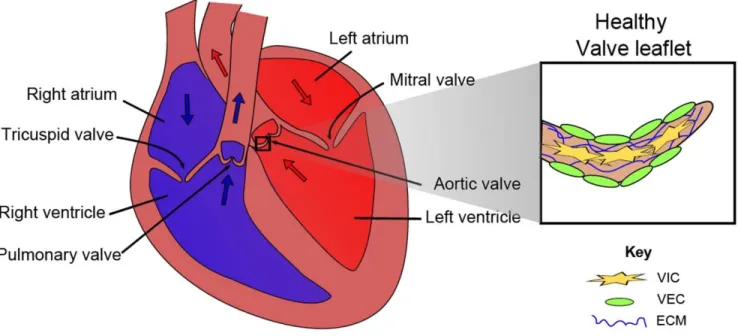
The aortic valve is a thin, tri-leaflet structure located at the base of the aortic root at the most distal portion of the left ventricular outflow tract. Functionally, the aortic valve serves to mediate the unidirectional flow of blood during the cardiac cycle, allowing flow from the left ventricle to the systemic vasculature during systole, and shutting to prevent backflow during diastole. The structure of the aortic valve is vital to maintaining its function. Human aortic valves consist of three histologic layers: 1) the fibrosa, which faces the aorta and is composed mostly of type I fibrillar collagen arranged circumferentially in parallel bundles in a matrix of elastin; 2) the spongiosa, which is the middle layer and is composed of glycosaminoglycans that act as shock absorbers for the valve; and 3) the ventricularis, which faces the left ventricle and is primarily composed of elastin fibers oriented radially [26].
Table 1 – Key abbreviations.
Figure 2.1 – Human heart and aortic valve leaflet anatomy. Adapted from Schroer AK and Merryman WD.
Mechanobiology of myofibroblast adhesion in fibrotic cardiac disease. Journal of Cell Science. 128 (2015): 1865- 75 [27].
Late stage CAVD is marked by obstruction of blood flow and aortic stenosis and has an estimated prevalence of 2% in patients between 70 and 80 years of age [28]. Prevalence of any aortic valve calcification is as high as 48% for those aged 75-76, and higher in 80–81 and 85–
86 year-old cohorts [29]. The incidence of this age-related disease is expected to grow dramatically in the next 25 years as the proportion of people over 65 in the United States nearly doubles [30]. Calcific aortic valve stenosis is the main indication for the over 100,000 valve replacements performed annually in the US and necessitates surgical intervention [31], which is currently the only therapeutic option [32]. Unfortunately, given the advanced age of many CAVD patients, this intervention carries with it a high rate of morbidity and mortality and is therefore not recommended in elderly or fragile patients, leaving many to suffer the ill effects of progressively worsening heart function as CAVD advances [33]. A better understanding of the biological mechanism driving the valvular calcification process might allow us to develop well-tolerated, non-invasive pharmacologic therapies.
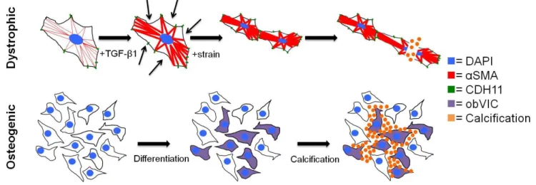
Once believed to be a passive process of degeneration, aortic valve calcification is now thought to be an active process of valvular remodeling mediated largely by the valve’s resident cells, AVICs [34]. AVICs are a heterogeneous population of fibroblast-like cells present in all three layers of the aortic valve and important in the structural maintenance of the valve, especially in the maintenance of the extracellular matrix (ECM) [35, 36]. Gross pathologic progression of CAVD is characterized by the formation of calcific nodules (CNs), which are cellular aggregates comprised of a mixture of calcium phosphate phases [37]. Two hypothetical mechanisms of CN formation exist: 1) TGF-β1 mediates activation of myofibroblasts, causing dystrophic calcification via apoptotic mechanisms [38], and 2) a population of AVICs spontaneously transdifferentiate into osteoblast-like cells and subsequently regulate mineralization (Figure 2.2) [39, 40]. In a study of human valves, 83% of the group demonstrated evidence of dystrophic calcification and 13% of those valves had mature lamellar bone and evidence of active bone remodeling [41]. It is unclear whether these processes occur simultaneously, sequentially, or independently. Therefore, we will selectively focus on elucidating the mechanism of the more prevalent dystrophic pathway [42].
Figure 2.2 – Proposed mechanisms of valve calcification. The dystrophic pathway is mediated by a TGF-β1 induced increase in αSMA and cadherin-11, which increases the cells’ contractility and strengthens their connections to each other. Under pathological strain, the increased and uneven tension tears cells apart, leading
to calcification via apoptosis. The osteogenic pathway proceeds by osteogenic differentiation into obVICs, likely from qVICs. These obVICs actively form mineralized deposits. Adapted from Bowler MA and Merryman WD. In vitro models of aortic valve calcification: solidifying a system. Cardiovascular Pathology. 24 (2015): 1-10. [25]
As a human ages, the aortic valve remodels: AVIC density and proliferation decrease, elastin content increases, and collagen fibers become more aligned. In CAVD, however, elastin is fragmented and overall content is decreased, while collagen content increases and becomes disorganized, contributing to valve leaflet thickening [43]. Remodeling of the ECM and subsequent stiffening that is characteristic of CAVD have been shown to regulate cellular processes [44]. For example, AVICs cultured in the presence of TGF-β1 on type I fibrillar collagen gels, fibrin-coated tissue culture plastic, or hydrogels of ~25 kPa formed osteogenic CNs, whereas nodules on ~120 kPa formed through the dystrophic pathway via myofibroblastic differentiation [45, 46]. This suggests that after the initiation of disease, a positive feedback between activation of AVICs and substrate stiffness exacerbates disease progression, at least in the case of dystrophic remodeling.
CDH11 and smooth muscle alpha-actin (αSMA) are two mechanically active proteins involved in dystrophic calcification. CDH11, a mechanosensitive transmembrane protein involved in cell-cell adhesion, is thought to be responsible for mediating much of the intercellular tension integral to the dystrophic pathway [47], and αSMA is involved in cell motility and intracellular contractility [48]. When these proteins are upregulated in the diseased state, individual AVICs experience increased and regionally heterogeneous tension and contraction, resulting in membrane tearing and apoptosis-mediated calcification. This cell death provides increased imbalance in cellular forces and a local increase in substrate stiffness, acting as a nucleation event in the formation of a CN [47, 49].
Both CDH11 and αSMA are overexpressed in diseased human valves, though previous in vitro work suggests that their relative expression levels are inversely regulated at the level of individual cells [50]. While cadherins are known to complex with a family of mechanical adapter proteins called catenins intracellularly, further downstream signaling events are poorly understood. A better understanding of CDH11 signaling and activation, and how it interacts with contractile proteins to mediate CNs, could uncover potential therapeutic targets for CAVD.
2.2 – Contribution of Aortic Valve Interstitial Cells to Calcific Aortic Valve Disease
Since the AVIC population is heterogeneous, the various subpopulations may contribute to CAVD pathogenesis differently depending on their function. While a definitive categorization of AVICs subpopulations has remained elusive, recent efforts to classify AVICs based on their observed phenotypic behavior has yielded five groups: embryonic progenitor
endothelial/mesenchymal cells, quiescent VICs (qVICs), activated VICs (aVICs), progenitor VICs (pVICs), and osteoblastic VICs (obVICs) [51]. VICs predominantly come from a population of endocardial cells lining the endocardial cushion which undergo endothelial to mesenchymal transformation (EMT) early in embryonic development. These cells are crucial in valve development and there is evidence that these progenitors participate in adult valve repair [51].
qVICs are responsible for maintaining physiological valve structure and function. While the exact function of qVICs is undefined, they are believed to regulate ECM homeostasis and inhibition of angiogenesis [51]. pVICs are considered valve stem cells and they are likely responsible for VIC proliferation in response to tissue injury. pVICs may originate from aortic valve endothelial cells (AVECs) that undergo an EMT-like process [51-53]. These EMT-related events are likely directly mediated by the mechanical forces present in the valve. In a recent study using chick explanted atrioventricular canals, EMT was found to occur preferentially in higher regions of strain [54].
This developmental process is likely recapitulated in an unregulated fashion during CAVD progression. This suggests that as the valve stiffens, more AVECs are transformed into pVICs and qVICs, setting the stage for subsequent activation.
aVICs are qVICs that have become myofibroblasts, characterized by αSMA expression and increased contraction. This activation occurs under pathological injury cues or abnormal mechanical stress via cytokines and growth factors produced by activated AVECs and macrophages [51]. aVICs are associated with increased ECM secretion and degradation, matrix metalloproteinase and tissue inhibitor of metalloproteinase expression, proliferation and migration, and secretion of cytokines including TGF-β1. If apoptotic pathways become abnormal, aVICs can lead to calcification; this is referred to as the dystrophic pathway. obVICs are VICs that have undergone osteoblastic differentiation and promote calcification in vitro. The addition
of organic phosphate to culture media induces this differentiation and subsequent calcification depends on the upregulation of alkaline phosphatase (ALP) activity. Adding bone morphogenic protein 2 (BMP2) and 25-hydroxycholesterol increases the rate of CN formation, as does TGF- β1, which induces calcification via an apoptotic mechanism [55]. BMP2 has been shown to be higher in stenotic human aortic valves [56] and upregulates osteogenic pathways involving Msx2 and Wnt signaling [57] and Runt-related transcription factor 2/core-binding factor subunit alpha- 1 (Runx2/CBFα1) [58]. It is likely that AVECs are regulating aVIC or obVIC function and that, given the presence in vivo of both BMP2 and TGF-β1, a combination of osteogenic and dystrophic pathways is occurring. Therefore, we are most concerned with the transitions to and behavior of obVICs and especially aVICs.
Porcine aortic VICs have also been categorized by morphology in vitro. After clonal expansion, Chen et al. defined four major subpopulations of VICs: 1) S-type are tightly connected, swirling fibroblast-like cells enriched for osteogenic progenitors (defined by presence of ALP in CNs), 2) loosely-packed fibroblast-like cells, 3) small, flat cells, and 4) large, flat cells [59]. More quantitative characterization of these subtypes was performed on porcine AVICs and pulmonary VICs (PVICs) via fluorescence-activated cell sorting (FACS). The percentage of VICs expressing most markers probed did not differ between AVIC and PVIC populations, though PVICs did express more SSEA4 (a marker of human embryonic stem cells, or pVIC-like) [60].
These characterizations may correlate well with some of the previously defined VIC subpopulations, but the contribution of CDH11-expressing VICs to calcification is unknown.
While AVICs are the primary cell type implicated in CAVD pathogenesis, it is likely that several other distinct native and non-native cell populations also play important roles. AVECs
sheath the surface of the leaflets and are oriented circumferentially and form a single cell monolayer, expressing Von Willebrand factor and nitric oxide (NO) [61-63]. Circulating cells have recently been implicated in the progression of calcification as well; elevated levels of endothelial progenitor cells with an osteoblastic phenotype and osteogenic precursor cells have been associated with severe and early heterotopic ossification, respectively [64, 65]. Early stages of CAVD develop lesions similar to those observed in atherosclerosis, suggesting a role for inflammatory signaling [66, 67]. Consistent with this observation are regularly observed elevated levels of macrophages and T-lymphocytes in human calcified aortic valves [41, 44, 68, 69].
These cell populations all contribute to CAVD progression, but it is likely that they influence AVIC behavior through the secretion of various bioactive agents.
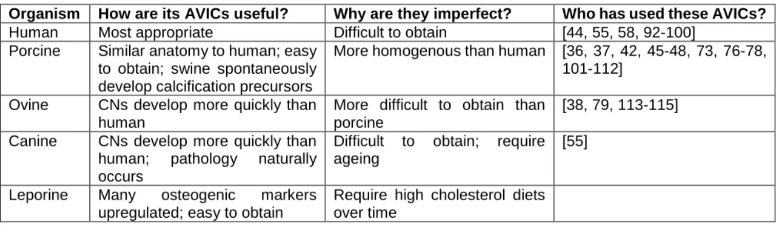
2.3 – The Appropriate Model Organism
The ideal in vitro model would use primary human AVICs, but availability is the chief limiter of using human-derived samples. The next best cell would retain all characteristics of the human cells important to CAVD. Since it is believed that the important mediators of calcification are AVICs, we can narrow our search to finding a species with AVICs comparable to human AVICs.
Non-human primates are a logical choice because of their genetic similarity. However, maintenance of these organisms requires more space, time, money, and permissions than other organisms. Likely for these reasons, non-human primate AVICs have not been isolated, though Macaca nemestrina aortic smooth muscle cells have been isolated to investigate proteoglycan expression [70]. Porcine hearts are both anatomically and physiologically similar to human hearts. The growth of the heart in swine from birth to four months is analogous to that in humans
from birth to mid-teens [71] and remodeling in atherosclerosis of micropigs closely resembles human pathology [72]. Interestingly, their valves contain the same αSMA-positive population of cells in the ventricularis [35]. Swine can also develop spontaneous valvular atherosclerotic lesions, a precursor to calcification [40, 73]. The first isolation of porcine AVICs noted that they appear more homogenous than murine or leporine VICs and had a high recovery rate after being frozen, leading to the extensive use of porcine AVICs in in vitro studies [74]. Though these cells are widely used and multiple research groups have reported calcification and mineralization, Cloyd et al. reported that porcine AVICs cultured in osteogenic media with TGF-β1 (which should activate both dystrophic and osteogenic pathways) did not form mineral deposits. They used Raman spectroscopy to show that even Alizarin Red-positive nodules did not exhibit mineralization [37]. While pig anatomy is highly similar to human anatomy, porcine AVICs in vitro is still a limited model. One important limitation specific to in vitro cell culture systems is the age of the cells. In 20% of long-term cell culture, AVICs become contact-inhibited monolayers and behave unstably [75]. Also, the metabolic activity of porcine AVICs was found to be passage number dependent [76]. Late-stage cultured AVICs demonstrated higher numbers of myofibroblasts [77, 78]. Thus, porcine AVICs are generally used no later than passage 7. Though porcine AVICs have limitations, they are the best available model.
Ovine AVICs have been shown to form CNs when treated with TGF-β1 within 72 hours, and to calcify, assayed via Alizarin Red staining, within two weeks [38, 79]. Canine AVICs were also considered early in the development of CAVD research [55]. Specifically, beagles demonstrate age-related changes in aortic valves, including calcification; changes were especially apparent in the fibroblasts, suggesting a similar mechanism to human calcification [80]. In vitro, canine AVICs spontaneously formed CNs containing hydroxyapatite over two to three weeks, compared to human AVICs developing nodules in about six weeks under the same
conditions [55]. Also, while an imperfect model, many similarities exist between canine and human myxomatous mitral valve disease, reinforcing the likeness between human and canine valves [81]. While canine AVICs were deemed very similar to humans’, they are not often used, likely as a function of convenience – dogs have longer life spans than small animal models and are not maintained at a large scale for another purpose, as pigs are for food. Rabbits are used for in vivo studies, but not as often in vitro, likely because they require high cholesterol diets to develop calcification [32, 82-84].
Mice are another popular model organism, perhaps because of their low cost, easy management, short life spans, and availability of genetic mutants. Murine cell lines can be easily immortalized, allowing for near indefinite expansion and use without regard for passage limitations. AVICs could be harvested from a variety of genetically-altered models such as ApoE-/-, Notch1+/-, and LDLr-/- [40, 85-90]. Though some of these models are the only ones to exhibit the hemodynamic effects of aortic valve stenosis, murine valvular structure is significantly different from human [40, 91]. Specifically, human valves have trilaminar structure, but murine valves only have a fibrosa and spongiosa [91]. While non-ideal, murine AVICs would provide a convenient model that facilitates genetic manipulation allowing for further exploration of CAVD mechanisms. A summary of the advantages and limitations of the AVICs derived from each model organism can be found in Table 2.1.
Table 2.1 – Examination of advantages and disadvantages associated with AVICs derived from common model organisms.
Organism How are its AVICs useful? Why are they imperfect? Who has used these AVICs?
Human Most appropriate Difficult to obtain [44, 55, 58, 92-100]
Porcine Similar anatomy to human; easy to obtain; swine spontaneously develop calcification precursors
More homogenous than human [36, 37, 42, 45-48, 73, 76-78, 101-112]
Ovine CNs develop more quickly than human
More difficult to obtain than porcine
[38, 79, 113-115]
Canine CNs develop more quickly than human; pathology naturally occurs
Difficult to obtain; require ageing
[55]
Leporine Many osteogenic markers upregulated; easy to obtain
Require high cholesterol diets over time
2.4 – Biochemical Cues Relevant to Calcific Aortic Valve Disease
A number of cytokines are known to modulate AVIC behavior, including inducing disease progression in vitro. Activation of the Wnt signaling pathway, involving Wnt3a, Wnt7a, and nuclear translocation of β-catenin, has been shown to promote calcification [116]. Wnt receptor LRP5 and β-catenin, factors in canonical Wnt signaling, also showed increased expression in diseased human aortic valves [117]. Both TGF-β1 and substrate stiffness have been shown to regulate signaling through MAPKs, p38 and extracellular signal-regulated kinase (ERK), both of which have been shown to promote the myofibroblast phenotype [118]. TNFα, interleukin1-β, and interleukin-6 (IL-6) have been shown to regulate Notch signaling [119], which enhances toll- like receptor 4 (TLR4) stimulation in human AVICs via nuclear factor kappa-B (NF-κB) [99].
Interestingly, IL-6 has been shown to be upregulated by CDH11 engagement [120]. In human AVICs, TNFα has been shown to accelerate calcification, assayed via ALP activity, Alizarin Red, and von Kossa [98]. Silencing TLR4 attenuates BMP2 expression, and stimulating TLR2 or TLR4 induces CN formation in human AVICs [97]. Receptor activator of NF-κB ligand (RANKL), a
surface-bound molecule of the TNF family, has also led to increased calcification in vitro [94, 121]. Together, these data support a role for inflammatory signaling in CAVD progression.
Porcine AVECs are able to inhibit AVIC calcification via NO secretion, inhibiting the differentiation to obVICs. Increasing the expression and activity of endothelial NO synthase (eNOS) in hypercholesterolaemic leporine aortic valves led to decreased calcification [122].
Additionally, blocking NO led to increased calcification even in 3D AVEC-AVIC co-culture [109].
Ex vivo culture of porcine aortic valve cusps in osteogenic media demonstrated significantly more CN formation on the fibrosa side than the ventricularis, which was exacerbated with NO inhibition. In healthy human valves, eNOS levels are much higher on the ventricularis than the fibrosa, further supporting the important protective effect of NO [109].
TGF-β1 is upregulated in diseased human valves and, when given exogenously in vitro, exacerbates nodule formation [38]. TGF-β1 has been shown to activate myofibroblasts in valves leading to increased αSMA expression via Smads and p38 [107, 123]. As these myofibroblasts become more contractile, they likely activate latent TGF-β1 from the ECM [124]. This positive feedback loop provides a strong potential mechanism for dystrophic disease progression. Some experiments have shown that fibroblast growth factor 2 (FGF-2) treatment can block nodule formation and matrix contraction of AVICs, effectively counteracting TGF-β1 treatment [79]. In addition, antagonism of 5 hydroxytryptamine receptor 2B (5HT2B), a TGF-β1-dependent cardiopulmonary serotonin receptor, has been shown to prevent myofibroblast differentiation and CN formation in porcine AVICs by inhibiting downstream TGF-β1-mediated signaling [123].
Another recent strategy is to target CDH11, a protein believed to mediate cell-cell tension in
CAVD and that has higher expression in calcified human valves; siRNA knockdown of CDH11 in vitro prevented TGF-β1-mediated CN formation [47].
2.5 – Mechanical Environment of the Aortic Valve
Many traditional CAVD in vitro studies have occurred in a static environment, but the valves exist in a dynamic mechanical state; this likely affects calcification mechanisms.
Interestingly, calcific lesions occur preferentially on the aortic side of the valve in the fibrosa, which is normally the stiffer of the two surfaces [125-127]. As the aorta stiffens with age, axial stiffening and circumferential compliance increase [128]; this results in higher mechanical loads placed on the circumferentially-aligned collagen fibers of the fibrosa, along which AVICs reside [129]. Also, an increase in transvalvular flow greater than 0.3 m/s per year is a clinical predictive marker for patients who might benefit from surgery, suggesting that increased flow contributes to pathological progression [130]. NO release by AVECs is regulated by flow; under laminar shear stress, NO is released and helps maintain valvular homeostasis via signaling to AVICs.
However, low and oscillating shear stress, as would occur on the aortic side of a diseased valve, inhibits this release [131]. Also, while the AVICs themselves are not directly exposed to fluid flow, it has been shown that flow alone can differentiate fibroblasts (the majority cell type of the AVIC population) into myofibroblasts [106]. This positive feedback of a stiffening valve that can no longer properly regulate its AVICs to maintain homeostasis is evidence of the importance of the dynamic environment on disease progression.
Substrate composition has also been shown to affect calcification. AVICs cultured on fibrin or tissue culture polystyrene exhibited significantly more CNs than on collagen, fibronectin,
or laminin [102]. In addition, the presentation of RGD to AVICs resulted in far more calcification than the presentation of YIGSR or DGEA. RGD, YIGSR, and DGEA are ECM-derived peptide sequences derived from fibronectin/fibrin/laminin/collagen, laminin, and collagen, respectively.
Their receptors are αvβ3/α5β1/α1β1 integrins, 67 kDa laminin receptor, and α2β1 integrin, respectively. Further investigation showed that disruption of the α5β1 integrin- or 67 kDa laminin receptor-mediated binding between AVICs and ECM results in increased calcification [102].
Fibronectin-coated tissue culture polystyrene suppresses calcification markers, while fibrin- coated tissue culture plastic enhances calcification as demonstrated by CN number, ALP activity, αSMA expression, CBFα1 expression, and calcium content via the o-cresolphthalein complexone method. However, both fibronectin and fibrin coating of soft hydrogels suppresses calcification [132]. This suggests that substrate stiffness may be more important than specific ECM component interactions. However, the method in which stiffness is modulated (i.e. by increasing crosslinking) is often coupled to the presentation of ECM components.
Several groups have begun probing CAVD progression using dynamic in vitro models.
Fisher et al. showed that CN formation is strain dependent and that strain drastically reduces the time to nodule formation – 48 hours versus three to 21 days [133]. At the tissue level, in a bioreactor under cyclic strain, porcine aortic valve cusps showed greater evidence of calcification under 15% (pathologic) strain than 10% strain (physiologic) [134]. In a related study of vascular calcification, 7% cyclic, equibiaxial strain yielded greater mineralization than unstrained calcifying vascular cells [135]. Strain alone is able to induce higher levels of myofibroblastic phenotype as measured by αSMA and collagen synthesis than untreated, unstrained cells, suggesting that strain exacerbates calcification via the dystrophic pathway [48]. In 3D cultures of porcine AVICs, osteogenic media alone was unable to induce calcification, but the addition of
mechanical stress via anchoring the gel led to significant calcification, as well as increases in αSMA, Runx2, and osteocalcin mRNA levels [109]. These studies demonstrate the critical role that mechanical stress and strain has on AVICs, and how such stress can lead to disease.
2.6 – Cadherin Signaling
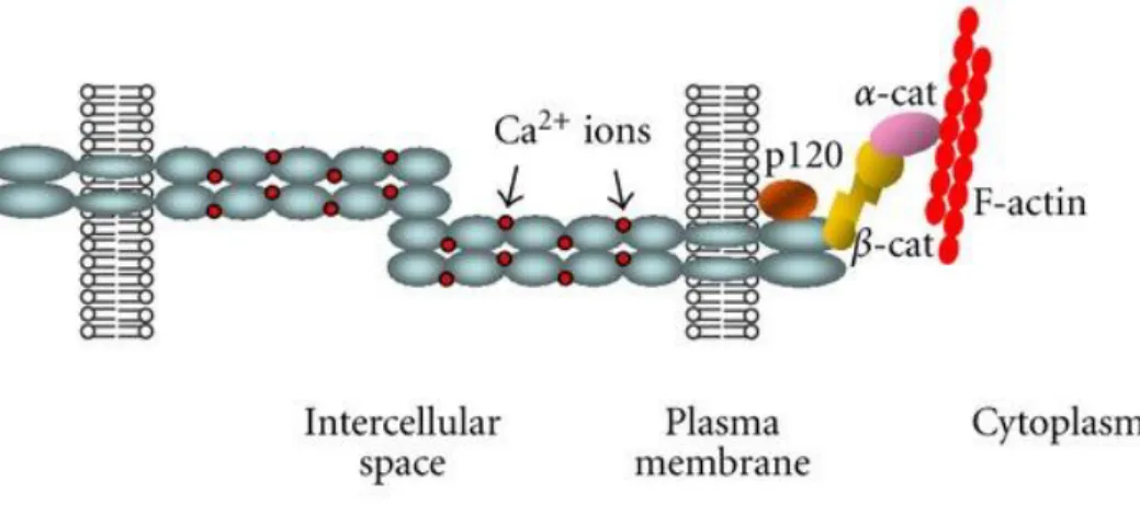
Cadherins are a family of single-pass transmembrane glycoproteins involved in calcium- mediated homotypic cell-cell adhesion. Their extracellular portion is comprised of highly homologous repeat domains of approximately 110 amino acids each and Ca2+ binding sites between each domain. Extracellular homotypic interactions can occur at the membrane of the same cell, termed cis interactions, or between neighboring cells, termed trans [136]. The intracellular region of cadherins is subdivided into a cytoplasmic binding domain and a juxtamembrane domain and is known to complex with β-catenin, p120-catenin, γ-catenin, and angiomotin. These provide connection to the cytoskeleton through α-catenin and are required for full strength homotypic bonds to persist. More functions of these associated proteins are discussed later but are poorly understood in the context of CAVD.
Five subgroups of cadherins exist. Type I classical cadherins (Cadherin-1, Cadherin-2, Cadherin-3, etc.) are defined by five extracellular domains, five Ca2+ binding domains, and short cytoplasmic domains. Type II atypical cadherins (Cadherin-5, Cadherin-7-12, etc.) are different from type I classical cadherins in that they lack an HAV (His-Ala-Val) adhesion recognition sequence [137]. Desmosomal cadherins bind to γ-catenin and desmoplakin and are linked to keratin intermediate filaments [138]. Flamingo cadherins have eight extracellular cadherin
regions, seven passes through the membrane, and their intracellular interactions are unknown [139]. Protocadherins are the largest subgroup and contain clustered and non-clustered types.
They are primarily observed in the developing nervous system, lack the adhesive sites of classical cadherins, and have unique loop structures [140].
Cadherins that complex with catenins, the classical subgroup, form a specialized type of adhesion junction called an adherens junction. β-catenin binds the cytoplasmic binding domain and regulates the extracellular bond strength while also providing physical interaction through α-catenin with the actin cytoskeleton [141]. Sequestration of β-catenin at the membrane also prevents its nuclear translocation and affects its involvement in canonical Wnt signaling and yes- associated protein 1 (YAP1). However, β-catenin can recruit RhoGEFs and/or compete with Arp2/3 [142]. α-catenin can regulate Wnt canonical signaling as well as Ras-MAPK and YAP1 [143]. p120-catenin binds the juxtamembrane domain and regulates cadherins’ persistence at the cell membrane. This is accomplished by p120-catenin masking an endocytotic signal as it is bound to the cadherin tail, therefore preventing internalization [144]. p120-catenin complexing with engaged cadherins locally down-regulates RhoA [145]. Also, when cadherins engage in a trans interaction, PIP3 accumulates, leading to local Rac1 activation [146]. Rho-associated protein kinase (ROCK) is required in epithelial cells for recruitment of myosin light chain II (MLC II) to adherens junctions [147]. p120-catenin normally inhibits transcriptional repressor KAISO and regulates Rho-GTPases and NF-κB signaling, so its sequestration at the membrane also has widespread effects on contractility [148]. Interactions between Wnt and p120-catenin signaling may prove relevant. p120-catenin complexing with cadherin-1 (CDH1) has been shown to control the sequestration of Wnt factor, glycogen synthase kinase-3 beta (GSK-3β) [149].
CDH11 has been shown to interact intracellularly with angiomotin in mammalian cells.
Angiomotin is a member of the Motin family and is involved in for cell polarity, migration, and the Hippo pathway. Deletion of the angiomotin binding domain of CDH11 resulted in weaker adhesion between L cells in an aggregation assay, providing evidence that angiomotin plays a role in regulating CDH11 binding strength. Deletion of the binding domain also demonstrated that angiomotin is required for CDH11-mediated migration [150]. Cadherins can also interact with receptor-type tyrosine kinases (RTKs) to promote many growth and proliferative signaling pathways; CDH1 associates with EGFR, cadherin-5 (CDH5) associates with VEGFR2, and cadherin-2 (CDH2) can stimulate FGFR signaling [148]. Collectively, these interactions provide a springboard for pathways to probe in AVICs.
Several cadherins have been observed to shed their extracellular domain (~75 kDa), leaving their transmembrane, juxtamembrane, and cytoplasmic domains available to subsequent cleavage by γ-secretase [151]. This shedding has been associated with diseases such as rheumatoid arthritis [151], pulmonary fibrosis [152], breast cancer [153], and the process of apoptosis [154, 155]. Different mechanisms have been reported for the initial cleavage of the cadherin ectodomain [152, 154, 156], but the enzyme responsible for CDH11 shedding has not yet been conclusively identified [151, 157]. The biological function of these shed ectodomains is not fully investigated, but there is some correlation with invasion and migratory behavior, and CDH11 engagement leads to upregulation of IL-6 secretion [151, 153, 156, 157]. Regardless, this phenomenon provides an interesting mechanism for paracrine signaling as well as a potential biomarker of disease.
Figure 2.3 – Classical cadherins forming an adherens junction between cells. Adapted from Gama, A. and F. Schmitt. Cadherin cell adhesion system in canine mammary cancer: a review. Veterinary medicine
international. (2012): 357187 [158].
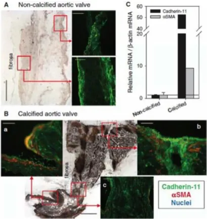
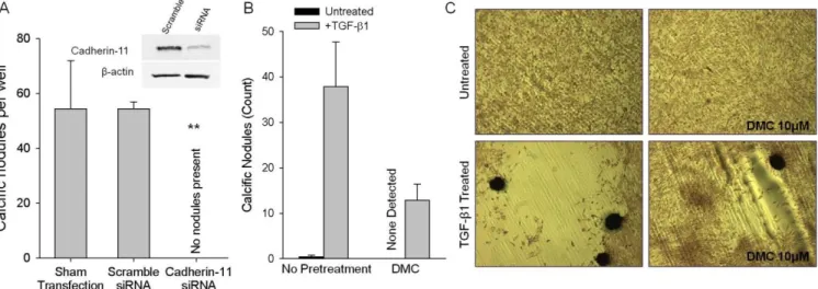
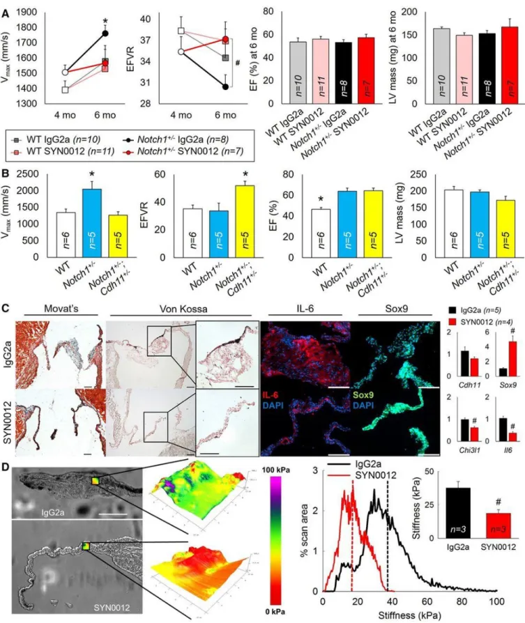
For this work, we focus on CDH11 because we have demonstrated its necessity for producing CNs in porcine AVICs in vitro and it is upregulated in human calcified valves (Figure 2.4). The Merryman Mechanobiology Laboratory has validated the importance of CDH11 expression and engagement in vitro as well. siRNA knockdown of CDH11 prevented the formation of CNs when porcine AVICs were subjected to TGF-β1 treatment and equibiaxial strain (Figure 2.5A) [47]. When AVICs were treated with dimethyl celecoxib (DMC), an inactive analog of the COX2 inhibitor, celecoxib, to block CDH11 homotypic interactions, the number of CNs formed was significantly reduced (Figure 2.5B-C) [159]. Additionally, a murine model of heritable CAVD, the Notch1+/- mouse, has higher CDH11 expression in its AVICs [160] and a CDH11 overexpression mouse demonstrates evidence of CAVD [161]. Together, these findings support an important role of CDH11 in CAVD.
Figure 2.4 – Calcified human valves display elevated CDH11 and αSMA. CDH11 and αSMA protein expression (A, B) and transcription (C) is upregulated in diseased human aortic valves. Adapted from Hutcheson JD, Chen J, Sewell-Loftin MK, Ryzhova LM, Fisher CI, Su YR, and Merryman WD. Cadherin-11 regulates cell-cell
tension necessary for calcific nodule formation by valvular myofibroblasts. Arteriosclerosis, Thrombosis, and Vascular Biology. 33 (2013). [47]
Figure 2.5 – CDH11 is required for CN formation. CDH11 expression knockdown (A) or blocking CDH11 engagement (B) each prevents CN formation (C). (A) is adapted from Hutcheson JD, Chen J, Sewell-Loftin MK, Ryzhova LM, Fisher CI, Su YR, and Merryman WD. Cadherin-11 regulates cell-cell tension necessary for calcific nodule formation by valvular myofibroblasts. Arteriosclerosis, Thrombosis, and Vascular Biology. 33 (2013). [47]
CDH11, or osteoblast-cadherin, was identified in 1994 in a murine osteoblastic line and differs from its human variant at only 17 amino acids [162]. It is an atypical classical cadherin, meaning it contains the five extracellular domain repeats, but no HAV sequence. In addition to expression in the heart, CDH11 has been identified at high levels in reproductive tissues as well as smooth muscle, lung, and cerebral cortex [163, 164]. CDH11 overexpression is associated with the early stages of breast cancer as well as gastrointestinal, brain, and central nervous system tumors. It is also a target for rheumatoid arthritis, a common inflammatory disease [165].
CDH11 is unique from other cadherins because of several key characteristics. Homotypic bonds formed by CDH11 are two-fold stronger than those formed by CDH2, another mesenchymal cadherin, and also far stronger than bonds formed by CDH1 and CDH5 [166].
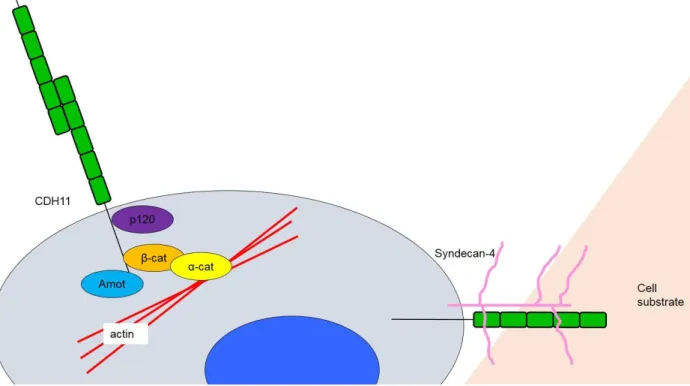
CDH11 is also the only cadherin known to participate in focal adhesions, making regulation of cell-substrate interactions as well as the classical cell-cell interactions relevant. In both primary human fibroblasts and neural crest cells, CDH11 (but not CDH2 or C-cadherin) was observed to localize to focal adhesions. Langhe et al. demonstrated the requirement of CDH11’s transmembrane and cytoplasmic domains in concert with the extracellular portion of syndecan- 4 to adhere to fibronectin [167]. Syndecan-4 is a transmembrane heparan sulfate proteoglycan of ~20 kDa that binds fibronectin and can regulate focal adhesion formation through Rho family GTPase and protein kinase C [168-170].
Figure 2.6 – CDH11 is a unique cadherin. CDH11 uniquely participates in both cell-cell and cell-substrate interactions.
2.7 – Notch1 Signaling
Mutations in Notch1 in humans have been correlated with an increased risk of aortic stenosis [171], and Notch1 has recently been shown to be silenced by long noncoding RNA H19 in cases of idiopathic CAVD [172], making it a relevant signaling pathway to disease. Notch signaling is a highly conserved system comprised of four notch receptors in mammals (Notch1- 4). The receptor is a single-pass transmembrane protein that binds to ligands Delta-like and Jagged and can then be cleaved via γ-secretase. This causes release of the intracellular domain which translocates to the nucleus and can affect cell maintenance, proliferation, and apoptosis [160]. Notch1 haploinsufficiency has also been shown to result in higher CDH11 expression in murine AVICs, providing another link between CDH11 and CAVD. Figure 2.7 summarizes recent findings in the crosstalk between Notch1 and CDH11 in the context of CAVD [160].
Figure 2.7 – Mechanism of Notch1+/- AVIC activation. Adapted from Chen J, Ryzhova LM, Sewell-Loftin MK, Brown CB, Huppert SS, Baldwin HS, and Merryman WD. Notch1 mutation leads to valvular calcification through enhanced myofibroblast mechanotransduction. Arteriosclerosis, Thrombosis, and Vascular Biology. 35 (2015).
[160]
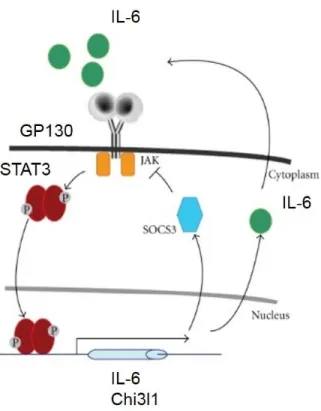
2.8 – Interleukin-6 Signaling
CDH11 engagement is known to increase the secretion of IL-6, suggesting an inflammatory role of CDH11 as well. IL-6 is a complex ~25 kDa cytokine, acting in both pro- and anti-inflammatory capacities. Typically secreted by macrophages, fibroblasts, and endothelial cells, IL-6 can also be secreted by osteoblasts, smooth muscle cells, and adipose tissue [173].
IL-6 complexes with IL-6Rα and glycoprotein 130 (GP130), resulting in a signaling cascade
through Janus kinases (JAKs) and signal transducers and activators of transcription (STATs) (Figure 2.8). Chitinase-3-like protein 1 (Chi3l1) is downstream of STAT3 and is a secreted inflammatory glycoprotein with unclear function, but association with a variety of cardiovascular diseases [174, 175]. It has also been shown to regulate IL-6-mediated STAT3 phosphorylation, suggesting positive feedback that could exacerbate inflammation [176]. IL-6 has been found to play a role in diseases such as rheumatoid arthritis, atherosclerosis, and prostate cancer [177- 179]. Many anti-IL-6 therapeutics are under investigation, but the first to be FDA-approved is tocilizumab for rheumatoid arthritis, Castleman’s disease, and systemic juvenile idiopathic arthritis [180-182].
Figure 2.8 – IL-6 signaling axis. IL-6 complexes with GP130 to signal through JAKs and phosphorylate STAT3.
STAT3 then causes the transcription of IL-6 and Chi3l1, which can itself cause phosphorylation of STAT3 through IL-6, providing a positive feedback loop of inflammatory signaling. Adapted from Ozawa Y, Kurihara T, Tsubota K,
and Okano H. Regulation of posttranscriptional modification as a possible therapeutic approach for retinal neuroprotection. Journal of Opthalmology. (2011): 506137. [183]
2.9 – Considerations for Evaluation of In Vitro Calcification
Calcium Assays
Evaluation of valve calcification can be separated into two categories: direct, in which the level of calcium or mineralization is directly measured, and indirect, in which markers of the proposed dystrophic and/or osteogenic pathways toward calcification are measured. Direct evaluation has the advantage of determining whether the assay leads to a pathological outcome functionally, whereas the indirect measurements yield more mechanistic information (Table 2.2).
Direct evaluation techniques include von Kossa staining [39, 47, 93, 94, 98, 101, 133, 184-186], Alizarin Red staining [37, 46, 47, 79, 84, 92, 97, 98, 101-103, 106-109, 111, 112, 115, 133, 186-188], energy-dispersive X-ray spectroscopy (EDS) [39, 189], Raman spectroscopy [37, 190, 191], scanning electron microscopy (SEM) [37, 84, 189], transmission electron microscopy (TEM) [37, 96, 189], atomic absorption [115, 192], arsenazo III [84, 133, 184], and o- cresolphthalein complexone [94, 102, 115] measurements. While these are all used as measures of calcification, not all are perfectly specific and thus are often used in concert. The gold standard for calcium detection is atomic absorption spectroscopy. Atomic absorption spectroscopy is based on the principle that different elements absorb different wavelengths of light and it works by atomizing the sample, sending light usually from a hollow cathode lamp of a specific wavelength through the vaporized sample, and measuring the amount absorbed [193].
Samples with increased mineralization content exhibit higher absorbance levels compared to controls.
Probably the most common measure of calcification, Alizarin Red, or 1,2-
magnesium, manganese, barium, strontium, and iron, forms complexes with the dye in a chelation process, and results in a birefringent stain. Calcium is usually in much higher concentration than the other elements, allowing the inference that the areas stained have calcium present. Alizarin Red is often used to stain CNs to verify their mineralization and to help quantify the nodule assay, either by making the nodules easier to count or by extracting the dye for more rigorous quantification. Typical methods for quantifying the amount of dye involve staining of the cells or tissue, washing extensively, extracting via acetic acid or cetylpyridinium chloride, neutralization with ammonium hydroxide, and colorimetric detection at 405nm or 550nm. The acetic acid-ammonium hydroxide method is three times more sensitive than the cetylpyridinium method and results in a better signal to noise ratio, especially for weakly stained samples [46, 194]. This method is also advantageous over Arsenazo III quantification because it has a higher and wider linear range of detection [194].
Von Kossa is another common stain for mineralization, especially in tissue sections. The stain works by reducing the calcium ions with light and replacing them with silver deposits that appear dark grey or black in tissue [195]. This method is not specific for calcium phosphates [196], though it has been suggested that the yellow precipitates are specific [197]. Von Kossa can be further confused if performed on a C57BL/6 mouse, which has melanocytes that appear black in the aortic valve. Thus, von Kossa is performed frequently in combination with Alizarin Red staining.
Calcium content can be measured more directly by various methods, but it is important to note that these methods all require lysing of the sample, meaning that calcium from mineralized areas or calcific lesions is not differentiated from intracellular calcium. Arsenazo III is a metallochromogen that complexes with calcium at pH 6.75 without interference from any
other cations commonly present in serum or plasma, and is measured at 650nm [198]. When compared with the o-cresolphthalein complexone method, accuracy and calibration stability increased [199]. The o-cresolphthalein complexone method involves a reaction of Ca2+ ions with o-cresolphthalein complexone in an alkaline solution (8-Hydroxyquinoline at pH 10.6) and reading the sample absorbance at 660nm [200]. While these methods do not have a range of detection as large as Alizarin Red quantified via the acetic acid-ammonium hydroxide method, they are still useful for samples with low levels of calcium.
Other elemental methods include SEM, TEM, and Raman spectroscopy. SEM yields topographical and compositional information about the sample’s surface with a resolution on the order of nanometers. It can be performed on fixed, dehydrated, and gold-/platinum-/or carbon- sputter-coated samples or in wet conditions via environmental SEM (ESEM) [201]. TEM yields information about the sample’s chemical identity based on how it absorbs electrons and has a resolution on the order of picometers [202]. It can be performed on fixed, dehydrated, and stained samples. EDS analysis allows one to determine particular elements and their proportions in the sample. It functions on the principle that different elements will absorb different energy X- rays and the amount absorbed corresponds to the amount of element present [203]. EDS can be performed during SEM and ESEM; the advantage to using ESEM is that the samples do not have to be coated and high accelerating voltages can be used. EDS performed during ESEM is better because of the lack of interference from the coating and because the lack of sample preparation yields more authentic data. EDS coupled with ESEM yields quantitative data as well as qualitative [201, 203]. Raman spectroscopy is unique in that it can be performed on live cells.
This allows calcification to be measured over time. Raman has also been shown to be an effective diagnostic for human heart valve calcification. Given the appropriate training data, an
algorithm based on spectral shifts could predict whether the tissue was calcified with 100%
sensitivity and specificity [190, 191].