THOMAS PHEU A Field Guide to the
^f Cidaroid Echinoids
^f of the Northwestern : Atlantic Ocean,
Gulf of Mexico, and the Caribbean Sea
SMITHSONIAN CONTRIBUTIONS TO ZOOLOGY • 1970 NUMBER 40
SERIAL PUBLICATIONS OF THE SMITHSONIAN I N S T I T U T I O N The emphasis upon publications as a means of diffusing knowledge was expressed by the first Secretary of the Smithsonian Institution. In his formal plan for the Insti- tution, Joseph Henry articulated a program that included the following statement:
"It is proposed to publish a series of reports, giving an account of the new discoveries in science, and of the changes made from year to year in all branches of knowledge not strictly professional." This keynote of basic research has been adhered to over the years in the issuance of thousands of titles in serial publications under the Smithsonian
imprint, commencing with Smithsonian Contributions to Knowledge in 1848 and continuing with the following active series:
Smithsonian Annals of Flight Smithsonian Contributions to Anthropology
Smithsonian Contributions to Astrophysics Smithsonian Contributions to Botany Smithsonian Contributions to the Earth Sciences
Smithsonian Contributions to Paleobiology Smithsonian Contributions to ^oology Smithsonian Studies in History and Technology
In these series, the Institution publishes original articles and monographs dealing with the research and collections of its several museums and offices and of professional colleagues at other institutions of learning. These papers report newly acquired facts, synoptic interpretations of data, or original theory in specialized fields. Each publica- tion is distributed by mailing lists to libraries, laboratories, institutes, and interested specialists throughout the world. Individual copies may be obtained from the Smith- sonian Institution Press as long as stocks are available.
S. DILLON RIPLEY Secretary
Smithsonian Institution
ZOOLOGY NUMBER 40
Thomas Pheian A F i e l d G u i d e to the
Gidaroid Echinoids of the Northwestern Atlantic Ocean,
Gulf of Mexico, and the Caribbean Sea
SMITHSONIAN INSTITUTION PRESS CITY OF WASHINGTON
1970
ABSTRACT
Phelan, Thomas. A Field Guide to the Cidaroid Echinoids of the Northwestern Atlantic Ocean, Gulf of Mexico, and the Caribbean Sea. Smithsonian Contributions to Zoology, 40:1-67. 1970.—Twelve species of cidaroid echinoids from the northwestern Atlantic Ocean, Gulf of Mexico, and the Caribbean Sea are described, compared, illustrated, and keyed for identification. The first description of the denuded test of Histocidaris nuttingi Mortensen is presented. A lectotype and paralectotype are selected for Histocidaris sharreri (A. Agassiz). Poriocidaris purpurata (Wyville Thompson), previously known from the eastern Atlantic, is reported for the first time from the Caribbean Sea.
Official publication date is handstamped in a limited number of initial copies and is recorded in the Institution's annual report, Smithsonian Year.
UNITED STATES GOVERNMENT PRINTING OFFICE WASHINGTON : 1969
For gale by the Superintendent of Documents, U.S. Government Printing Office Washington, D.C. 20402 - Price $1.25 (paper cover)
Thomas Phelan A Field Guide to the Cidaroid Echinoids of the Northwestern Atlantic Ocean,
Gulf of Mexico, and the Caribbean Sea
Introduction
The quest for knowledge of the sea and the study of marine organisms have been attracting an ever in- creasing number of biologists, students, and serious hobbyists. The primary interest may not be systema- tics, but the identity of specimens often is essential to other works. Identification is frequently hampered by lack of readily available literature. Indeed, many of the descriptions are scattered through out-of-print publications. This paper is the first of a series to pro- vide researchers who are not echinoid specialists with a field guide to the species of echinoids known to inhabit the northwestern Atlantic Ocean, Gulf of Mexico, and the Caribbean Sea. Twelve species of cidaroid echinoids are compared and keyed for identification.
The differences between some of these species are not as distinct as suggested in the literature. Therefore, in addition to presenting a key for identification, the variations in form that cause difficulty in identifica- tion are discussed. This study is based on material in the United States National Museum (USNM) and the Museum of Comparative Zoology (MCZ), Harvard.
Thomas Phelan, Division of Invertebrate Paleontology, Smithsonian Institution, Washington, D.C. 20560
Acknowledgments
The author thanks Dr. David Pawson and Miss Maureen E. Downey of the Division of Echinoderms, United States National Museum for the use of speci- mens and a friendly exchange of ideas; Professor Barraclough Fell, Museum of Comparative Zoology, Harvard, for the loan of specimens. I am sincerely grateful to Dr. Porter M. Kier for valuable advice and encouragement all of which were generously given.
Diagnostic Features of Cidaroid Echinoids
Echinoids are of many forms, each representing adap- tation to a particular niche in the marine environ- ment. Some echinoids are almost entirely radially sym- metrical; others have strong bilateral features yet re- tain some radial symmetry. Echinoids with the mouth and anus at opposite poles of a vertical axis and almost exclusively radially symmetrical are commonly called sea urchins or regular echinoids. The cidaroids are sea urchins with distinctive features that readily set them apart from other kinds.
The more or less globular main portion of the skele- ton termed the test or corona is composed of twenty vertical columns of calcareous plates. In the cidaroids and most other sea urchins these plates are rigidly joined together. The vertical columns of plates are arranged in similar pairs to form ten distinct sections.
Five sections of the test consist of perforate plates.
These alternate with five sections composed of imper- forate plates (Plate 1: figure 7; Plate 2: figures 1, 2).
Two columns of perforate plates form one ambulac- rum. In the cidaroids each ambulacral plate bears one pair of pores to accommodate a tube foot. The pores are arranged in a single vertical sinuate series (Plate 2: figures 1, 2). The single series of pore pairs and simple plates are not restricted to the cidaroids.
Other sea urchins commonly have simple plates, but many noncidaroids have compound plates with several pore pairs per plate. The pore pairs of noncidaroids are arranged in single series, multiple series, or in a series of arcs. Toward the ambulacral midline adjacent to each pore pair there is a marginal tubercle on the cidaroid echinoids that also forms a vertical series (Plate 2: figure 6), small inner tubercles are scattered or arranged in series between the two series of mar- ginal tubercles. There are no large primary tubercles with accompanying large primary spines in the ambulacra.
Each of the five sections consisting of two vertical columns of imperforate plates is termed an interambu- lacrum. Interambulacral plates of cidaroid echinoids have one large primary tubercle per plate which is crenulate (Plate 11: figures 1, 3) or noncrenulate (Plate 17: figure 5). This tubercle commonly bears a large primary spine. The largest spines are at or just above the equator or ambitus of the test (Plate 4: fig- ure 6; Plate 9: figure 2). Noncidaroid echinoids bear one or more large primary spines per interambu- lacral plate. A large, smooth, somewhat circular area devoid of surface ornamentation is present in the region around each primary tubercle. This consists of the boss or expanded base of the primary tubercle and the sur- rounding somewhat depressed ring or areole, which serves for primary spine muscle attachment (Plate 2:
figure 1). Beyond the areole the plate is covered with small secondary tubercles. Those bordering the areole are slightly larger than the others, commonly forming a distinct scrobicular ring (Plate 6: figures 3, 4). The tubercles of the scrobicular ring carry scrobicular spines, specialized secondary spines (Plate 5: figure 6).
Among the noncidaroids only the salenioid echinoids have a similar interambulacral plate. The salenioids are distinguished by other features of the test which are discussed in the following paragraphs.
At the apex of the test is a complex of plates called the apical system (Plate 2: figures 4, 8). There are five
ocular plates, one situated at the adapical end of each ambulacrum. Each ocular plate bears an ocular or terminal pore. Five additional plates each with a geni- tal pore are situated at the adapical ends of the inter- ambulacra. The genital plates are larger than the ocu- lars, and the genital pore is also larger than the terminal pore. One of the genital plates has a porous sievelike area, the madreporite, commonly slightly larger than the other genital plates. These ten plates are collec- tively termed the oculogenital ring.
The oculogenital ring surrounds a flexible mem- brane, the periproct, in which the anus is located.
Periproct outline and plate arrangement are important diagnostic features. The periproct of a cidaroid echi- noid is always pentagonal and bears plates decreasing in size toward the centrally located anus (Plate 2:
figures 4, 8). This is a distinct feature of the cidaroids.
The salenioids that most closely resemble the cida- roids have the anal opening off-center due to the pres- ence of a large suranal plate absent in the cidaroids.
There is a large opening on the underside or ad- oral side of the denuded test. Prior to the loss of tissue this opening is covered with plates and a flexible membrane, the peristome (Plate 1: figures 7, 8; Plate 11: figure 2), with the mouth located in the center.
In cidaroid echinoids imbricating ambulacral plates,
B
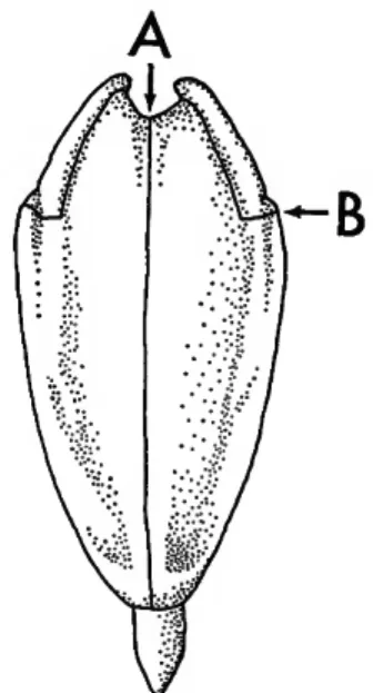
FIGURE 1.—The top of the junction of pyramid halves (A) is higher than the lowest part of the epiphyses (B) in a cidaroid lantern. The reverse is true in noncidaroid lanterns. See PLATE 1: figures 2, 3.
and to some extent interambulacral plates, extend across the peristome to the mouth (Plate 1: figure 7).
Interambulacral plates bordering the peristome of cidaroids internally bear apophyses (Plate 1: figure 5), the main supports for the masticating device com- monly called the Aristotle's lantern or simply the lan- tern (Plate 1: figure 2). Noncidaroid sea urchins have as the main lantern supports auricles that are internal projections from the ambulacral plates (Plate 1: figure 6).
The cidaroid lantern contains five jaws, termed pyr- amids, each with an unkeeled tooth (Plate 1: figure 1).
A keel is a projection on the inner surface of the tooth that extends throughout the length of the hard portion (Plate 1: figure 4). Noncidaroids have un- keeled or keeled teeth.
Each pyramid (Figure 1; Plate 1: figure 2) consists of two demipyramids which join along the dental slide. The junction surface is called the symphysis.
A notch on each side of the top of a pyramid accom- modates a lantern member termed an epiphysis. In
cidaroid echinoids the uppermost part of the junc- tion of pyramid halves (Figure 1A), symphysis, is higher than the upper limit of the lateral wings at the junction with the epiphyses (Figure 1B). The re- verse is true on noncidaroid sea urchins (Plate 1:
figure 3).
Cidaroids have no gills, but all other rigid-test sea urchins including the salenioids have gills accompa- nied by a notch in each basicoronal interambulacral plate. The notch opens toward the peristome. Gill notches may be deep, as on Tripneustes ventricosus
(Lamarck) (Plate 1: figure 8), or very shallow, as in Arbacia punctulata (Lamarck). The absence of gill notches is a diagnostic feature of the cidaroids (Plate 1: figure 7).
A list of significant cidaroid and noncidaroid char- acters is provided for quick reference. Some of the cidaroid features listed are not restricted to cidaroids;
Those restricted to cidaroids are in boldface type.
Only a few significant noncidaroid characters are presented.
list of Significant Characters
Test:
(General)
Ambulacra:
Cidaroid Rigid, regular sea urchin form (periproct within apical system) lacking gill notches
Plates simple
Pore pairs in single series Sinuate to some extent Marginal tubercle adjacent to each pore pair
Noncidaroid
Interambulacra:
Apical System:
Peristome:
Teeth:
Pyramids:
One primary tubercle and spine per plate
Primary tubercles perforate Apophyses are the prominent lantern supports
Periproct pentagonal Covered with a regular series of imbricating plates in the ambu- lacra, and to some extent in the interambulacra
Unkeeled
Upper limit of symphysis is higher than the top of the lateral wings at the junction with the epiphyses
Rigid, regular sea urchin form (periproct within apical system) with gill notches
Flexible, regular sea urchin form Rigid, irregular echinoid form (periproct outside apical system) Plates compound
Pore pairs in multiple series, or in a series of arcs
Large primary tubercles and spines
Auricles are the prominent lantern supports
More than one primary tubercle and spine per plate
Primary tubercles imperforate
Suranal plate
One pair of perforate plates in line with each ambulacrum
Keeled
The top of the lateral wings at the junction with the epiphyses is
higher than the upper limit of the symphysis
The Diagnostic Value of Gidaroid Pedicellariae Pedicellariae, small pincerlike organs, defend the ech- inoid from small organisms, especially pelagic larvae seeking a place for attachment The pedicellariae are scattered all over the test among the spines (Plate 8:
figure 8). Most pedicellariae consist of three jawlike valves at the distal end of a flexible stalk which contains a calcareous supporting rod. Pedicellariae with valves containing venomous glands are termed globiferous pedicellariae (Figures 2-6; Plate 19: figures 6, 7) and are recognized by the presence of a chamber within each valve to accommodate the venom gland. This chamber has an opening (Figure 2B) at the distal end of the valve blade. The relative size of the blade open- ing, its position, and tooth configuration are diagnos- tically important.
The term tridentate is applied to nonvenomous pedi- cellariae commonly with three relatively long-bladed valves (Figure 7; Plate 15: figure 1). One species discussed in this paper has only two valves in each of its tridentate pedicellariae (Plate 10: figure 2). Only the large globiferous (Figures 2-6) and large trident- ate pedicellariae (Figure 7) are used in this work as aids in identification.
FIGURE 3.—The large globiferous pedicellariae of Tretocidaris have a large single end tooth and a very small blade opening.
A short closed space is between the blade opening and the end tooth.
FIGURE 2.—The large globiferous pedicellariae of Cidaris and Calocidaris have a large single end tooth (A) and a large blade opening ( B ) . A short closed space is between the blade opening and end tooth.
FIGURE 4.—The large globiferous pedicellariae of Eucidaris and Stylocidaris lack a large single end tooth at the tips of the blades. The blade opening is terminal.
FIGURE 5.—The large globiferous pedicellariae of Stereoci- daris lack a large single end tooth at the tips of the blades.
The blade opening is subterminal. See Figure 6.
FIGURE 6.—The two uppermost teeth of the blade opening are commonly larger than the other teeth surrounding the blade opening on the large globiferous pedicellariae of stereocidaris. These two larger teeth commonly coalesce giv- ing the illusion of a single end tooth, but there is no closed space between these teeth and the blade opening as in Cidaris and Calocidaris.
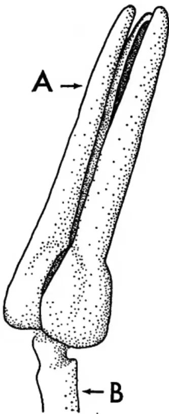
FIGURE 7.—A three-valve tridentate pedicellaria. The blade (A) is the slender upper portion of each valve. The valves lack the venom chambers present in globiferous pedicellariae.
The valves of a pedicellaria are attached to a calcareous sup- porting rod ( B ) .
B
Diagnostic features of Cidaroid Pedicellariae
No globiferous pedicellariae
Histocidaris Poriocidaris
Large globiferous pedicellariae have a single large end tooth, closed space between end tooth and blade opening
Blade opening large, Figure 2
Cidaris Calocidaris
Blade opening very small, Figure 3
Trelocidaris
No large single end tooth
Blade opening terminal, Fig- ure 4
Stylocidaris Eucidaris
Blade opening subterminal, Figures 5, 6
Stereocidaris
Diagnostic features of the large tridentate pedicellariae on the histocidarids
Valves very large and broad, Plate 12:
figure 5
H. sharreri
Valves very large, long, and slender, Plate 15:
figure 1
H. nuttingi
All pedicellariae composed of two highly compressed valves, Plate 10:
figure 2
Poriocidaris purpurata
Techniques
The large globiferous pedicellariae of Cidaris and Calocidaris are best studied with the valves separated and the tissue removed. This is easily accomplished using a solution of 10 parts water to 1 part household bleach, and the process takes approximately 15 minutes to complete. The valves should then be rinsed in water.
The calcite lattice structure of an echinoid is trans- lucent and therefore does not photograph well in re- flected light. In order to overcome this problem, the
dry specimens were coated with red ink, but alizarin crimson watercolor or any low-sediment dye can be used. After the ink dried thoroughly, the specimen was lightly coated with ammonium chloride from a "smoke generator" (Teichert, 1948, p. 102). In Plate 1: figure 8, only the area around the edge of the peristome was treated. Notice how vividly the gill slits stand out in the photograph compared to the tubercles in the lower right-hand corner.
Key to the Cidaroid Echinoids of the Northwestern Atlantic Ocean, Gulf of Mexico, and the Caribbean Sea
la. Tubercles strongly crenulate, crenulation on some or most tubercles even below the ambitus;
peristomial ambulacra! plates with an internal prolongation (Plate 10: figure 3 ) ; glo- biferous pedicellariae lacking; oral primary spines very strongly serrate, distinctly curving toward mouth Histocidaris, Poriocidaris 2 b. Not as above 3 2a. All pedicellaria bivalve (Plate 10: figure 2), oral primaries with blunt rounded tips (Plate 11:
figure 6). Primary spines white, uppermost primaries of some specimens have a long brown or purple collar, spines cylindrical or gently tapering. Secondary spines brown or purplish brown; ambulacral inner tubercles sparse (Plate 12: figures 6, 7 ) ; few interambulacral secondary tubercles (Plate 11: figure 3) Poriocidaris purpurata (Wyville Thomson)
Key to the Cidaroid Echinoids of the Northwestern Atlantic Ocean, Gulf of Mexico, and the Caribbean Sea—Continued
b. Oral primary spines have a slender tip (Plate 15: figure 6 ) ; large tridentate pedicellariae with long slender blades (Plate 15: figure 1 ) ; all spines white or nearly so; test commonly white; marginal tubercles of two sizes; larger form distributed in a zigzag pattern with accompanying larger marginal spines (Plate 14: figure 5 ) ; ambulacral midzone with an abundance of inner tubercles Histocidaris nutlingi Mortensen c. Oral primaries rather short with blunt broadly rounded tips (as in P. purpurata, Plate 11:
figure 6 ) ; primary spines white, secondaries brown (Plate 13: figures 1-3); tridentate pedicellariae with large broad valves (Plate 12: figure 5 ) ; ambulacrum with very few inner tubercles (Plate 12: figures 8, 9, 11); interambulacral plate with few secondary tubercles (Plate 14: figures 1, 2) Histocidaris sharreri (A. Agassiz) 3a. Large globiferous pedicellariae with distinct end tooth (Figures 2, 3) 4 b. Large globiferous pedicellariae without the single end tooth (Figures 4-6) 6 4a. Blades of large globiferous pedicellariae with large opening (Figure 2 ) ; areoles usually large, deep; upper tubercles of some specimens weakly crenulate 5 b. Blades of large globiferous pedicellariae with very small opening (Figure 3 ) ; areoles shallow, upper side of upper tubercles strongly crenulate (Plate 21: figure 4 ) ; primary spine with light and dark bands, spinules larger on upper than underside of primary spines (Plate 21:
figures 5-7; Plate 22: figures 1, 2) Tretocidaris bartletti (A. Agassiz) 5a. Test relatively globular interambulacral midzone slightly sunken (Plate 2: figures 2, 8 ) ; extrascrobicular tubercles of upper interambulacral plates in horizontal rows separated by furrows (Plate 2: figures 1, 2 ) ; primary spines very long smooth (lacking spinules) and shiny (Plate 2: figures 3, 5; Plate 3: figures 1, 4, 7 ) ; scrobicular (Plate 2: figure 7) and marginal spines slender, long, with pointed tips Calocidaris tnicans (Mortensen) b. Test commonly flattened above (Plate 4: figure 6 ) ; many uppermost tubercles rudimentary (Plate 5: figures 1, 2 ) ; scrobicular ring of tubercles inconspicuous, extrascrobicular tubercles of upper interambulacral plates in horizontal rows separated by furrows (Plate 5:
figures 1, 2 ) ; primary spines fairly smooth not shiny, usually white, cylindrical or tapering, spinules noticeable on adoral primary spines but commonly reduced or lacking on those of the ambitus and above, large primary spines directed out horizontally, tip commonly slightly flaring to form a small hollow "hoof (Plate 4: figure 1)
Cidaris abyssicola (A. Agassiz) c. Test slightly flattened above; few rudimentary tubercles, areoles very large; extrascrobicular area very limited, secondary tubercles very small (Plate 6: figure 3 ) ; primary spines with well-developed spinules, those above the ambitus commonly flared to paddle shape (Plate 8:
figures 1 , 2 ) ; oral primaries commonly with moderate serration
Cidaris blakei (A. Agassiz) d. Test slightly flattened above, few rudimentary tubercles, areoles large, extrascrobicular area with fairly large secondary tubercles (Plate 6: figure 4 ) ; sutures not naked; primary spines (Plate 10: figure 1) 2 to 2.5 times the horizontal diameter of the test, commonly swollen beyond the short collar (Plate 8: figure 7 ) , tapering to the tip, rows of spinules well developed; a few of the largest spines on some specimens have a flat widening of the tip (Plate 8: figure 6), oral primary serration slight to lacking
Cidaris rugosa (H. L. Clark) 6a. Test low flattened, very few rudimentary tubercles; ambulacra midline and all interambulacral sutures very naked; 5 to 7 interambulacral plates per row; primary spines 1.5 to 2 times the horizontal diameter of the test, round slender, only very gently tapering toward the tip, almost cylindrical, many directed upward but not vertically; moderately developed rows of spinules 7 b. Test low, adapically flattened (Plate 22: figure 5 ) ; midline of ambulacra not naked; inter- ambulacra with 5 to 7 plates in a row, interambulacral transverse sutures not naked, interambulacral midline sutures with only a slight trace of nakedness; primary spines 1.5 to 2 times the horizontal diameter of the test, directed out horizontally, well-developed rows of spinules, uppermost row commonly forming a conspicuous crest (Plate 22:
figures 8, 9) Stereocidaris ingolfuma Mortensen 359-863—69-
Key to the Gidaroid Echinoids of the Northwestern Atlantic Ocean, Gulf of Mexico, and the Caribbean Sea—Continued
c. Test commonly flattened adapically; ambulacra! midline not naked; interambulacra with 9 to 12 plates per column, areoles rather small, shallow (Plate 17: figure 5 ) , inter- ambulacral sutures not naked; primary spines stout, short, slightly longer than the hori- zontal diameter of the test, but may be less, primary spine spinules are wartlike, a dense covering of hair commonly hides the spine shaft, crownlike tip (Plate 16: figures 2-4;
Plate 17: figures 1-3). Scrobicular spines with very broad blunt tip (Plate 16: figure 1).
Eucidaris tribuloides (Lamarck) 7a. Round granules scattered rather uniformly on all apical system plates (Plate 19: figure 1 ) ; ambulacral midline and all interambulacral sutures very naked and white (Plate 19:
figure 3; Plate 18: figure 6 ) ; scrobicular and marginal spines with reddish midline stripe (Plate 19: figure 3 ) ; primary spines on some specimens banded (Plate 18: figure 6;
Plate 20: figure 4) Stylocidaris affinis (Philippi) b. Naked reddish-brown ring in apical system, granules elongate, directed away from both
sides of the reddish-brown ring (Plate 19: figures 2, 5 ) ; ambulacral midline and all inter- ambulacral sutures very naked and reddish-brown (Plate 19: figure 4 ) ; scrobicular spines white without midline stripe (Plate 19: figure 4) Stylocidaris lineata Mortenscn
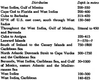
Distribution
Only a general listing of distribution is provided be- cause collecting has been insufficient and reports have
been hampered by inaccuracies due to misidentifica- tion of specimens.
Species Calocidaris micans Cidaris abyssicola Cidaris blakei Cidaris rugosa Eucidaris tribuloides Histoddaris nuttingi Histocidaris sharreri Porioeidaris purpurata Stereocidaris ingolfiana Stylocidaris affinis
Stylocidaris lineata Tretocidaris bartletti
Distribution West Indies, Gulf of Mexico Cape God to Florida and West Indies Cuba to Barbados
32°N off U.S. east coast, south through West Indies
Throughout the West Indies, Gulf of Mexico, and Bermuda
Cuba to Antigua Leeward Islands
South of Ireland to the Canary Islands and Caribbean Sea
North Atlantic Denmark Strait to Cape Verdes and the Caribbean Sea
Bermuda, West Indies, Caribbean Sea, and Gulf of Mexico, eastern Atlantic and the Mediter- ranean Sea
West Indies
West Indies, Caribbean
Depth in meters 200-330 200-400 315-420 130-540 littoral to 450 320-415
?—570—?
750-1800 300-1750 30-1000
100-500 140-625
Calocidaris micans (Mortensen) PLATE 2: FIGURES 1-8; PLATE 3: FIGURES 1-7
Calocidaris micans.—For a complete synonymy see Morten- sen, 1928, p. 312; for an additional description see Mor- tensen, 1910, p. 2.
The test of Calocidaris micans is relatively high, up to 0.75 of the horizontal diameter of the test. I t is gen- erally globular in appearance when viewed from the side; the test is not flattened above or below (Plate 2 :
figure 2). T h e peristome and apical system maintain the regular curving of the sides. The basicoronal plates are not incurving at the edge of the peristome.
The ambulacral interporiferous zone in the ambital region contains four series of almost equal-size tuber- cles; an inner tubercle and a slightly larger marginal tubercle on each plate (Plate 3 : figure 6 ) . On com- plete specimens the ambulacral midline is only scarcely naked.
The interambulucral midzone is sunken as there is
a sloping of the extxascrobicular area from the areole to the midline suture. Extrascrobicular tubercles are arranged in almost horizontal rows separated by distinct furrows. Areoles are large, the scrobicular ring reaching the upper and lower edges of all but the up- permost plates. Areole confluence is generally limited to the two lower plates of each column. On some specimens the upper side of the upper tubercules and spines are slightly crenulate (Plate 2: figures 1,5). The scrobicular ring is somewhat inconspicuous, because the scrobicular tubercles are only slightly larger than the adjacent extrascrobicular tubercles.
The primary spines are without doubt the most diagnostic feature of C. micans. They are almost cylindrical, gently tapering at the tip, smooth and shiny, completely lacking hairs and spinules. Primary spines above the ambitus have approximately 16 nar- row glassy zones running the length of the shaft. These are so slightly elevated above the rest of the spine that they are not noticeable to the unaided eye. Below the ambitus these glassy zones form inconspicuous glassy ridges completely lacking spinules. All other cidaroids develop rather conspicuous spinules on the primary spines below the ambitus; even those that have rather smooth spines above the ambitus. The most spectacular specimens of C. micans have primary spines up to three times the horizontal diameter of the test
(Plate 3: figures 1, 7), but on others they are com- monly one and a half times the horizontal diameter of the test. The primary spines are almost white, but there is a reddish-brown pigment in the collar, slight olive color in the neck and shaft, and a trace of reddish brown toward the tip. Specimens give the overall ap- pearance of being white, but close observation reveals a slight olive tint.
COMPARISON WITH OTHER SPECIES.—Indentification difficulties would most likely be limited to differentia- tion between C. micans and some specimens of Cidaris abyssicola. Cidaris abyssicola is noticeably flattened adapically and somewhat adorally as there is a slight incurving of the test at the edge of the peristome.
The primary spines are fusiform or cylindrical, white, somewhat chalky in appearance. Spinules are common- ly small, even lacking on some spines. The primary spines of some specimens are very smooth and some- what shiny. The various kinds of spines may be present on a single specimen. Below the ambitus of C. abyssi- cola the primary spines develop rows of spinules even on specimens with smooth shiny upper spines. This
contrasts with C. micans which lacks spinules even on primary spines below the ambitus. Spines below the ambitus on C. micans bear low smooth glassy ridges that extend the length of the shaft.
The form of C. abyssicola that Mortensen (1910, p. 13) described as C. abyssicola var. teretispina (Plate 5: figure 3) has slender, usually cylindrical, smooth, and quite shiny primary spines and a faint olive pig- ment. The primary spines at and above the ambitus are therefore quite indistinguishable from those of the shorter spine specimens of C. micans. Below the am- bitus the primary spines bear spinules as on other speci- mens of C. abyssicola. The scrobicular and secondary spines are very similar to the slender pointed ones of C. micans. The slender smooth spine specimens of C.
abyssicola commonly have some primary spines terminating with a "hollow-hoof tip similar to those found on some spines of the more typical specimens
(Plate 4: figure 1).
I have not observed Mortensen's variety C. abyssi- cola teretispina with a globular test similar to that of C. micans. The more typical C. abyssicola with stouter white spines has been observed with a rather globular test and elevated apical system (Plate 5: figure 2).
Rudimentary tubercles occur on fairly large inter- ambulacral plates on C. abyssicola (Plate 5: figure 1), but they are apparently limited to smaller plates on C.
micans (Plate 2: figure 8).
DISTRIBUTION.—West Indies and Gulf of Mexico at depths of 200-330 meters.
Cidaris abyssicola (A. Agassiz)
PLATE 1: FIGURE 7; PLATE 4 : FIGURES 1-6; PLATE 5 : FIGURES 1 - 6
Cidaris abyssicola—For a complete synonymy see Mortensen, 1928, p. 301; for additional descriptions see Mortensen, 1910, p. 13, and 1928, p. 301.
The test of Cidaris abyssicola is commonly rather low with a flattened apical system and peristome (Plate 4:
figure 6). The basicoronal plates are slightly incurving at the edge of the peristome. At the ambitus the pe- riphery is circular with no sunken area in the inter- ambulacra. The flattened test is not a reliable diagnos- tic feature as some specimens are quite globular with elevated apical system (Plate 5: figure 2) and gently curving sides instead of the strongly arched condition found in the more flattened specimens.
The ambulacra are slightly sinuate, and the marginal
10
tubercles are in regular series. The inner tubercles are variously arranged on different specimens. There may be one small inner tubercle below center on each ambulacral plate or as many as three inner tubercles are common. The arrangement with three inner tuber- cles is generally one above the other adjacent to the marginal tubercle and the third below the center of the plate near the midline suture. The lower edge of the plate commonly bears several small granules. The inner tubercles tend to form in uniform series, but in some specimens they are rather irregularly arranged.
Specimens with inner tubercles in regular series high on the test may have them in a jumble near the peri- stome (Plate 5: figures 4,5). The arrangement of inner tubercles is not a diagnostic one for this species. It is more significant that the interporiferous zone is rather crowded with tubercles, while not obscuring the mid- line as in Stereocidaris ingolfiana.
The upper plates of the interambulacra of most specimens are slightly higher than wide and many bear rudimentary primary tubercles (Plate 5: figure 1). The areoles are moderately large and the scrobic- ular rings reach the upper and lower edges of the plates everywhere below the ambitus. The areoles are confluent in the two or three lowermost plates of a column. Extrascrobicular tubercles are in more or less horizontal rows separated by furrows and in much greater abundance above than below the ambitus.
The primary spines are directed out horizontally as spokes around the hub of a cartwheel (Plate 4: figure 6). The large primary spines are commonly rather smooth, spinules are small or lacking. These spines are generally white, even chalky in appearance and from 1 to 1.5 times the horizontal diameter of the test, cylindrical or fusiform. A "hollow hoof is com- mon on spines with unbroken tips (Plate 4: figure 1).
Below the ambitus the primary spines develop more prominent spinules.
The test and spines are generally white, but touches of pink and olive are common. The collar of the pri- mary spines is generally the darkest portion of the specimen. Scrobicular spines of some specimens have a slight olive midline, but not a stripe or as distinct as the red midline stripe of Stylocidaris affinis.
COMPARISON WITH OTHER SPECIES.—Cidaris abys-
sicola is rather distinct and should not be difficult to identify, but since some specimens bear superficial re- semblance to Calocidaris micans a comparison is nec- essary. A more detailed comparison is given under Calocidaris micans. The large primary spines of C.
micans are very smooth and shiny. Most specimens of Cidaris abyssicola have fairly smooth but not shiny large primary spines, but there are some specimens of C. abyssicola that have more slender rather shiny spines with a slight olive tint. These specimens of C. abyssi- cola can be distinguished from specimens of C. micans by examination of the primary spines below the am- bitus with a hand lens. Spinules can be observed on these spines of C. abyssicola, but only longitudinal glassy ridges are present on the primary spines below the ambitus on C. micans. The oral primary spines of C. abyssicola generally have distinctly serrate edges.
The oral primaries of C. micans lack serrations but have well-developed longitudinal ridges and thin edges (Plate 3: figure 3). The midzones of the interambu- lacra of C. micans are sunken. They are not sunken on C. abyssicola.
DISTRIBUTION.—East coast of the United States south of Cape Cod, including Florida, and the West Indies at depths of 200 to 400 meters.
Cidaris blakei (A. Agassiz)
PLATE 6: FIGURES 1, 3, 5 ; PLATE 7: FIGURES I, 6 - 8 ; PLATE 8: FIGURES 1-5, 8; PLATE 9: FIGURES 1, 2
Cidaris blakei.—For a complete synonymy see Mortensen, 1928, p. 307; for additional descriptions see A. Agassiz, 1883, p. 10, and Mortensen, 1928, p. 307.
The test of Cidaris blakei is moderately to slightly flat- tened above and below (Plate 7: figure 1), and its height is approximately two-thirds the horizontal di- ameter. The test is round at the ambitus, the inter- ambulacral midzones are not sunken. There is a slight incurving of the basicoronal plates at the edge of the peristome.
The ambulacra are distinctly sinuate above the am- bitus, less sinuate below. This is apparently due to the very large areoles on the interambulacral plates above the ambitus. The pore pairs are slightly oblique and the ambulacral marginal tubercles are high on each plate, tending to crowd above the inner edge of the in- ner pore that is low on the plate. Small granules are common on the lower edge of the plates below the marginal tubercles which are in uniform series. Ad- jacent to the marginal tubercle is an inner tubercle low on the plate. This inner tubercle and the marginal tubercle lie in an oblique position very nearly parallel to the oblique position of the pore pair of the same plate. If an additional inner tubercle is present on the
NUMBER 40
plate, its position is commonly at random but crowd- ing the midline edge of the plate. Although I have not observed it, Mortensen (1928, p. 307) reported the additional inner tubercles as being in a uniform series. The inner tubercles adjacent to the marginal tubercles are in a uniform series.
The areoles are large and occupy a major portion of the interambulacral plates. Above the ambitus the areoles are very large and only very newly introduced plates have rudimentary tubercles. The areoles of the two or three lowermost plates are commonly con- fluent. Crenulation is not diagnostic but is weakly present in the uppermost tubercles of some specimens.
The scrobicular ring of tubercles is very distinct. These tubercles are considerably larger than the almost granular secondary tubercles (Plate 6: figure 3).
The uppermost primary spines of some specimens resemble the fan-shaped leaves of the ginkgo tree (Plate 8: figures 1, 2; Plate 9: figure 2). On such specimens the great widening of the spine tip is progressively decreased with descending position of the spines toward the adoral side. Spines below the am- bitus are quite slender and lack the widened tip. The variation among specimens is remarkable and ranges from those as shown in Plate 8 to specimens possessing only slender-pointed tipped spines. Indeed, specimens with little or no widening of the spine tip are common.
Some slender spines bear a very small flared tip, like a funnel cone. The spinules are easily visible but not thorny. The oral primary spines are rather distinctly serrate. The secondary spines are small, slender, and almost pointed, and contrast with the broader more bluntly tipped scrobicular spines.
The slender, only slightly flattened, almost pointed marginal spines of the ambulacra, slender extra- scrobicular spines, and the almost granular size of the extrascrobicular tubercles are very diagnostic and are the most significant features of specimens lacking the fan-shaped spines. The very large areoles are impor- tant features also, but C. rugosa has large areoles and resembles some specimens of C. blakei.
The color was reported by A. Agassiz (1883, p. 12) as being brilliant vermilion. Most of the color has been lost from specimens stored in alcohol or dried. The spines may have been white, for they show no trace of color in the shaft.
COMPARISON WITH OTHER SPECIES.—Cidaris blakei
and Cidaris rugosa have been collected together at several localities and can be easily identified when they
bear their highly characteristic spines (Plate 8: figures 1, 2, 7), but some specimens of these species have only slender primary spines similar to those of Stylocidaris affinis (Plate 20: figure 4). Specimens that lack the distinctive spines may be identified by examining the extrascrobicular area of the interambulacral plates, the granular tubercles of the apical system, and the tips of the marginal spines. The extrascrobicular area of C.
blakei occupies a smaller percentage of the plate and is covered with more delicate secondary tubercles than on C. rugosa (Plate 6: figures 3, 4). The granular tubercles of the apical system of C. blakei are also smaller and less prominent than those of C. rugosa
(Plate 6: figures 5,6).
The inner tubercles of the ambulacra are commonly in more uniform series in C. blakei. On specimens of C. rugosa with a single inner tubercle on each ambula- cral plate a partial vertical overlap of tubercles occurs and a single zigzag series is formed along the midline (Plate 7: figure 4). Large specimens of C. rugosa com- monly have inner tubercles of the ambulacra one above the other adjacent to the marginal tubercles of a single plate (Plate 7: figure 9). This condition may exist on a majority of the plates with additional inner tubercles toward the midline. These specimens tend to show a uniform series of inner tubercles. The inner tubercle arrangement is variable and therefore of little help in identification.
The marginal spines of C. rugosa are noticeably broader and more bluntly tipped than those of C.
blakei (Plate8: figures 8,9).
DISTRIBUTION.—Cuba to Barbados in the West In- dies at depths of 315 to 420 meters.
Cidaris rugosa (H. L. Clark)
PLATE 6: FIGURES 2, 4, 6; PLATE 7: FIGURES 2-5, 9; PLATB 8: FIGURES 6, 7, 9; PLATE 10: FIGURE 1
Cidaris rugosa.—For a complete synonymy see Mortensen, 1928, p. 305; for additional description see H. L. Clark, 1907, p. 210.
The test of Cidaris rugosa is moderately to slightly flat- tened above and below (Plate 7: figure 2). Height of the test is approximately two-thirds the horizontal diameter. Only rarely are the basicoronal plates incur- ving at the edge of the peristome. The test is round at the ambitus with no sunken interambulacral midzone.
The ambulacra are moderately sinuate; only slightly more noticeably so above than below the ambitus. Pore pairs are in a slightly oblique position on the plates and
12
the marginal tubercles are higher than center, but the tubercles only tend to crowd above the inner pores adorally. The marginal tubercles form a uniform series and bear fairly broad, noticeably flattened, bluntly tipped spines (Plate 8: figure 9). Small granules are common below the marginal tubercles. There is no naked area in the midzone between the rows of mar- ginal tubercles. When only one inner tubercle is present on each ambulacral plate, it occupies most of the area between the marginal tubercle and the midline suture.
The appearance of crowding is evident. In the above condition the inner tubercles partially overlap each other along the midline suture forming a single zigzag series (Plate 7: figure 4). On larger specimens addi- tional inner tubercles are present. Adjacent to the mar- ginal tubercle there are commonly two inner tubercles, one above the other (Plate 7: figure 9). These tend to form into a uniform series. An additional inner tubercle is commonly present on the plate crowding the midline and the vertical pair of inner tubercles.
This arrangement on C. rugosa gives the appearance of a more uniform series of inner tubercles than on those with a single inner tubercle in a zigzag series. The lack of any naked area in the midzone is the most significant feature of the interporiferous zone. The variability of inner tubercle arrangement exempts their number and position from being a good diagnostic feature.
Each interambulacral plate has a large areole which occupies a major portion of the plate. Rudimentary tubercles are limited to newly introduced plates. The scrobicular rings are prominent and extend to the upper and lower edges of the plates except on the uppermost plates of the test The two lowermost plates commonly have confluent areoles. The extrascrobic- ular tubercles are prominent, standing out on the plate as rugged little knobs. This feature markedly dis- tinguishes the interambulacral plates of C. rugosa from those of C. blakei (Plate 6: figures 3, 4). The small granular tubercles of the apical system are also prom- inent (Plate 6: figure 6).
Primary spines of the more typical specimens are from 2 to 2.5 times the horizontal diameter of the test; markedly swollen just above the collar, round, and tapered to the tip. The spinules are prominent but not thorny (Plate 8: figure 7). The swollen area above the collar is absent on some specimens, and the spines then have a more slender appearance. Some of
the longer spines on some specimens have a flattened slightly expanded tip resembling the less expanded spines of C. blakei (Plate 8: figure 6). The oral pri- maries vary from nonserrated to moderately serrated.
The marginal, scrobicular, and extrascrobicular spines are flattened, rather broad and blunt tipped (Plate 8:
figure 9). The scrobicular spines may be so blunt as to appear truncated.
The color is lost from most dried or alcohol speci- mens but was reported as more or less rose red or brick red adapically by H. L. Clark (1907, p. 210).
COMPARISON WITH OTHER SPECIES—Cidaris rugosa
is compared with C. blakei in detail following the de- scription of that species. Cidaris rugosa is readily distinguished from Stylocidaris affinis, Stylocidaris line- ata, and Tretocidaris bartletti by the lack of naked su- tures, especially along the ambulacral midline. The large globiferous pedicellariae differ also. Cidaris ru- gosa has a large opening below the large end tooth, S.
affinis and S. lineata lack an end tooth. Tretocidaris bartletti has a large end tooth but below it is a very small opening. Neither C. rugosa nor Stereocidaris in- golfiana has naked ambulacral sutures; S. ingolfi- ana lacks the large single end tooth on the large globif- erous pedicellariae, commonly has a crest or wing on the upper side of the primary spines, and a more jum- bled arrangement of ambulacral inner tubercles.
Cidaris rugosa is readily distinguished from C. abys- sicola by the large number of rudimentary tubercles and smoother primary spines of C. abyssicola.
DISTRIBUTION.—From 32 degrees off the United States east coast through the Greater Antilles and Les- ser Antilles as far as Barbados. It is known from depths of 130-540 meters.
Eucidaris tribuloides (Lamarck)
PLATE 1: FIGURES 1, 2, 5 ; PLATE 16: FIGURES 1-5; PLATE 17: FIGURES 1-7; PLATE 18: FIGURES 1-3
Eucidaris tribuloides.—For a complete synonymy and addi- tional description see Mortensen, 1928, p. 400.
The test of Eucidaris tribuloides is moderately flattened above and below (Plate 17: figure 5). There is a mod- erate incurving of the basicoronal plates at the edge of the peristome. The test at the ambitus is commonly cir- cular, but numerous specimens are subpentagonal and a small percentage pentagonal. The radius in the pen-
tagonal specimens is greatest in the interambulacral midzone and shortest in the ambulacra. The flattening of the test above and below is relatively more promi- nent on small specimens than on large ones of 50 mm or more. The apical system is commonly slightly larger than one-third of the horizontal diameter of the test and approximately the same size as the peristome. On small to medium-size specimens the apical system may be noticeably smaller than the peristome. The madre- porite is commonly enlarged and prominent on de- nuded specimens.
The ambulacra are only slightly sinuate (Plate 17:
figures 4, 5). Marginal tubercles are prominent, the bosses on some specimens contiguous and transversely oval, arranged in uniform series. Inner tubercles crowd the interporiferous zone and the crowding exists re- gardless of the presence of one, two, or three inner tubercles per plate. Two inner tubercles per plate are the most common, but there is only a very slight tend- ency for the inner tubercles to form in series.
Interambulacral plates in the region of the ambitus are approximately twice as wide as high and have shal- low small areoles. The tubercles of the scrobicular ring are noticeably more prominent than the extrascrobicu- lar tubercles of the interambulacral midzone (Plate 17:
figure 5). There is less difference in size between the scrobicular tubercles and the extrascrobicular tubercles bordering the ambulacra (Plate 17: figures 4, 5). In the interambulacral midzone the extrascrobicular tu- bercles are in somewhat horizontal rows separated by furrows. The large number of primary tubercles in vertical series is an important diagnostic feature (Plate 17: figure 5). A specimen 35 mm in diameter com- monly has 9 to 10 primary tubercles in a vertical series.
The primary spines are short, from about half the horizontal diameter of the test to only slightly larger than the horizontal diameter (Plate 16: figures 2-4).
They are fairly stout, either cylindrical or noticeably tapered. The tip of the spine (Plate 17: figure 1) has a crown formed by lamellae, each of which is at the tip end of a row of warts (Plate 17: figures 1, 2). At the center of the crown is a small prominence. Except when very small, specimens lack pointed spinules. The shaft of each spine is covered by a dense spongy cover- ing of anastomosing hairs. The extent of development of this covering varies between individuals. Under water those with a very dense covering appear to have
a coat of fur (Plate 17: figure 3). The neck of the primary spines is very short and very difficult to ob- serve on nondenuded specimens. Oral primaries are oval in transverse outline, only slightly different from transitional and ambital primaries. The scrobicular spines are broad, straight sided. The tips are very blunt and concave, almost shovellike on some specimens
(Plate 16: figure 1).
The large globiferous pedicellariae lack an end tooth and are similar to the kind shown in figure 4.
The specimens are brown, commonly darkest at the collar and tips of the marginal and scrobicular spines.
The brown pigment of the primary spines is commonly obscured by the gray or white covering of anastomosing hairs. The denuded test is olive; the areoles white or almost so.
Eucidaris tribuloides is usually found in fairly shal- low water, but its depth range extends into the upper limits of the distribution of Stylocidaris affinis. The two species are commonly collected at the same station, especially off the west coast of Florida. Very small specimens of both species are strikingly similar but can be differentiated by the following features.
Both species have light and dark banding of the primary spines with well-developed thorns. Close ex- amination of the dark bands of E. tribuloides will re- veal that some of the thorns have already developed into typical warts (Plate 18: figure 3). Observe the dark bands nearest the base of the spines as this is where the first warts appear. As the individual grows, wart development progresses more rapidly in the dark bands than the light ones, and a stage is reached where the thorns remain only in the light bands (Plate 18:
figures 1-3). These also change to warts and the color banding usually leaves. The crownlike tip develops on at least one primary spine even on extremely small in- dividuals of E. tribuloides (Plate 18: figures 1-3). One or two very broad tipped large scrobicular spines will be present even on the very small specimens (Plate 18:
figure 2).
The thorns on the primary spines of S. affinis remain and, as the spines grow, simply become the spinules.
The scrobicular spines of even the smallest specimens of S. affinis have the prominent reddish midline stripe
(Plate 18: figure5).
DISTRIBUTION.—West Indies, Gulf of Mexico, and Bermuda. Littoral to 450 meters.
14
Histocidaris nuttingi Mortensen
PLATE 14: FIGURES 3 - 5 ; PLATE 15: FIGURES 1-7
Histocidaris nuttingi.—For a complete synonymy sec Morten- sen, 1928, p. 98; for additional descriptions see Morten- sen, 1926, and 1928, p. 98.
The test is broad and quite flattened adapically (Plate 15: figure 7), widest high on the test, well rounded above the ambitus, more tapering adorally with slight to no incurving of the basicoronal plates. The horizon- tal outline is round.
The marginal tubercles are in series but not of the normal size gradient. Size and arrangement of these tubercles and the attached spines are important diag- nostic features. A denuded ambulacrum viewed from the horizontal position shows larger tubercles alternat- ing down the two marginal series in a zigzag pattern among smaller tubercles of the series (Plate 14: figure 5). This zigzag pattern is present on all four speci- mens available for study. The larger marginal tuber- cles bear proportionately longer slender spines. The smaller marginal tubercles are very nearly equal in size to the inner tubercles. So abundant are the inner tubercles that they and their slender spines obscure the midline suture.
Ambulacral plates of the peristome bear the internal prolongation common among the histocidarids (Plate 10: figure 3).
The widest interambulacral plates are approxi- mately twice as wide as high. Crenulation is strong even on tubercles below the ambitus. At the ambitus and above, the large shallow areoles crowd the adoral transverse plate sutures distorting the curvature of the lower side of the areole and scrobicular ring (Plate 14:
figure 4). So severe is the crowding that some of the scrobicular tubercles are actually projections of the lower edge of the plate situated in line with the suture.
The extrascrobicular area on each side of the scro- bicular ring bears numerous secondary tubercles. Those on the midline suture side of the plates are noticeably arranged in nearly horizontal rows (Plate 14: figure 4).
The apical system is quite flat and has an abundance of tubercles scattered over its surface. Tubercles on the genital plates are most dense around the genital pores.
The very large genital pores of the MCZ specimen in Plate 14: figure 3 suggest that it is a female. This spec- imen (MCZ 7729a) is approximately 75 mm in diam- eter and is almost the same size as the USNM speci- men collected by RV Oregon at station 6699. The
USNM specimen has much smaller genital pores which are difficult to observe due to the density of secondary spines. A few very small plates are on the genital pore membranes of the female MGZ 9927a. Most of the small plates bear tubercles the same size as the tuber- cles on the genital plates.
The primary spines are quite varied on each individ- ual as can be seen in Plate 15: figures 2-6. Some of the spines are remarkably long. A medium-size specimen 35 mm in horizontal diameter (MCZ 7731) bears a spine 150 mm in length. The ratio of spine length to test diameter is greater in medium-size specimens than in large ones. The shaft of the spines is smooth, shiny and white, cylindrical to gently tapering to the tip.
Thorns are commonly scattered along the shaft at inter- vals of about 5 mm (Plate 15: figures 2, 3) and arranged in three or four rows which are not easily recognized unless viewed from the end of the spine.
The spines may terminate in a rather blunt tip or especially on the larger specimens the tips may be swollen or flared due to the growth of narrow expanded longitudinal ridges limited to the tip region (Plate 15:
figure 4). Transitional spines are strongly serrate and curve adorally (Plate 15: figure 5). Oral spines of the basicoronal plates taper to a relatively slender tip, curve adorally, and are markedly serrate (Plate 15: figure 6).
The large tridentate pedicellariae are diagnostic.
They are very large and easily seen with the unaided eye and have long slender blades (Plate 15: figure 1).
The tips of the blades commonly terminate with slightly different lengths (Plate 15: figure 1). Mor- tensen (1926, p. 6) suggested that this is probably due to the tips breaking and regrowth. As with other his- tocidarids, H. nuttingi possesses no globiferous pedicel- lariae.
The medium-size specimens are all white, but the larger specimens have white spines and some yellowish to brownish pigment in the test.
The three histocidarids are compared and discussed following the description of Poriocidaris purpurata (Wyville Thomson).
Mortensen (1926) wrote his original description of H. nuttingi without actual observation of the single specimen available at the time. Dr. Nutting of the University of Iowa furnished Mortensen with photo- graphs, spines, pedicellariae, and a description of the specimen. The holotype is presumed lost, as I have been informed by Dr. Jerry J. Kollros, Chairman of the Department of Zoology, University of Iowa, that
NUMBER 40
the specimen is not at the Museum. Fortunately spec- imens identified as Histocidaris nuttingi Mortensen are in the collections—three in MCZ and one in USNM. All four were studied and the identification as H. nuttingi is correct. The very long slender blades of the large tridentate pedicellariae, the slender oral spines, white test and spines, very long thorny primary spines, and the abundance of ambulacral inner tuber- cles and spines agree with Mortensen's description.
One discrepancy exists in the illustrated oral spine of Mortensen's original description and the oral spines observed on the basicoronal plates of the specimens studied. The spine illustrated by Mortensen (1926, pi.
1: figs. 1, 2) is relatively longer and is identical to the transitional spine of the third interambulacral plate from the peristome. A spine from this position is shown in Plate 15: figure 5, and an oral spine from a basicoronal plate is shown in Plate 15: figure 6.
The width of the base end of the shaft is quite variable, but all have a slender tip.
I believe that Nutting sent a transitional spine to Mortensen and not a true basicoronal oral spine. A close look at Mortensen's illustration of the full adoral view (Mortensen, 1926, Plate 4: figure 11), shows transitional and oral spines with the same shape as those on the specimens at the Museum of Comparative Zoology and the United States National Museum.
DISTRIBUTION.—Cuba to near Antigua at a depth of 328 to 411 meters. So few specimens are available that the distribution of Histocidaris nuttingi is very inadequately known.
Histocidaris sharreri (A. Agassiz)
PLATE 12: FIGURES 1-5, 8-11; PLATE 13: FIGURES 1-3;
PLATE 14: FIGURES 1, 2
Histocidaris sharreri.—For a complete synonymy and addi- tional description see Mortensen, 1928, p. 86.
The test is almost globular, moderately elevated at the apical system (Plate 13: figure 1; Plate 14: figure 1), and only very slightly flattened adorally.
The ambulacra are moderately sinuate, and have large marginal tubercles in uniform series. The most significant feature of the ambulacra is the near naked interporiferous zones (Plate 12: figures 9, 11). Many plates lack even a single inner tubercle. I believe that the deep ambulacral midline suture which Mortensen (1929, p. 87) suggests as being diagnostic of this spe- cies is only an irregularity or growth deformity of the
specimen he studied as it is not continuous on the specimen. The upper portion of Plate 12: figure 11 shows the region of the ambulacrum illustrated by Mortensen (1928, pi. 68: fig. 4). Peristomial ambulac- ral plates bear an internal prolongation as in other histocidarids (Plate 10: figure 3).
Each large intermediately deep areole of the inter- ambulacral plates is surrounded by a prominent scro- bicular ring which crowds the plate sutures. The extra- scrobicular area is therefore very limited, with few secondary tubercles and spines (Plate 14: figure 1).
The primary tubercles are strongly crenulate even on tubercles below the ambitus (Plate 14: figures 1, 2).
There is a marked difference in the relative size of the genital pores of the two specimens studied. The very large genital pores of the smaller specimen, MCZ 253 (Plate 12: figure 1), indicate that it is a female.
The smaller genital pores of MCZ 362 (Plate 12:
figure 10) suggest that it is a male. Tubercles of the genital plates are most abundant around the genital pores, and on the female form a distinct ring around the large pores. Small tubercles are also scattered along the periproctal edge of the genital plates.
The two specimens were too few to permit determi- nation of the normal variation in primary spine form and only the lower portion of two large primary spines remain attached to MCZ 253. The length of the upper- most primary spines is approximately 1.5 times the horizontal diameter of the test. These spines lack or rarely possess thorns. The tips flare out (Plate 12:
figure 4) due to radial expansion of the longitudinal ridges. At the ambitus the tips differ in that the ridges expand to form an elongate swollen tip (Plate 12:
figures 2, 3). At or just below the ambitus the primary spines develop two rows of serrations which become progressively larger on transitional spines approaching the peristome (Plate 13: figures 1, 3). Below the ambi- tus the spines curve adorally. The curvature increases with adoral position. The oral primaries are broadly rounded at the tip, transversely flat adorally, and con- vex adapically. The spines are strongly serrate and curve longitudinally toward the mouth. Specimen MCZ 362 has suffered considerable loss of spines but a drawing in Agassiz (1883, pi. 3) shows the spine forms and position. The tips of spines from this speci- men are shown in Plate 12: figures 2—4. Only the lower portion of two primary spines remain attached to MCZ 253. Well-developed thorns (Plate 12: figure 1) occur on the shafts of these broken spines. There
are no thorns on the primary spines of MCZ 362. The presence or absence of thorns is a normal variant in the histocidarids and their absence on MCZ 362 is not significant.
The large tridentate pedicellariae are the most diag- nostic feature. They are very large, and easily observed with the unaided eye, and have three rather long broad blades (Plate 12: figure 5). No globiferious pedicel-
lariae occur in any species of the histocidarids.
The specimens are brown with a small amount of yellow pigment. The primary spines are white with a yellow-brown collar.
The three histocidarids are compared and discussed in detail following the description of Poriocidaris pur- pur ata (Wyville Thomson).
COMMENTS ON THE HISTORY AND CONFUSION ASSO- CIATED WITH Histocidaris sharreri.—The echinoid that is currently recognized as Histocidaris sharreri was named Porocidaris sharreri in the original description (A. Agassiz, 1880, p. 71). Three specimens, two large males and a small female, were mentioned in this de- scription, but it was not until the more complete description (A. Agassiz, 1883, p. 12, pi. 3; pi. 4: figs.
1, 2), again only mentioning the three specimens, that drawings of this species were published. Mor- tensen (1903, pp. 23, 28) recognized that more than one species was represented in the descriptions and illustrations of A. Agassiz (1880, 1883). The cur- rent name of the species that Mortensen separated from H. sharreri is Calocidaris micans. In A. Agassiz
(1883) only the illustration in Plate 3 and a portion of the description on pages 12 and 13 represent the species currently recognized as H. sharreri. The speci- men that A. Agassiz (1883) illustrated on plate 3 is MCZ 362.
Following the listing of MCZ 362 in the catalog of recent echinoid type specimens (Downey, 1968, p. 62) is the entry " ( = Calocidaris micans)." Downey in personal communication stated that it was not in- tended to indicate that this specimen (MCZ 362) was a specimen of Calocidaris micans. Specimen MCZ 362 is herein selected as the lectotype of H. sharreri, and specimen MCZ 253 is designated a paralectotype.
The two specimens in the Museum of Comparative Zoology (MCZ 362 and MCZ 253) are the large male and small female mentioned by A. Agassiz (1880, 1883). The third specimen mentioned by Agassiz was later renamed Calocidaris micans.
DISTRIBUTION.—Leeward Islands: Nevis and St.
Kitts. The distribution of all histocidarids of the north- western Atlantic, Caribbean, and Gulf of Mexico is inadequately known.
Poriocidaris pur pur ata (Wyville Thomson)
PLATE 10: FIGURES 2 - 8 ; PLATE 11: FIGURES 1—6; PLATE 12:
FIGURES 6, 7
Poriocidaris purpurata.—For a complete synonymy and ad- ditional description see Mortensen, 1928, p. 104.
The test is noticeably flattened adapically and adorally (Plate 11: figure 3), with well-rounded sides. Some specimens are widest high on the test, others widest at the midpoint. Commonly there is no incurving of the basicoronal plates at the edge of the peristome. A few specimens exhibit a slight tendency toward incurving.
The ambulacra are moderately sinuate, marginal tubercles in a uniform series, inner tubercles lacking or very scattered in the upper portion of the ambu- lacra (Plate 12: figures 6, 7). Below the ambitus the tubercles are more abundant but normally only one or rarely two per plate. Plates lacking inner tubercles are common throughout the ambulacra. Poriocidaris purpurata possesses the internal prolongation (Plate
10: figure 3) on the peristomial ambulacral plates common to the histocidarids.
The shallow areoles are very large, the scrobicular ring circling near all the plate sutures on plates at and above the ambitus (Plate 11: figures 1, 3). Only a few extra scrobicular secondary tubercles occur on these plates. Below the ambitus the extrascrobicular areas at each side of the areoles are relatively larger and are accompanied by a proportionate increase in secondary tubercles (Plate 11: figure 2). Primary tubercles are crenulate even below the ambitus but strongly crenulate on the uppermost two or three of each series.
Primary spines are 2 to 2.5 times the horizontal diameter of the test, are cylindrical or slightly taper- ing. A few specimens with a few well-developed thorns on some spines have been collected from the eastern Atlantic. Normally numerous very small ridges of spinules run the length of the shaft. The spinules are directed strongly toward the tip of the spine. The col- lar on the uppermost primary spines of some speci- mens extends for a considerable distance up the spine.
Some spines with long collars have a swelling in the collar. Purple to brownish pigment is common in the collar but the shaft is usually white.