HAND-OUT MATAKULIAH
BIOLOGI SEL
WAHIDAH MAHANANI RAHAYU, S.T.P., M.Sc.
PROGRAM STUDI TEKNOLOGI PANGAN
UNIVERSITAS AHMAD DAHLAN
MACROMOLECULE:
PROTEIN
Wahidah Mahanani R., S.T.P., M.Sc.
TEKNOLOGI PANGAN UNIVERSITAS AHMAD DAHLAN
Empat kelompok makromolekul
• Semua makhluk hidup tersusun dari 4 kelompok molekul biologis berukuran besar:
• Karbohidrat
• Lipid
• Protein
• Asam nukleat
• Makromolekul molekul berukuran besar yang tersusun dari ribuan atom yang saling berikatan kovalen
• Struktur molekulernya menentukan fungsinya
Beda struktur beda fungsi biologisnya
2
Perbandingan antarmakromolekul
Beragam jenis
Protein
• Protein meliputi beragam jenis struktur beragam fungsi
• Protein menyusun lebih dari 50% massa kering dari sebagian besar sel
• Protein berfungsi sebagai katalis, penyangga struktural, penyimpan, transpor, komunikasi antarsel, pergerakan, dan pertahanan dari serangan benda asing.
4
Fungsi katalisis
Enzim
5
Enzymatic proteins
Enzyme
Example: Digestive enzymes catalyze the hydrolysis of bonds in food molecules.
Function: Selective acceleration of chemical reactions
More About Enzymes
6
• Enzim jenis protein yang berfungsi sebagai katalis untuk mempercepat reaksi biokimiawi
• Enzim dapat melakukan fungsinya secara berulang
Fungsi penyimpanan
7
Storage proteins
Ovalbumin
Amino acids for embryo Function: Storage of amino acids
Examples:
1. Casein, the protein of milk, is the major source of amino acids for baby mammals.
2. Plants have storage proteins in their seeds.
3. Ovalbumin is the protein of egg white, used as an amino acid source for the developing embryo.
Komunikasi/koordinasi antarorgan
Protein Hormonal
8
Hormonal proteins
Function: Coordination of an organism’s activities Example: Insulin, a hormone secreted by the
pancreas, causes other tissues to take up glucose, thus regulating blood sugar concentration
High
blood sugar
Normal blood sugar Insulin
secreted
Mekanisme pertahanan
9
Defensive proteins
Virus
Antibodies Bacterium Function: Protection against disease
Example: Antibodies inactivate and help destroy viruses and bacteria.
Transportasi
10
Transport proteins
Transport protein
Cell membrane Function: Transport of substances
Examples: Hemoglobin, the iron-containing protein of vertebrate blood, transports oxygen from the lungs to other parts of the body. Other proteins transport
molecules across cell membranes.
Protein Reseptor
11
Signaling molecules
Receptor protein
Receptor proteins
Function: Response of cell to chemical stimuli
Example: Receptors built into the membrane of a nerve cell detect signaling molecules released by other nerve cells.
Protein Struktur
60 m
Collagen
Connective tissue
Structural proteins
Function: Support Examples:
1. Keratin is the protein of hair, horns, feathers, and other skin appendages.
2. silk fibers insects and spiders use them to make their cocoons and webs, respectively.
3. Collagen and elastin proteins provide a fibrous framework in animal connective tissues.
Protein susunan monomer Asam Amino
• Asam amino molekul organik yang tersusun dari
gugus karboksil
& gugus amino
• Asam amino berbeda
sifat berdasarkan variasi rantai samping
disebut gugus R
13
Amino group
Carboxyl group
carbon
Rantai samping (guugs R)
Polipeptida
• Polipeptida polimer tidak bercabang yang disusun dari sekitar 20 asam amino
• protein merupakan molekul dengan fungsi biologis yang terdiri dari 1 atau beberapa polipeptida
14
Nonpolar side chains; hydrophobic Side chain
Glycine (Gly or G)
Alanine (Ala or A)
Valine (Val or V)
Leucine (Leu or L)
Isoleucine (Ile or I)
Methionine (Met or M)
Phenylalanine (Phe or F)
Tryptophan (Trp or W)
Proline (Pro or P)
Asam amino Hidrofobik:
Tidak larut air!
16
Hidrofilik:
larut air
17
Hidrofilik bermuatan!
Ikatan Peptida
• Asam amino dihubungkan oleh ikata peptide
• 1 rantai polipeptida merupakan rantai asam amino yang bisa saja tersusun dari ribuan monomer
• Masing-masing polipeptida memiliki
sekuen/urutan asam amino linier yang unik, dengan ujung karboksil (carboxyl end/C-
terminus) dan ujung amino (amino end/N- terminus)
18
Pembentukan ikatan Peptida
19
20
Pembentukan ikatan Peptida
Struktur dan Fungsi Protein
• At first, all we have is a string of AA’s bound with peptide bonds.
• Once the string of AA’s interacts with itself and its environment (often aqueous), then we have a
functional protein that consists of one or more
polypeptides precisely twisted, folded, and coiled into a unique shape
• The sequence of amino acids determines a protein’s three-dimensional structure
• A protein’s structure determines its function
21
4 level Struktur Protein
• Primary structure consists of its unique sequence of amino acids
• Secondary structure, found in most proteins, consists of
coils and folds in the polypeptide chain
• Tertiary structure is
determined by interactions among various side chains (R groups)
• Quaternary structure results when a protein consists of multiple polypeptide chains
22
Primary Structure
• Primary structure, the sequence of
amino acids in a protein, is like the order of letters in a long word
• Primary structure is determined by
inherited genetic information
Secondary Structure
• The coils and folds of secondary structure result from hydrogen
bonds between repeating constituents of the
polypeptide backbone
• Typical secondary
structures are a coil called an helix and a folded
structure called a pleated sheet
24
Secondary Structure
Tertiary Structure
• Tertiary structure is determined by interactions between R groups, rather than interactions
between backbone constituents
• These interactions between R groups include actual ionic bonds and strong covalent bonds
called disulfide bridges which may reinforce the protein’s structure.
• IMFs such as London dispersion forces (LDFs a.k.a. and van der Waals interactions), hydrogen bonds (IMFs), and hydrophobic interactions (IMFs) may affect the protein’s structure
26
Tertiary Structure
27
Quaternary Structure
• Quaternary structure results when two or more polypeptide chains form one
macromolecule
• Collagen is a fibrous protein consisting of three polypeptides coiled like a rope
28
Quaternary Structure
• Hemoglobin is a globular protein consisting of four polypeptides: two alpha and two beta
chains
29
• A slight change in primary structure can affect a
protein’s structure and ability to function
• Sickle-cell disease, an inherited blood disorder,
results from a single amino acid substitution in the
protein hemoglobin
30
“Normal” Red Blood Cells
Sickle-Cell Disease:
Perubahan di Primary Structure
Sickle-Cell Disease:
Perubahan di Primary Structure
• A slight change in primary structure can affect a protein’s structure and ability to function
• Sickle-cell disease, an inherited blood disorder, results from a single amino acid substitution in the protein hemoglobin
31
Sickle-Cell Disease:
A change in Primary Structure
32
Apa yang menentukan struktur Protein?
• Selain struktur primer, sifat fisik dan kimiawi protein dapat mempengaruhi struktur
• Perubahan lingkungan pH, konsentrasi
garam, suhu, dan faktor-faktor lain perubahan struktur protein
• Perubahan struktur asli protein
denaturasi
• Protein yang terdenaturasi inaktif secara biologis
33
Denaturasi:
pemutusan ikatan atau kerusakan struktur
34
Asam Nukleat
• Nucleic acids store, transmit, and help express hereditary information
• The amino acid sequence of a polypeptide is programmed by a unit of inheritance called a gene
• Genes are made of DNA, a nucleic acid made of monomers called nucleotides
35
Two Types of Nucleic Acids
• There are two types of nucleic acids
– Deoxyribonucleic acid (DNA)
– Ribonucleic acid (RNA)
• DNA provides directions for its own replication
• DNA directs synthesis of
messenger RNA (mRNA) and, through mRNA, controls protein synthesis
• Protein synthesis occurs on ribosomes
36
Figure 5.25-1
Synthesis of
mRNA mRNA
DNA
NUCLEUS
CYTOPLASM 1
Figure 5.25-2
Synthesis of
mRNA mRNA
DNA
NUCLEUS
CYTOPLASM mRNA
Movement of mRNA into cytoplasm 1
2
Figure 5.25-3
Synthesis of
mRNA mRNA
DNA
NUCLEUS
CYTOPLASM mRNA
Ribosome
Amino acids Polypeptide
Movement of mRNA into cytoplasm Synthesis of protein 1
2
3
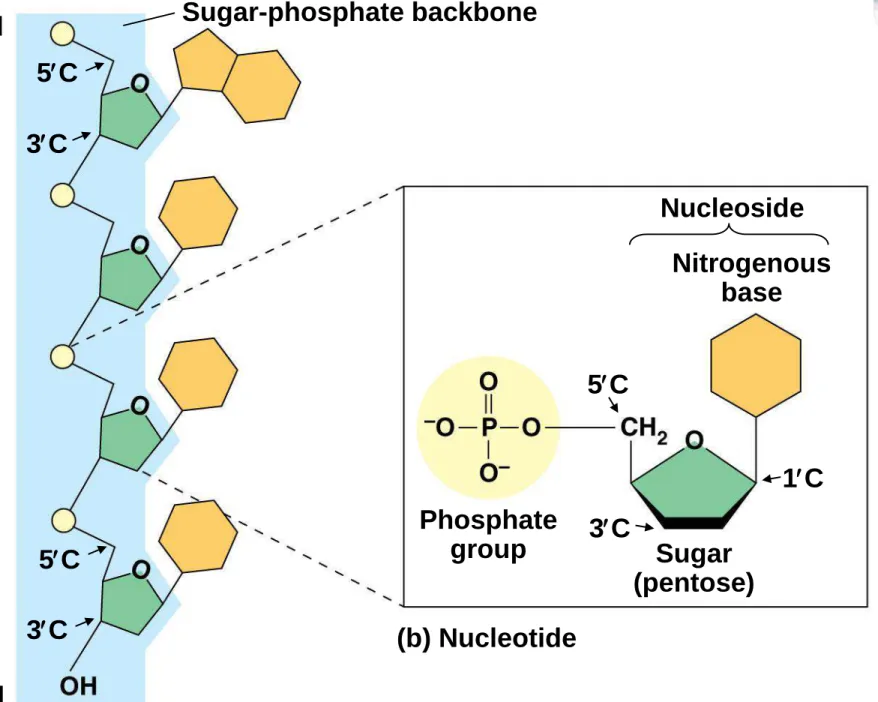
The Components of Nucleic Acids
• Each nucleic acid is made of monomers called nucleotides
• Each nucleotide consists of a nitrogenous base, a pentose sugar, and one or more phosphate groups
40
Figure 5.26ab
Sugar-phosphate backbone 5 end
5C 3C
5C 3C 3 end
(a) Polynucleotide, or nucleic acid
(b) Nucleotide Phosphate
group Sugar
(pentose) Nucleoside
Nitrogenous base 5C
3C
1C
Figure 5.26c
Nitrogenous bases
Cytosine (C)
Thymine (T, in DNA)
Uracil (U, in RNA)
Adenine (A) Guanine (G)
Sugars
Deoxyribose (in DNA)
Ribose (in RNA) (c) Nucleoside components
Pyrimidines
Purines
• There are two families of nitrogenous bases – Pyrimidines (cytosine, thymine, and uracil)
have a single six-membered ring
– Purines (adenine and guanine) have a six- membered ring fused to a five-membered ring
• In DNA, the sugar is deoxyribose; in RNA, the sugar is ribose
44
The Devil is in the Details
• Adjacent nucleotides are joined by covalent bonds that form between the —OH group on the 3 carbon of one nucleotide and the phosphate on the 5 carbon on the next
• These links create a backbone of sugar-phosphate units with
nitrogenous bases as appendages
• The sequence of bases along a DNA or mRNA polymer is unique for each gene
45
• RNA molecules usually exist as single polypeptide chains
• DNA molecules have two polynucleotides
spiraling around an imaginary axis, forming a double helix
• In the DNA double helix, the two backbones run in opposite 5→ 3 directions from each other, an arrangement referred to as antiparallel
• One DNA molecule includes many genes
46
• The nitrogenous bases in DNA pair up and form hydrogen bonds: adenine (A) always with
thymine (T), and guanine (G) always with cytosine (C)
• Called complementary base pairing
• Complementary pairing can also occur between two RNA molecules or between parts of the
same molecule
• In RNA, thymine is replaced by uracil (U) so A and U pair
47
Sugar-phosphate backbones
Hydrogen bonds
Base pair joined
by hydrogen bonding
Base pair joined by hydrogen
bonding
(b) Transfer RNA (a) DNA
5 3
3 5
Link to Evolution
• The linear sequences of nucleotides in DNA molecules are passed from parents to offspring
• Two closely related species are more similar in DNA than are more distantly related species
• Molecular biology can be used to assess evolutionary kinship
49
MACROMOLECULE:
LIPID
Wahidah Mahanani R., S.T.P., M.Sc.
TEKNOLOGI PANGAN UNIVERSITAS AHMAD DAHLAN
• Diagram of the
composition of an E. coli cell
Lipid Lipid terdapat pada tumbuhan, hewan, manusia, dan mikroorganisme.
Lipid merupakan zat lemak yang berperan dalam berbagai sel hidup.
Tersusun atas unsur karbon (CH), hidrogen (H), dan
oksigen (O), serta kadang kala ditambah fosfor (P) serta nitrogen (N).
Disimpan sebagai sumber energi sekunder dan sebagian lain bertindak sebagai komponen penting dari membran sel.
Lipid terdiri dari beberapa jenis, yang terpenting adalah lemak, fosfolipid, dan steroid.
Lemak
• Penyusun lemak sintesis dehidrasi antara molekul gliserol dan asam lemak.
• Gliserol adalah rangka karbon yang memiliki tiga
gugus alkohol. Rumus empirisnya adalah C3H4(OH)3.
• Asam lemak merupakan rantai karbon yang panjang yang memiliki gugus karboksil.
• Rantai karbon yang memiliki banyak ikatan hidrogen asam lemak jenuh.
• Tidak jenuh jika atom-atom karbonnya memiliki ikatan rangkap lebih dari satu.
• To make a fat molecule, the hydroxyl groups on the glycerol backbone react with the carboxyl groups of fatty acids in a dehydration synthesis reaction.
• This yields a fat molecule with three fatty acid tails bound to the glycerol backbone via ester linkages (linkages containing an oxygen atom next to a
carbonyl, or C=O, group).
• Triglycerides may contain three identical fatty acid tails, or three different fatty acid tails (with different lengths or patterns of double bonds).
• Lemak identik dengan minyak hewani dan minyak nabati yang terutama terdiri dari gliserida.
• Lemak merupakan ester yang terbentuk melalui
reaksi tiga molekul asam lemak dan sebuah molekul gliserol.
• Lemak bersifat tidak mudah menguap, tidak larut dalam air, terasa berminyak atau licin ketika
disentuh, dan berbentuk padat pada suhu kamar.
Beberapa jenis lemak
Fungsi
• Lebih dari 90 persen lemak diperoleh dari sekitar 20 jenis tumbuhan dan hewan. Lemak berfungsi sebagai cadangan makanan atau sumber energi di dalam
tubuh.
Steroid
• Steroid merupakan senyawa turunan lipid yang tidak terhidrolisis.
• Steroid berfungsi sebagai hormon, seperti hormon seks, hormon adrenal kortikal, asam empedu, sterol.
• Contoh-contoh steroid antara lain adalah kolesterol, esterogen, dan testosteron.
Fosfolipid
• Fosfolipid merupakan jenis lemak majemuk.
• Fosfolipid merupakan lipid yang berjumlah banyak (sebagai lesitin atau fosfatidietanolamin) yang di
dalamnya asam fosfat serta asam lemak diesterifikasi menjadi gliserol dan terdapat dalam semua sel hidup serta dalam plasma membran
• Fats have received a lot of bad publicity, and it’s true that eating large amounts of fried foods and other “fatty”
foods can lead to weight gain and cause health problems.
However, fats are essential to the body and have a number of important functions.
• For instance, many vitamins are fat-soluble, meaning that they must be associated with fat molecules in order to be effectively absorbed by the body.
• Fats also provide an efficient way to store energy over long time periods, since they contain over twice as much energy per gram as carbohydrates, and they additionally provide insulation for the body.
• Like all the other large biological molecules, fats in the right amounts are necessary to keep your body (and the bodies of other organisms) functioning correctly.
• Beberapa fungsi fosfolipid antara lain adalah:
• Lesitin membawa lemak dalam aliran darah dari satu jaringan ke jaringan lainya;
• Fosfatidiletanolamin berperan dalam proses pembekuan darah;
• Fosfolipid merupakan komponen utama dinding sel
phospholipid bilayer
Cell wall
SINTESIS ASAM NUKLEAT
Wahidah Mahanani R., S.T.P., M.Sc.
TEKNOLOGI PANGAN UNIVERSITAS AHMAD DAHLAN
PENGERTIAN
• Asam nukleat senyawa kimia polimer nukleotida yang terdapat di dalam inti sel (Nukleus),
• Asam nukleat biopolymer berbobot molekul tinggi
unit monomernya mononukleotida.
• Asam nukleat terdapat pada semua sel hidup dan bertugas untuk menyimpan dan mentransfer
genetic, kemudian menerjemahkan informasi ini secara tepat untuk mensintesis protein yang khas bagi masing- masing sel.
• berperan dalam penyimpanan & pemindahan informasi genetik yang berhubungan dengan pewarisan sifat turunan.
STRUKTUR &
NOMENKLATUR
LINGKARAN FURANOSA
STRUKTUR &
NOMENKLATUR
1 2 1 2
1. Lingkar pirimidina 2. Lingkar imidazole
• NUKLEOSIDA vs NUKELOTIDA?
Nucleoside = Nucleotide =
Nitrogenous base – ribose
Nitrogenous base – ribose – phosphate
SIFAT-SIFAT ASAM NUKLEAT
• Stabilitas asam nukleat
Ketika melihat struktur tangga berpilin molekul DNA atau struktur sekunder RNA, sepintas akan terlihat bahwa struktur tersebut menjadi stabil karena
adanya ikatan hidrogen.
PADAHAL penentu stabilitas struktur asam nukleat terletak pada interaksi penempatan (stacking
interactions) antara pasangan-pasangan basa.
Permukaan basa yang bersifat hidrofobik
menyebabkan molekul-molekul air dikeluarkan dari sela-sela perpasangan basa sehingga perpasangan tersebut menjadi kuat.
SIFAT-SIFAT ASAM NUKLEAT
• Pengaruh asam
• Di dalam asam pekat dan suhu tinggi, misalnya
HClO4 dengan suhu lebih dari 100ºC, asam nukleat akan mengalami hidrolisis sempurna menjadi
komponen-komponennya. Namun, di dalam asam mineral yang lebih encer, hanya ikatan glikosidik
antara gula dan basa purin saja yang putus sehingga asam nukleat dikatakan bersifat apurinik.
SIFAT-SIFAT ASAM NUKLEAT
• Pengaruh alkali
Peningkatan pH akan menyebabkan perubahan struktur guanin dari bentuk keto menjadi bentuk enolat karena molekul tersebut kehilangan sebuah
proton menyebabkan terputusnya sejumlah ikatan hidrogen sehingga pada akhirnya rantai ganda DNA mengalami denaturasi.
Hal yang sama terjadi pula pada RNA. Bahkan pada pH netral sekalipun, RNA jauh lebih rentan terhadap hidrolisis bila dibadingkan dengan DNA karena
adanya gugus OH pada atom C nomor 2 di dalam gula ribosanya.
SIFAT-SIFAT ASAM NUKLEAT
• Denaturasi kimia
Sejumlah bahan kimia diketahui dapat menyebabkan denaturasi asam nukleat pada pH netral.
Contoh yang paling dikenal adalah urea (CO(NH2)2) dan formamid (COHNH2). Pada konsentrasi yang relatif tinggi, senyawa-senyawa tersebut dapat
merusak ikatan hidrogen. Artinya, stabilitas struktur sekunder asam nukleat menjadi berkurang dan rantai ganda mengalami denaturasi.
SINTESIS ASAM NUKLEAT
• SINTESIS PURIN via jalur de Novo dan SALVAGE
• SINTESIS PRIMIDIN via jalur de Novo dan SALVAGE
• TAKE A LOOK AT THIS FIRST
JALUR DE NOVO
• Sintesis nukleotida dimulai dengan prekursor metaboliknya:
- asam amino
- ribosa-5-fosfat, - CO2,
- unit satu karbon.
JALUR DE NOVO
Nukleus fosfat penyusun purin dan pirimidin berasal dari 5-Phospho- D-ribosyl- 1-pyrophosphate (PRPP).
PRPP berasal dari Ribosa 5 fosfat + ATP.
Ribosa 5 fosfat berasal dari HMP shunt.
PRPP fosfo ribosil 1 amin
Enzim amidofosforibosil transferase dengan bantuan glutamin sebagai pendonor NH3 melewati 10 rangkaian reaksi akan membentuk IMP. IMP adenilosuksinat dan xantilat.
Adenilosuksinat AMP xantilat GMP.
JALUR SALVAGE
• Jalur Salvage: sintesis nukleotida dengan daur ulang dari basa bebas atau nukleosida yg dilepaskan dari pemecahan asam nukleat.
• Disini PRPP akan diubah menjadi purin-ribonukleotida.
• Contohnya
Adenin + PRPP adenilat + Ppi.
PREKURSOR
FIRST COMMITED STEP
ADDITION OF GLYCINE
FORMYLATION
AMIDOTRANSFERASE
IMIDAZOLE RING CLOSURE
CARBOXYLATION
CARBON MOVING
ASPARTATE ADDITION
FUMARATE SUBSTRACTION
2
NDFORMYLATION
Finally… INOSINATE
Finally… INOSINATE
Regulatory control of purine
biosynthesis
PURINE SALVAGE
PURINE CATABOLISM
& SALVAGE
PYRIMIDINE BIOSYNTHESIS
Hal-hal penting dalam sintesis de novo pirimidine:
• cincin pirimidine disintesis terpisah dr gula ribosa nya
• Daur pirimidine de novo tidak bercabang produk akhir dr daur adalah UMP yang mrpkn bahan dari CMP
• Reaksi pertama pembtkan karbamoyl aspartate dr asp dan carbomoyl-P titik regulasi yg penting dlm daur tsb
• Aspartat transcarbomoylase (ATCase) diaktivasi oleh ATP dan dihambat oleh CTP sbg produk akhir
Degradasi pirimidin
REPLIKASI DNA &
SINTESIS PROTEIN
Wahidah Mahanani R., S.T.P., M.Sc.
TEKNOLOGI PANGAN UNIVERSITAS AHMAD DAHLAN
CHECK THIS OUT FIRST!
3
Replication of DNA.
The Central Dogma (F. Crick):
DNA replication DNA transcription mRNA translation Protein (genome) (transcriptome) (proteome) Expression and transfer of genetic information:
Replication: process by which DNA is copied with very high fidelity.
Transcription: process by which the DNA genetic code is read and transferred to messenger RNA (mRNA). This is an intermediate step in protein expression
Translation: The process by which the genetic code is converted to a protein, the end product of gene expression.
The DNA sequence codes for the mRNA sequence, which codes for the protein sequence
“It has not escaped our attention that the specific pairing we have postulated immediately suggests a possible copying mechanism for the genetic material.” Watson & Crick
Replication
Replisomes: assemblies of “enzyme factories”.
Component Function Helicase
Primase
Clamp protein DNA polymerase Ligase
Unwinds the DNA double helix Synthesizes primers
Threads leading strand Joins assembled nucleotides Joins Okazaki fragments in lagging strand
5
DNA is replicated by the coordinated efforts of a number of proteins and enzymes.
For replication, DNA must be unknotted, uncoiled and the double helix unwound.
Topoisomerase: Enzyme that unknots and uncoils DNA Helicase: Protein that unwinds the DNA double helix.
DNA polymerase: Enzyme that replicates DNA using each strand as a template for the newly synthesized strand.
DNA ligase: enzyme that catalyzes the formation of the phosphodiester bond between pieces of DNA.
DNA replication is semi-conservative: Each new strand of DNA contains one parental (old, template) strand and one
daughter (newly synthesized) strand
9
Unwinding of DNA by helicases expose the DNA bases
(replication fork) so that replication can tak e place.
Helicase hydrolyzes ATP in order to break the hydrogen bonds Between DNA strands
DNA replication
(Fig. 26.8, p. 1192)
animation: http://www.hhmi.org/biointeractive/dna/DNAi_replication_vo2.html
Replication
Separation of the two original strands and synthesis
of two new daughter strands using the original strands as templates.
By breaking H-bonds
12
DNA Polymerase: the new strand is replicated from the 5’ 3’
(start from the 3’-end of the template)
DNA polymerases are Mg2+ ion dependent
The deoxynucleotide 5’-triphosphate (dNTP) is the reagent for nucleotide incorporation
3’-hydroxyl group of the growing DNA strand acts as a nucleophile and attacks the -phosphorus atom of the dNTP.
13
Replication of the leading strand occu rs continuously in the
5’ 3’ direction of the new strand.
Replication of the lagging strand occurs discontinuously. Short
DNA fragments are initially synthesize d and then ligated together.
DNA ligase catalyzes the formation of the phosphodiester bond
between pieces of DNA. (Fig. 26.8, p. 1192)
Unwinds the DNA double helix.
- Replication of DNA starts with unwinding of the double helix.
- Unwinding can occur at either end or in the middle.
- Attach themselves to one DNA strand and cause separation of the double helix.
Helicases
Catalyze the synthesis of primers.
Primers:are short nucleotides (4 to 15).
- They are required to start the synthesis of both daughter strands.
- Primases are placed at about every 50 nucleotides in the lagging strand synthesis.
Primases
It catalyzes the formation of the new strands.
- It joins the nucleoside triphosphates found in the nucleus.
DNA Polymerase
- A new phosphodiester bond is formed between the 5’-phosphate of the nucleoside triphosphate and the 3’-OH group of the new DNA strand.
Ligase
In formation of lagging strand, small fragments (Okazaki) are join together by ligase enzyme.
Gene expression: activation of a gene to produce a specific protein.
Protein Synthesis
Only a small fraction (1-2%) of the DNA in a chromosome contains genes.
Base sequence of the gene carries the information to produce one protein molecule.
Change of sequence New protein
• Sintesis Protein – Transkripsi dan Translasi
• Sintesis protein merupakan dasar untuk mempelajari bagaimana informasi genetik di dalam DNA diekspresikan dalam makhluk hidup.
• Dalam istilah genetik sering dikenal dengan yang namanya sentral dogma.
Sentral dogma merupakan serangkaian alur informasi dari DNA yang diterjemahkan melalui RNA kemudian menjadi protein di dalam tubuh makhluk hidup.
• Sintesis protein memiliki sumber informasi di DNA dalam bentuk gen. Gen tersebut berupa rangkaian kode-kode basa nitrogen. Informasi dalam gen akan diterjemahkan dalam bentuk mRNA. mRNA kemudian akan
digunakan untuk merangkai asam amino yang didapatkan dari luar dan dalam tubuh.
• Sintesis protein terjadi pada organel yang dinamakan dengan ribosom.
Sintesis protein sangat memerlukan keberadaan RNA, yaitu suatu rantai tunggal basa nitrogen dengan backbone yang sama dengan DNA. Adapun pembagian jenis-jenis RNA secara lengkap adalah sebagai berikut.
• a. mRNA (messenger RNA / RNA duta)
RNA duta merupakan RNA yang dibuat oleh proses yang dinamakan dengan transkripsi pada inti sel.
Peranan mRNA adalah membawa informasi genetik yang ada pada DNA menuju ribosom. Informasi yang terdapat pada mRNA berupa kodon yang tersusun
secara triplet, misalkan UCA, UCU, atau AAG. Kodon
tersebut dibuat triplet atau tiga-tiga karena 4 pangkat 3 hasilnya 64, yang kombinasi hurufnya diatas 20.
• b. tRNA (transport RNA / RNA transfer)
• RNA transfer merupakan RNA yang berperan untuk membawa asam amino dari sitoplasma menuju
ribosom saat terjadi sintesis protein. tRNA disintesis di salah satu bagian inti sel secara langsung. Dalam proses pentransferan asam amino, tRNA
memerlukan energi yang berasal dari pemecahan molekul ATP menjadi ADP + Pi.
• Tahap-Tahap Sintesis Protein
• Sintesis protein dibagi menjadi dua tahapan utama, yaitu transkripsi dan translasi. Transkripsi secara garis besar merupakan proses pembuatan mRNA dari DNA dalam inti sel. mRNA tersebut lalu bergerak menuju ribosom. Setelah itu, proses translasi, yang meliputi penerjemahan dan perangkaian asam amino,
berlangsung di ribosom.
Transcription: synthesis of mRNA (messenger RNA)
protein Tr anscription Tr anslation
DNA replication
DNA mRNA
Revers e trans criptase RNA replication
Translation Gene expression
Reverse transcription
1. Transkripsi – Pemindahan informasi dari DNA ke mRNA
Proses ini sebenarnya merupakan awal mula informasi pada DNA dipindahkan menuju protein pada makhluk hidup.
Transkripsi diawali dari pemutusan ikatan H pada DNA oleh protein-protein pengurai DNA. Proses tersebut mengakibatkan terbukanya rantai DNA pada berbagai tempat.
Terbukanya rantai DNA memicu RNA polimerase melekat ke daerah yang dinamakan dengan promotor. RNA polimerase selanjutnya melakukan sintesis molekul mRNA dari arah 3′ DNA, sedangkan pada mRNA dimulai dari ujung 5′ menuju 3′.
Dari kedua rantai DNA, hanya salah satu rantai yang akan diterjemahkan menjadi mRNA.
Rantai DNA yang diterjemahkan menjadi protein rantai sense atau DNA template atau DNA cetakan, Rantai pasangannya DNA antisense.
Dari DNA template inilah mRNA akan membentuk
rantai berpasangan dengan basa-basa yang ada pada DNA sense.
Komponen untuk pembuatan mRNA t nukleotida trifosfat, seperti ATP, GTP, UTP, dan CTP.
Fungsi RNA polimerase katalis reaksi penempelan nukleotida triposfat sehingga terbentuk rantai. Energi yang digunakan untuk menjalankan reaksi tersebut berasal dari masing-masing
nukleotida trifosfat yang kaya akan energi.
Ketika sintesis mRNA berakhir mRNA yang terbentuk
selanjutnya akan dipindahkan dari inti menuju ribosom,
kemudian diterjemahkan menjadi protein di ribosom.
Transcription
Genetic information is copied from a gene in DNA to make a mRNA.
Begins when the section of a DNA that contains the gene to be copied unwinds.
Polymerase enzyme identifies a starting point to begin mRNA synthesis.
Transcription
The DNA splits into two strands:
Template strand: it is used to synthesize RNA.
Coding Strand (Informational strand): it is not used to synthesize RNA.
- Transcription proceeds from the 3’ end to the 5’ end of the template.
- When mRNA is released, the double helix of the DNA re-forms.
(informational strand, non-template strand)
Direction of transcription
Transcription
- G – A – A – C – T -
- C – U – U – G – A -
Section of bases on DNA (template strand):
Complementary base sequence in mRNA:
RNA Polymerase
Polymerase enzyme moves along the unwound DNA, forming bonds between the bases.
C is paired with G, T pairs with A But A pairs with U (not T).
Sample Problem 22.6
From the template strand of DNA below, write out the mRNA and informational strand of DNA sequences:
Template strand: 3’—C T A G G A T A C—5’
mRNA: 5’—G A U C C U A U G—3’
Informational 5’—G A T C C T A T G—3’
strand:
Transcription
Translation
mRNA (as a carrier molecule) moves out of the nucleus and goes to ribosomes.
tRNA converts the information into amino acids.
Amino acids are placed in the proper sequence.
Proteins are synthesized.
Gene expression
Overall function of RAN’s in the cell: facilitate the task of synthesizing protein.
Genetic code: language that relates the series of nucletides in mRNA to the amino acids specified.
Genetic code
• The sequence of nucleotides in the mRNA determines the amino acid order for the protein.
• Every three bases (triplet) along the mRNA makes up a codon.
• Each codon specifies a particular amino acid.
• Codons are present for all 20 amino acids.
Genetic code
A
U
U C
C
A
Leu Leu Leu Leu
Val Val Val Val
Ser Ser Ser Ser Pro Pro Pro Pro Thr Thr Thr Thr Ala Ala Ala Ala
Arg Arg Arg Arg
Gly Gly Gly Gly UUU
UUC
Phe Phe Leu Leu
Ile Ile Ile AU U AU C AU A
U AU U AC
Tyr Tyr
CAU CAC
His His Gln Gln AAU
AAC
As n As n Lys Lys A sp A sp Glu Glu
Trp Cys Cys
Ser Ser Arg Arg UCU
UCC UCA UCG
G
U C A G CUU
CUC CUA CUG
CCU CCC CCA CCG
CGU CGC CGA CGG
U C A G
G
ACU ACC ACA ACG
U C A G U C A G GU U
GU C GU A GU G
GCU GCC GCA GCG
GGU GGC GGA GGG
S top
5' 3'
UUA UUG
Met*
AU G
U AA U AG
Stop Stop
CAA CAG
AAA AAG GAU GAC GAA GAG
U GA U GG U GU U GC
A GU A GC A GA A GG
*AUG s ign als tran slation initiation as w ell as codin g for Met
Genetic code
• 64 condons are possible from the triplet combination of A, G, C, and U.
• UGA, UAA, and UAG, are stop signals.
(code for termination of protein synthesis).
• AUG has two roles:
1. Signals the start of the proteins synthesis (at the beginning of an mRNA).
2. Specifies the amino acid methionine (Met) (in the middle of an mRNA).
•Codons are written from the 5’ end to the 3’ end of the mRNA molecule
tRNA (transfer RNA)
A G U
U C A
Codon on mRNA
Anticodon loop
tRNA translates the codons into specific amino acids. Serine - It contains 70-90 nucleotides.
- The 3’ end, called the acceptor stem and always has the nucleotide ACC and a free OH group that binds a specific amino acid.
- Anticodon: a sequence of three
nucleotides at the bottom of tRNA, which
is complementary to three bases in an mRNA and it can identify the needed amino acid.
Transcription
Translation
• mRNA attaches to smaller subunit of a ribosome.
• tRNA molecules bring specific amino acids to the mRNA.
• Peptide bonds form between an amino acid and the end of the growing peptide chain.
• The ribosome moves along mRNA until the end of the codon (translocation).
• The polypeptide chain is released from the ribosome and becomes an active protein.
Protein synthesis
Sometimes several ribosomes (polysome) translate the same strand of mRNA at the same time to produce several peptide chains.
Termination
Ribosome encounters a stop condon.
No tRNA to complement the termination codon.
An enzyme releases the complete polypeptide chain from the ribosome.
A
U
U C
C
A
Leu Leu Leu Leu
Val Val Val Val
Ser Ser Ser Ser Pro Pro Pro Pro Thr Thr Thr Thr Ala Ala Ala Ala
Arg Arg Arg Arg
Gly Gly Gly Gly UUU
UUC
Phe Phe Leu Leu
Ile Ile Ile
AU U AU C AU A
U AU U AC
Tyr Tyr
CAU CAC
His His Gln Gln AAU
AAC
As n As n Lys Lys A sp A sp Glu Glu
Trp Cys Cys
Ser Ser Arg Arg UCU
UCC UCA UCG
G
U C A G CUU
CUC CUA CUG
CCU CCC CCA CCG
CGU CGC CGA CGG
U C A G
G
ACU ACC ACA ACG
U C A G U C A G GU U
GU C GU A GU G
GCU GCC GCA GCG
GGU GGC GGA GGG
S top
5' 3'
UUA UUG
Met*
AU G
U AA U AG
Stop Stop
CAA CAG
AAA AAG GAU GAC GAA GAG
U GA U GG U GU U GC
A GU A GC A GA A GG
*AUG s ign als tran slation initiation as w ell as codin g for Met A
U
U C
C
A
Leu Leu Leu Leu
Val Val Val Val
Ser Ser Ser Ser Pro Pro Pro Pro Thr Thr Thr Thr Ala Ala Ala Ala
Arg Arg Arg Arg
Gly Gly Gly Gly UUU
UUC
Phe Phe Leu Leu
Ile Ile Ile AU U AU C AU A
U AU U AC
Tyr Tyr
CAU CAC
His His Gln Gln AAU
AAC
As n As n Lys Lys A sp A sp Glu Glu
Trp Cys Cys
Ser Ser Arg Arg UCU
UCC UCA UCG
G
U C A G CUU
CUC CUA CUG
CCU CCC CCA CCG
CGU CGC CGA CGG
U C A G
G
ACU ACC ACA ACG
U C A G U C A G GU U
GU C GU A GU G
GCU GCC GCA GCG
GGU GGC GGA GGG
S top
5' 3'
UUA UUG
Met*
AU G
U AA U AG
Stop Stop
CAA CAG
AAA AAG GAU GAC GAA GAG
U GA U GG U GU U GC
A GU A GC A GA A GG
*AUG s ign als tran slation initiation as w ell as codin g for Met Amino acids form the three-dimensional structure (active protein).
Translation
There are 3 stages in translation:
1. Initiation begins with mRNA binding to the ribosome.
2. Elongation proceeds as the next tRNA molecule delivers the next amino acid, and a peptide bond forms between the two amino acids.
Translation
3. Termination: Translation continues until a stop codon (UAA, UAG, or U